Implications
Treating bovine mammary epithelial cell line (BME-UV1) with (-)-epigallocatechin-3-gallate (EGCG) or hydroxytyrosol (HTyr) exerted anti-oxidative and anti-inflammatory effects in bovine mammary epithelial cells. The results of this study emphasized that these polyphenols provide dual protection in bovine cells. In particular, HTyr was able to reduce oxidative stress by inhibiting reactive oxygen species production, whereas EGCG was able to attenuate inflammatory responses by decreasing the expression of pro-inflammatory genes, suggesting that these compounds might be a potential natural alternative to be used in dairy cattle as a supplement for reducing the development of oxidative and inflammatory processes related to parturition or as topical treatments for the control of bovine mammary gland inflammation.
Introduction
Polyphenols are a group of secondary metabolites widely found in different plant organs such as fruits and leaves and their related processing products. Many studies have demonstrated that daily consumption of polyphenols-rich beverages and food such as green tea and olive oil is associated with a low incidence of several diseases including cancer, diabetes, obesity, leukemia, Parkinson’s and cardiovascular diseases (Visioli et al., Reference Visioli, Poli and Galli2002; Mota et al., Reference Mota, Landim, Targino, da Silva, da Silva and Pereira2015). The beneficial health properties of green tea and olive oil consumption are related mainly to EGCG and HTyr, respectively, two phenolic compounds characterized by a wide range of biological activities (Visioli et al., Reference Visioli, Poli and Galli2002; Mota et al., Reference Mota, Landim, Targino, da Silva, da Silva and Pereira2015). (-)-Epigallocatechin-3-gallate is the most abundant catechin phenolic compound found in Camellia sinensis L. leaves; and it is chemically characterized by the presence of two pyrogallol moieties (Figure S1). (-)-Epigallocatechin-3-gallate is responsible for much of the health-promoting properties of green tea including the anti-oxidative, anti-inflammatory, anti-obesity, anti-diabetic and cardio-protective effects (Chen et al., Reference Chen, Xu, Li, Du, Chen, Liu, Peng, Wang and Song2015; Mota et al., Reference Mota, Landim, Targino, da Silva, da Silva and Pereira2015). Recently, green tea extracts rich in EGCG have gained great attention as supplements and pharmaceutics to prevent serious diseases including cancer and neurodegenerative diseases (Tomas-Barberan and Andres-Lacueva, Reference Tomas-Barberan and Andres-Lacueva2012). Hydroxytyrosol is a low-molecular weight phenol characterized by a catechol moiety (Figure S1). It is found in olive oil products, for example, extra-virgin olive oil and table olives, which are important components of the Mediterranean diet (Bernini et al., Reference Bernini, Gilardini Montani, Merendino, Romani and Velotti2015). It originates mainly from the enzymatic hydrolysis of oleuropein during the maturation of the olives, processing and storage of olive oil (Gambacorta et al., Reference Gambacorta, Tofani, Bernini and Migliorini2007). During olive oil processing, a considerable amount of HTyr converges in olive oil by-products such as olive oil wastewaters, which represents a valuable source of HTyr (Bernini et al., Reference Bernini, Gilardini Montani, Merendino, Romani and Velotti2015). Studies have demonstrated that HTyr exhibits antimicrobial, antitumoral, antioxidant and anti-inflammatory activities together to beneficial effects on the cardiovascular system (Bernini et al., Reference Bernini, Merendino, Romani and Velotti2013; Hu et al., Reference Hu, He, Jiang and Xu2014).
Oxidative stress plays a key role in the onset or progression of numerous human and animal diseases. High-yielding dairy cows undergo this deleterious process mainly during the peripartum period (Bernabucci et al., Reference Bernabucci, Ronchi, Lacetera and Nardone2005; Castillo et al., Reference Castillo, Hernández, Valverde, Pereira, Sotillo, Alonso and Benedito2006). For dairy cows, the transition period is characterized by important physiological changes that affect the health and production performance of the animals. Among tissues and organs, mammary gland, liver, muscles and adipose tissue are the most involved and more susceptible to oxidative stress (Abuelo et al., Reference Abuelo, Hernández, Benedito and Castillo2015). Substantial evidence confirms that increased oxidative stress and inflammatory response during the pre-partum and early lactation period may contribute to several metabolic and infectious diseases (retained placenta, ruminal acidosis, laminitis, ketosis, fatty liver, metritis, etc.) in dairy cattle (Contreras and Sordillo, Reference Contreras and Sordillo2011; Abuelo et al., Reference Abuelo, Hernández, Benedito and Castillo2015), in particular, oxidative stress contribute to mastitis pathogenesis during the transition period of dairy cows (Aitken et al., Reference Aitken, Karcher, Rezamand, Gandy, VandeHaar, Capuco and Sordillo2009). Recently, scientific attention has been oriented toward the use of naturally occurring products. Feed supplementation of antioxidants and anti-inflammatory compounds could potentially improve the health status and performance of animals. However, the therapies used have not achieved this consistently.
Despite the great number of studies describing the antioxidant and anti-inflammatory activities of both EGCG and HTyr in humans (Bernini et al., 2013) and monogastric animals (Mota et al., Reference Mota, Landim, Targino, da Silva, da Silva and Pereira2015; Hu et al., Reference Hu, He, Jiang and Xu2014), there are no studies, to the best of our knowledge, in the published literature that investigate EGCG and HTyr effects in ruminants. The finding of natural products to be used in dairy cattle, particularly during the peripartum period, as a supplement or as topical treatments could be useful in helping to reduce risk for diseases. On the basis of this lack of literature, the aim of this study was to evaluate both the anti-oxidative and anti-inflammatory effects of EGCG and HTyr in bovine mammary epithelial cell line and to compare their antioxidant and anti-inflammatory in vitro efficacy.
Material and Methods
Chemicals
Reagents and solvents of high purity were supplied by Sigma-Aldrich (Milan, Italy). Green tea leaves extract (Teavigo®) was manufactured by Taiyo Green Power under licence from Taiyo Kagaku Co., Ltd., and Swiss-based DSM Nutritional Products. Silica gel (200-300 mesh) and silica gel F254 plates were purchased from Merck (Milan, Italy). Hydroxytyrosol was synthesized according to a patented procedure optimized in our laboratories and purified by flash column chromatography (Bernini et al., Reference Bernini, Mincione, Barontini and Crisante2008).
Chemical Characterization of Teavigo® and Hydroxytyrosol
A pure sample of EGCG was found in a green tea extract commercially available (Teavigo®) while fresh HTyr was synthetized in our laboratories by an ecofriendly and efficient procedure based on the selective oxidation of tyrosol with 2-iodobenzoic acid, followed by a reduction with sodium dithionite (Figure S2; Bernini et al., Reference Bernini, Mincione, Barontini and Crisante2008). Finally, HTyr was purified by flash column chromatography on silica gel. Commercial tea extract Teavigo® and synthetic HTyr were characterized by nuclear magnetic resonance spectra (1H-NMR and 13C-NMR) using a spectrometer Avance III 400 MHz Bruker (Germany) dissolving 10 mg of each sample in 0.5 ml of dimethylsulfoxide-d6 and acetone-d6, respectively. Chemical shifts were expressed in parts per million (δ scale) and coupling constants in Hertz. Nuclear magnetic resonance spectra (Figure S3) evidenced the high purity both of EGCG found in Teavigo® (Imperatori et al., Reference Imperatori, Barlozzari, Scardigli, Romani, Macrì, Polinori, Bernini and Santi2018) and synthetic HTyr (Bernini et al., Reference Bernini, Mincione, Barontini and Crisante2008).
Cell culture
For this present study, a clonal bovine mammary epithelial cell (BME-UV1, RRID:CVCL_W716), obtained from a pregnant, lactating cow’s mammary gland and established by stable transfection with a plasmid encoding the thermolabile large T-antigen (Zavizion et al., Reference Zavizion, van Duffelen, Schaeffer and Politis1996), has been chosen as a valid model to study the bovine mammary epithelial metabolism. The BME-UV1 cells respond to epidermal growth factor and the insulin-like growth factor I, which are associated to growth and development of the mammary gland and show a morphology typical of luminal epithelial cells, in a single layer and of polygonal shape (Arévalo Turrubiarte et al., Reference Arévalo Turrubiarte, Perruchot, Finot, Mayeur and Dessauge2016). On the basis of their similarity with bovine lactating mammary epithelial cells, the BME-UV1 have been widely used as in vitro models to investigate milk production in dairy cows (Arévalo Turrubiarte et al., Reference Arévalo Turrubiarte, Perruchot, Finot, Mayeur and Dessauge2016).
Bovine mammary epithelial cell lines used in this study were kindly provided by Professor Antonella Baldi (Department of Health, Animal Science and Food Safety, University of Milan, Italy). The cells were incubated in a humidified air with 5% CO2, at 37°C, until 80% confluence. Cells were cultured in 75 cm2 tissue culture flasks (Costar, Corning, NY, USA), in a culture medium of DMEM-F12, RPMI-1640 and NCTC-135 (5:3:2 by volume), complemented with 10% fetal bovine serum, 0.1% lactose, 0.1% lactalbumin hydrolysate, 1.2 mM glutathione, 1 μg/ml insulin, 5 μg/ml transferrin, 1 μg/ml hydrocortisone, 0.5 μg/ml progesterone, 10 μg/ml L-ascorbic acid and antibiotics (penicillin 100 IU/ml; streptomycin 100 μg/ml). Culture medium and supplements were purchased from Sigma-Aldrich (Milano, Italy). For this study, BME-UV1 cells at passage number between 39 and 41 were used (Dipasquale et al., Reference Dipasquale, Basiricò, Morera, Primi, Tröscher and Bernabucci2018). The dissociation of the cell monolayer and subcultures was carried out every 2 or 3 days.
Experimental design
In order to test the antioxidant activities of EGCG or HTyr on bovine mammary gland, BME-UV1 cells were re-suspended in complete culture medium to a concentration of 5x10⁵ cells/ml and dispensed in culture flasks and 96-wells tissue culture plates. After 24 h, the medium was removed and replaced with routine culture medium and 50 μM EGCG or HTyr for 48 h. As a control to each, experiment cells not exposed to EGCG or HTyr were used.
(-)-Epigallocatechin-3-gallate and HTyr were first dissolved into aqueous buffer (culture medium) and from the stock solution followed other dilutions in complete culture medium prior to perform biological experiments. The concentration of EGCG and HTyr used in the experiments was established after screening tests and on the base of observations on BME-UV1, which showed little cytotoxicity toward EGCG or HTyr using concentrations higher than 50 μM after 24-h treatment (data not shown) and of observations on other cells (Peng et al., Reference Peng, Moritz, Han, Giddings, Lyon, Kollman, Winey, Yates, Agard, Drubin and Barnes2015; Karamese et al., Reference Karamese, Guvendi, Karamese, Cinar, Can, Erol, Aydin, Gelen and Karakus2016). Among the tested concentrations, 50 μM for both EGCG and HTyr has been chosen being the upper limit dose that did not cause cytotoxicity (data not shown). Cell viability after 48 h from addition of EGCG or HTyr was determined. Also, the content of reduced glutathione (GSH) and oxidized form of glutathione (GSSH) γ-glutamylcysteine ligase activity (γGCL) and reactive oxygen species (ROS) and malondialdehyde (MDA) concentration were determined. Reduced glutathione and oxidized form of glutathione ratio was calculated.
To test the potential protection of EGCG or HTyr against hydrogen peroxide (H2O2)-induced oxidative stress, cells were treated with EGCG or HTyr at non-toxic concentrations (50 μM) for 48 h as described above, followed by incubation at 37°C for 3 h with a single dose application (50 μM) of H2O2 and the content of ROS concentration were determined.
To test the preventive efficacy of EGCG or HTyr against induced inflammation and indirect oxidative stress by lipopolysaccharides (LPS; from Escherichia coli 055:B5, Sigma-Aldrich), cells were pretreated with compounds of interest as described above followed by incubation at 37°C for 3 h with a single dose application (20 μM) of LPS, to match acute inflammation. Gene expression of pro- and anti-inflammatory cytokines was determined. Reactive oxygen species were also determined in cells. The concentration 50 μM of H2O2 and 20 μM of LPS were selected on a previous preliminary cytotoxicity study and on results from other published studies (Basiricò et al., Reference Basiricò, Morera, Dipasquale, Tröscher and Bernabucci2017; Dipasquale et al., Reference Dipasquale, Basiricò, Morera, Primi, Tröscher and Bernabucci2018; Mastrogiovanni et al., Reference Mastrogiovanni, Bernini, Basiricò, Bernabucci, Campo, Romani, Santi and Lacetera2018).
For each experiment, at least three replicates were performed and were repeated at least twice.
Cell viability
For cell viability assay, cells were seeded into 96-wells microplates at an optimal density (5x10⁵ cells/ml) and were grown with EGCG or HTyr for 48 h. Cell viability was assayed using the Cell Proliferation Kit II (XTT test: sodium 30-[1-(phenylaminocarbonyl) 4-tetrazolium]-bis (4-methoxy-6-nitro) benzene sulfonic acid hydrate; Roche Applied Science, Indianapolis, IL, USA) according to the manufacturer’s instructions. Briefly, for the XTT test, to each well, after 48 h of EGCG or HTyr exposure, was added 50 μl of a mixture of two reagents, XTT labeling reagent and electron-coupling reagent (50:1). Cells are then placed back into the incubator for 24 h at 37°C. At the end of the incubation period, the absorbance was measured on a plate reader (Tecan Sunrise™) at 450 nm. Results were expressed as optical density.
Thiol redox status
For assessing the thiol redox status of BME-UV1 cells after EGCG or HTyr treatment, the GSH, the GSSH and the activity of γ-glutamate cysteine ligase were determined (Basiricò et al., Reference Basiricò, Morera, Dipasquale, Tröscher, Serra, Mele and Bernabucci2015). Briefly, for the determination of GSH and GSSG, adherent cells were detached using trypsin/EDTA solution and centrifuged at 4 500 × g at 4°C for 5 min. The pellet was re-suspended in 200 μl of PBS and lysed by two cycles of sonication (100 W for 30 s), the samples were centrifuged (15 000 × g for 5 min at 4°C) and stored at -80°C until analysis. In cell extracts, GSH/GSSH ratio was determined by colorimetric assays (BioAssay Systems, Hayward, CA, USA). Optical density was measured by a spectrophotometer (Tecan Sunrise™) at 405 for GSH/GSSG.
The γGCL activity was determined by a fluorescence assay as described by Chen et al. (Reference Chen, Xu, Li, Du, Chen, Liu, Peng, Wang and Song2015). Briefly, BME-UV1 cells were detached and centrifuged as described above. The cells were re-suspended in TES/SB buffer (wt/vol, 1/4), and were sonicated (100 W for 60 s), centrifuged (15 000 × g for 5 min at 4°C) and supernatants were collected. The supernatants were centrifuged again at 15 000 × g at 4°C for 20 min and the protein concentrations were determined using a BCA Protein Assay Kit from Pierce (Rockford, IL, USA), with bovine serum albumin as standard. For the γGCL activity assay, aliquot of supernatants (30 μl) were mixed with 30 μl of γGCL reaction cocktail and incubated at 37°C for 5 min. Cysteine solution was then added, and the mixtures were incubated at 37°C for 13 min and stopped. After placing on ice for 20 min, the mixtures were centrifuged at 2000 × g at 4°C for 10 min. Aliquot of each supernatant containing γ-glutamylcysteine (γGC) was added to a 96-well plate designed for fluorescence detection. For each assay, 20 μl of γGC standards was added to generate a standard curve. Next, 180 μl of 2,3-naphthalenedicarboxyaldehyde (NDA) was added to each well. Following incubation, the formation of NDA-γGC was measured (472 nm excitation/528 nm emission) using a fluorescent plate reader (Multimode Detector DTX 880, Beckman Coulter Inc., Fullerton, CA). The production of γGC in each sample was calculated using the standard curve. Values were expressed in μM/min/μg of total proteins.
Measurement of reactive oxygen species production
After treatments, cells were washed twice with phosphate buffer saline (Lonza, Swiss) and incubated with 20 μM 2’,7’-dichlorodihydrofluorescin diacetate probe (D6883 Sigma-Aldrich) in PBS at 37°C for 40 min as reported by Basiricò et al. (Reference Basiricò, Morera, Dipasquale, Tröscher, Serra, Mele and Bernabucci2015). Fluorescence was measured at 485 nm (excitation) and 535 nm (emission) wavelengths on a microplate reader (Multimode Detector DTX 880, Beckman Coulter Inc., USA).
Measurement of malondialdehyde production
For the determination of MDA, lipid peroxidation indicator, after treatments adherent cells were detached using trypsin/EDTA solution and centrifuged at 4500 × g for 5 min at 4°C. The pellet was re-suspended in 200 μl of PBS and lysed by two cycles of sonication (100 W/ 30 s), centrifugation at 15 000 × g for 5 min at 4°C, and samples stored at -80°C until analysis. In cell extracts, MDA concentrations were determined by colorimetric assays (Abcam, Cambridge, UK), CA). Optical density was measured by a spectrophotometer at 540 nm (Basiricò et al., Reference Basiricò, Morera, Dipasquale, Tröscher, Serra, Mele and Bernabucci2015).
RNA isolation and real-time PCR
The gene expression of bovine pro (tumor necrosis factor (TNF)-α, interleukin (IL)-1β and IL-6) and anti-inflammatory IL-10, cytokines that may play important roles in the mammary inflammatory response, were assayed in BME-UV1 cultured under the conditions described above and were carried out by real-time-PCR. All primers and probes sequences used, were previously reported by Dipasquale et al. (Reference Dipasquale, Basiricò, Morera, Primi, Tröscher and Bernabucci2018).
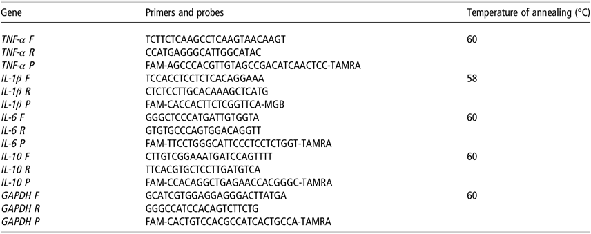
In order to isolate total RNA, BME-UV1 cells were seeded in cell culture flasks at the concentration of 5x10⁵ cells/ml in complete medium (Thermo Fisher Scientific, Waltham, USA) and treated as described above. Total RNA was isolated by QIAzol Lysis Reagent (79306 Qiagen, Hilden, Germany), according to the manufacturer’s instructions and stored at –80°C. RNA was quantified using Quant-iT RNA assay Kit (Invitrogen, Carlsbad, CA, USA) and fluorescence was measured at excitation/emission of 644/673 nm. One microgram of total RNA was reverse transcribed using a Quantitect reverse transcription kit (Qiagen, Hilden, Germany) in a total volume of 20 μl on a PCR Express thermal cycler (Hybaid, Ashford, UK). Quantitative probes real-time PCRs were performed following the manufacturer’s recommendations using LightCycler® 2.0 (Roche, Roche Applied Science, Indianapolis, IL, USA). To account for possible variation related to complementary DNA input or the presence of PCR inhibitors, the endogenous reference gene glyceraldehyde 3-phosphate dehydrogenase was simultaneously quantified for each sample, and data were normalized accordingly. In each PCR run, the cDNA samples were amplified in triplicate. Table 1 shows the specific characteristics of primers used for the real-time PCR. The relative quantification of the mRNAs was performed using the ΔΔCT method.
Deoxyribonucleic acid sequences of bovine sense and antisense primers and probes used for real-time PCR analysis

TNF-α = tumor necrosis factor-α; IL-1β = interleukin-1β; IL-6 = interleukin-6; IL-10 = interleukin-10; GAPDH = glyceraldehyde 3-phosphate dehydrogenase; F = forward; R = reverse; P = probe.
Statistical analysis
All data of the experiment are presented as least-squares means and SEM. The data were analyzed with ANOVA, using a general linear model (GLM procedure of Statistica-7 Software package, Stat Soft, Inc., USA). The model included the fixed effect of treatment (control, EGCG and HTyr), replicates as random effect and the error term. The significance of the differences was assessed by the Fisher’s Least significant difference (LSD) test and significance was declared at P < 0.05.
Results
Effect of epigallocatechin-3-gallate and hydroxytyrosol on cell viability
As shown in Figure S4, cells were still 100% viable at the concentration used (50 μM) at the end of the incubation period as determined by the XTT assay and no differences were observed between treatments. This assay indicated that the exposure to the different compounds did not show any cytotoxic effect.
Effect of epigallocatechin-3-gallate and hydroxytyrosol on thiol redox status
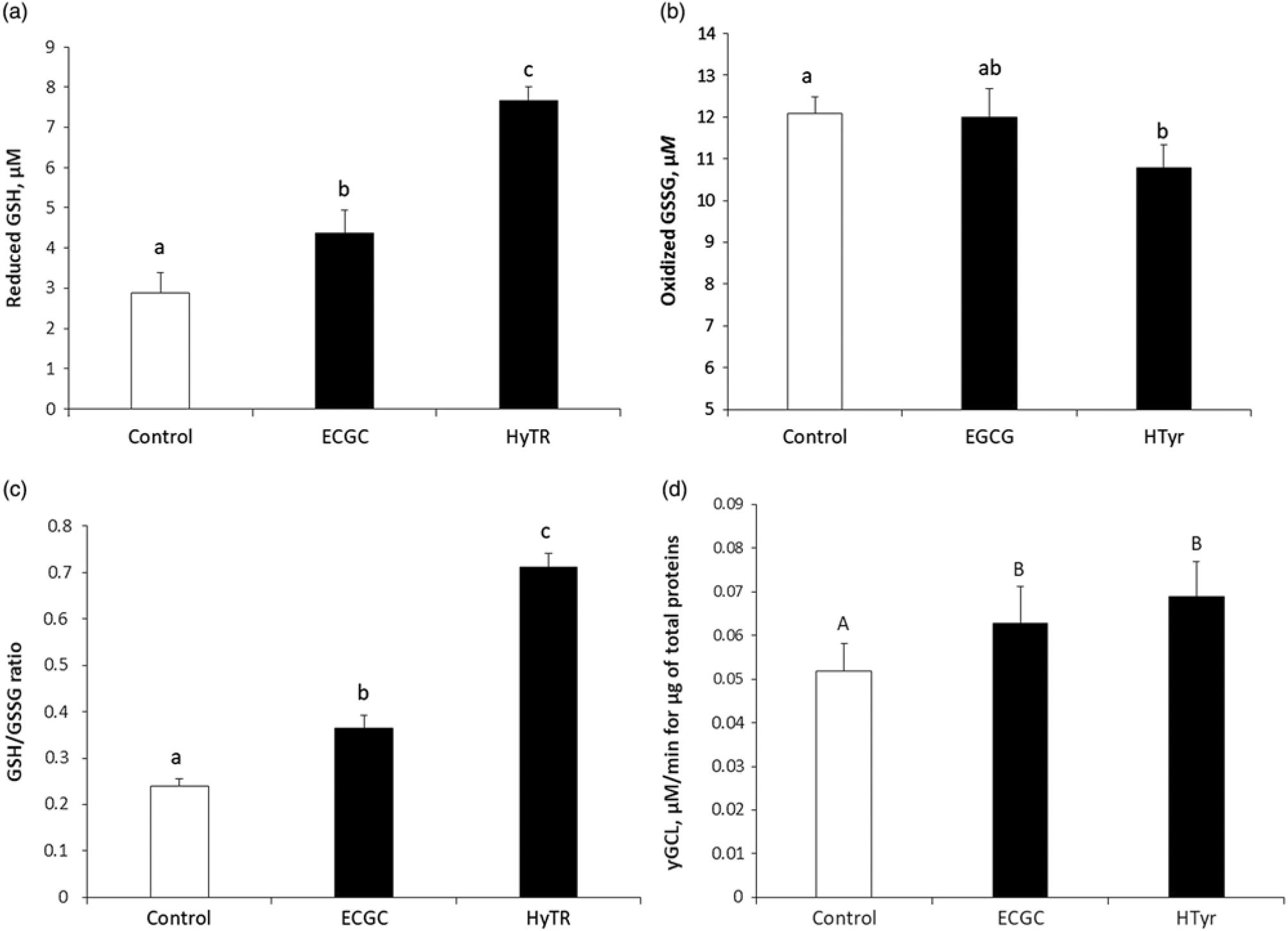
Oxidized and reduced glutathione: Figure 1 shows changes of non-enzymatic antioxidants like GSH. Compared to the control, an increase of reduced GSH was observed in cells treated with 50 μM EGCG (P < 0.05) or with HTyr (P < 0.01) (Figure 1a). Among treatments, values of reduced GSH were significantly higher (P < 0.05) in cells treated with HTyr compared to EGCG. Oxidized GSH level of cells treated with EGCG was not significantly different from that of control cultures, but it was higher than that observed in HTyr-treated cells (Figure 1b). The ratio GSH/GSSG showed the same trend of reduced GSH; in particular, HTyr showed an increase (P < 0.05) of GSH/GSSG ratio compared with EGCG (Figure 1c).
Intracellular concentration of reduced glutathione (GSH; a), oxidized glutathione (GSSG; b), and GSH-to-GSSG ratio (c) and γ-glutamate cysteine ligase (d) activity in bovine mammary epithelial cells after 48 h of exposure to 50 µM (-)-epigallocatechin-3-gallate (EGCG) or hydroxytyrosol (HTyr). The transparent bar represents the control (cells not treated) and the black bars represent cells treated with EGCG or HTyr. Data were analyzed by one-way ANOVA test and are reported as least squares means ± SEM (n = 6). Significant differences between treatments are represented by different letters (a, b = P < 0.05; A, B = P < 0.01). Fisher’s LSD test has been used to evaluate differences between treatments.

γ-Glutamyl-cysteine ligase activity: After the 48 h, EGCG or HTyr treatments induced a strong antioxidant response by increasing (P < 0.01) the intracellular γGCL activity compared with the control (Figure 1d). However, no differences were observed between the two treatments (Figure 1d).
Intracellular reactive oxygen species and malondialdehyde production
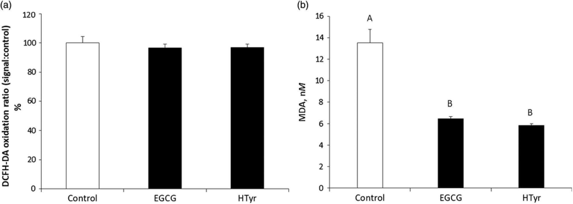
Intracellular ROS production as established by dichlorofluorescein fluorescence flow cytometry analysis was measured in cells supplemented with EGCG or with HTyr (Figure 2a). No differences of ROS production were observed between the treatments and the control.
Intracellular production of reactive oxygen species by 2’,7’-dichlorodihydrofluorescin diacetate probe (DCFH-DA) (a) and malondialdehyde (MDA; b) in bovine mammary epithelial cells after 48 h of 50 µM (-)-epigallocatechin-3-gallate (EGCG) or hydroxytyrosol (HTyr) exposure. The transparent bar represents the control (cells not treated) and the black bars represent cells treated with EGCG or HTyr. Data were analyzed by one-way ANOVA test and are reported as least squares means ± SEM (n = 6). Significant differences between treatments are represented by different letters (A, B = P < 0.01). Fisher’s LSD test has been used to evaluate differences between treatments.

Cell concentrations of MDA, lipid peroxidation indicator, are shown in Figure 2b. The treatment of cells with EGCG or with HTyr reduced (P < 0.01) the MDA level compared with untreated cells. No differences of MDA concentrations were observed between the two treatments.
Intracellular reactive oxygen species production after hydrogen peroxide and lipopolysaccharide challenge
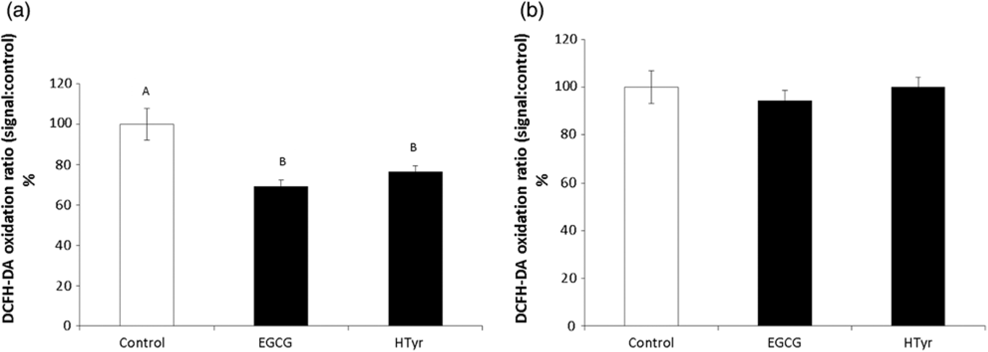
The potential of EGCG and HTyr to protect against H2O2-induced oxidative stress and indirect oxidative stress promoted by an inflammatory response elicited by LPS stimulation, was assessed by ROS test after exposure for 3 h to H2O2 or LPS. As shown in Figure 3a, cells supplemented with EGCG or HTyr and stimulated with H2O2 showed a decreased ROS production (P < 0.01) compared to the control. After pre-treatment with EGCG or HTyr and stimulation with LPS, BME-UV1 showed a ROS production similar to the control (Figure 3b). (-)-Epigallocatechin-3-gallate and HTyr were able to enhance cell resistance against induced H2O2 oxidative stress at 48 h.
Intracellular production of reactive oxygen species by 2’,7’-dichlorodihydrofluorescin diacetate probe (DCFH-DA) in bovine mammary epithelial cells after 48 h of 50 µM (-)-epigallocatechin-3-gallate (EGCG) or hydroxytyrosol (HTyr) supplementation and 3 h of 50 µM H2O2 (a) and lipopolysaccharide (20 μM) treatment (b). The transparent bar represents the control (cells not treated) and the black bars represent cells treated with EGCG or HTyr. Data were analyzed by one-way ANOVA test and are reported as least squares means ± SEM (n = 6). Significant differences between treatments are represented by different letters (A, B = P < 0.01). Fisher’s LSD test has been used to evaluate differences between treatments.

Quantification of messenger RNA expression of pro- and anti-inflammatory cytokines
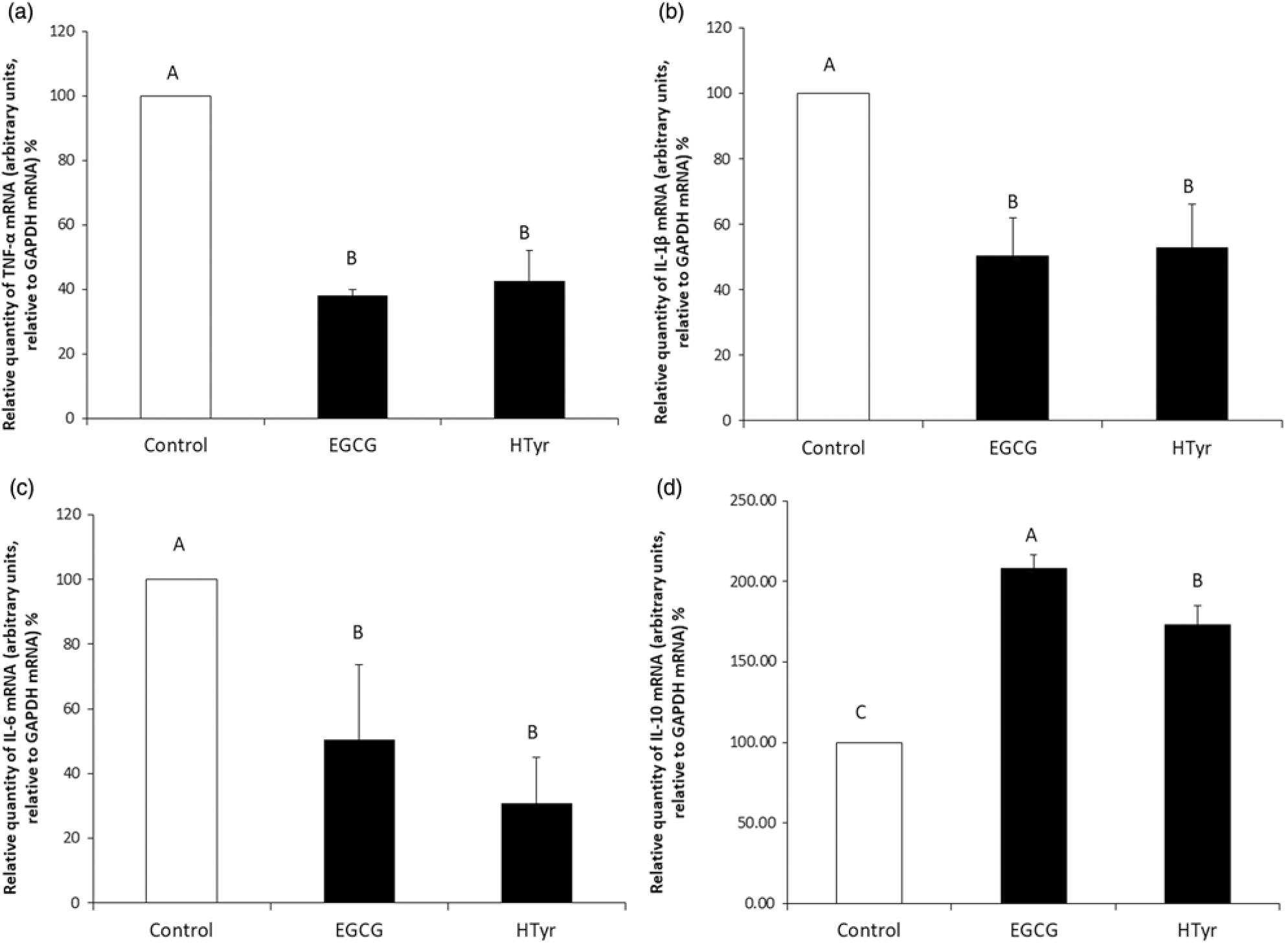
In Figure 4 mRNA expression of TNF-α, IL-1β, IL-6 and IL-10 (A, B, C and 336 D, respectively) are shown. Pro-inflammatory cytokines mRNA was lower (P < 0.01) in cells treated with EGCG or HTyr compared with the control. No differences were observed between treatments (Figure 4a, b and c). On the other hand, gene expression of the anti-inflammatory cytokine (Figure 4d) was markedly higher in cells treated with EGCG or HTyr compared to the control. Overall, IL-10 gene expression showed greater levels (P < 0.01) in cells treated with EGCG compared with HTyr.
Messenger RNA abundance of tumor necrosis factor-α (TNF-α; a), interleukin-1 (IL-1β; (b), IL-6 (c) and IL-10 (d) in bovine mammary epithelial cells after 48 h of 50 µM (-)-epigallocatechin-3-gallate (EGCG) or hydroxytyrosol (HTyr) supplementation, and after 3 h of lipopolysaccharide treatment (20 μM). The transparent bar represents the control (cells not treated) and the black bars represent cells treated with EGCG or HTyr. Data were analyzed by one-way ANOVA test and are reported as least squares means ± SEM (n = 6). Significant differences between treatments are represented by different letters (A, C = P < 0.01). Fisher’s LSD test has been used to evaluate differences between treatments. GAPDH = glyceraldehyde 3-phosphate dehydrogenase.

Discussion
Solid scientific evidences clearly demonstrate the healthy bioactivity properties of polyphenols (Tomas-Barberan and Andres-Lacueva, Reference Tomas-Barberan and Andres-Lacueva2012). Thus, attention in phenolic compounds has increased greatly and the main interest is focused on finding naturally occurring antioxidants and anti-inflammatory compounds for nutraceuticals uses to reduce or, if possible, replace current pharmacological treatments.
(-)-Epigallocatechin-3-gallate and HTyr are two of the major simple phenols present in green tea leaves and in olive oils, respectively. Many studies in animals and humans demonstrated that the bioavailability of phenols after ingestion is dose-dependent (Visioli et al., Reference Visioli, Galli, Bornet, Mattei, Patelli, Galli and Caruso2000; Vilaplana-Pérez et al., Reference Vilaplana-Pérez, Auñón, García-Flores and Gil-Izquierdo2014) and that when circulating in plasma they exert systemically their biological effects (Bernini et al., Reference Bernini, Merendino, Romani and Velotti2013; Kim et al., Reference Kim, Quon and Kim2014).
In dairy cows, Wein et al. (Reference Wein, Beyer, Gohlke, Blank, Metges and Wolffram2016) showed that intraduodenal administration (10, 20 and 30 mg/kg BW) of green tea extract resulted in increased plasma concentrations of epicatechin, epigallocatechin, epigallocatechin gallate in a dose-dependent manner. In contrast, after intraruminal application, almost none of the catechins contained in the green tea extract were detected in plasma samples.
The goal of this study was to evaluate both the anti-oxidant and anti-inflammatory effects of EGCG or HTyr in BME-UV1 using chemical and cellular assays, and to compare their different efficacies in mammary tissue.
As reported by Vilaplana-Pérez et al. (Reference Vilaplana-Pérez, Auñón, García-Flores and Gil-Izquierdo2014), there is no scientific evidence relating to the physiological concentrations of HTyr after olive oil ingestion by humans, but some authors have suggested that it could be between 10 and 100 μM (Warleta et al., Reference Warleta, Sánchez Quesada, Campos, Allouche, Beltrán and Gaforio2011). In this experiment, the concentration of EGCG and HTyr tested was in the range of their possible physiological plasma concentrations. In our experimental conditions, treating BME-UV1 with 50 μM of EGCG or HTyr did not affect cell viability, in relation to the exposure times. These results agree with those achieved in human mammary epithelial cells MCF10A, breast cancer cells MDA-MB-231 and MCF7 (Warleta et al., Reference Warleta, Sánchez Quesada, Campos, Allouche, Beltrán and Gaforio2011) and PC12 rat adrenal pheochromocytoma cell line (Peng et al., Reference Peng, Moritz, Han, Giddings, Lyon, Kollman, Winey, Yates, Agard, Drubin and Barnes2015) in which a 1 to 100 μM treatment of HTyr did not alter the cell cycle or induce apoptosis.
In this study, the individual exposure to 50 µM of each of the two polyphenolic compounds for 48 h strengthened the cellular defences against oxidative damage compared with control cells. Our results showed that cell supplementation with EGCG or HTyr increased the concentration of reduced GSH and the activity of γGCL while decreasing the MDA level. In particular, GSH levels were upregulated differently by HTyr, both with an induction of γGCL and with a reduction of GSSG production as demonstrated by the GSH/GSSH ratio. These results pointed out a differential antioxidant activity of the two compounds in BME-UV1 cells and showed that HTyr acts as a more efficient free radical scavenger than EGCG. Indeed Kim et al. (Reference Kim, Quon and Kim2014) reported that individual polyphenols have distinct specific molecular targets in various tissues with different efficacies and bioavailability. Interestingly, to date, for the first time the cytoprotective effects of EGCG and HTyr in bovine mammary gland were observed, and this is in agreement with literature data showing the antioxidant potential property of polyphenols compound in other species/experimental models (Bernini et al., Reference Bernini, Merendino, Romani and Velotti2013; Kim et al., Reference Kim, Quon and Kim2014; Vilaplana-Perez et al., Reference Vilaplana-Pérez, Auñón, García-Flores and Gil-Izquierdo2014; Karamerse et al., Reference Karamese, Guvendi, Karamese, Cinar, Can, Erol, Aydin, Gelen and Karakus2016). Similar results were obtained also by Warleta et al. (Reference Warleta, Sánchez Quesada, Campos, Allouche, Beltrán and Gaforio2011) in an in vitro study on human breast cells. Moreover, HTyr protects retinal pigment epithelial cells against this acrolein-induced oxidative stress by the induction of phase II detoxifying enzymes as the γ-glutamyl cysteine ligase (γ GCL) (Liu et al., Reference Liu, Sun, Zhu, Jia, Li, Jia, Wang, Weber, Long and Liu2007). Phase II enzymes perform a variety of vital cellular functions important for protecting against oxidative damage. Above all, γ-GCL controls the production of GSH, the major endogenous antioxidant thiol, capable of neutralizing reactive oxygen and nitrogen species that are constant dangers to the integrity of mammalian DNA and lipids (Ramos-Gomez et al., Reference Ramos-Gomez, Kwak, Dolan, Itoh, Yamamoto, Talalay and Kensler2001). Chen et al. (Reference Chen, Xu, Li, Du, Chen, Liu, Peng, Wang and Song2015) in an in vivo study reported that in mammary gland of rats, the treatment of EGCG (100 or 25 mg kg-1 day-1) decreased MDA concentration compared to the untreated model group. Studies in ARPE-19 human retinal pigment epithelial cells have shown that HTyr protects cells from oxidative damage induced by acrolein and endogenous end-product of lipid oxidation in age-related macular lesions (Zhu et al., Reference Zhu, Liu, Feng, Hao, Shen, Li, Sun, Sharman, Wang, Wertz, Weber, Shi and Liu2010). Tuzcu et al. (Reference Tuzcu, Sahin, Karatepe, Cikim, Kilinc and Sahin2008) reported that EGCG is a powerful antioxidant against lipid peroxidation and observed in thermo-neutral and heat stressed birds (Japanese quail) that MDA concentrations in serum and liver decreased in both groups as dietary EGCG supplementation increased. Therefore, the increased level of reduced GSH, the induction of γGCL and reduced MDA concentration indicate that EGCG or HTyr supplementation improved the antioxidant status of bovine mammary cells.
Measurement of basal and H2O2-induced ROS production was used as an additional test to evaluate and compare the potential protection of two different phenolic compounds against oxidative stress, which indeed were able to reduce intracellular ROS concentration in bovine mammary epithelial cells after H2O2-induced ROS production. Our findings pointed out a protective effect of HTyr and EGCG against H2O2-induced ROS production. These results agree with those achieved by HTyr in human mammary epithelial (MCF10A) cells (Warleta et al., Reference Warleta, Sánchez Quesada, Campos, Allouche, Beltrán and Gaforio2011). Moreover, Peng et al. (Reference Peng, Moritz, Han, Giddings, Lyon, Kollman, Winey, Yates, Agard, Drubin and Barnes2015) reported that HTyr efficiently scavenges free radicals in vitro and displays cytoprotection against oxidative stress-induced damage in PC12 cells. The ROS concentration after EGCG or HTyr treatment on LPS-exposed cells did not differ from the control cells in terms of oxidative stress. This might be explained by LPS incubation time, probably not enough to disturb intracellular redox balance and excessive ROS accumulation.
Moreover, the study examines the role of some pro- and anti-inflammatory markers to verify the effect of EGCG or HTyr on the inflammatory response of BME-UV1 cells. Under our experimental conditions, the exposure to 50 µM of EGCG or HTyr for 48 h induced a cell protection by inhibiting the trigger of the inflammatory process induced by LPS, known as a potent endotoxin able to induce inflammatory responses and to promote the synthesis and secretion of a variety of inflammatory cytokines (Li et al., Reference Li, Zhang, Cao, Zhang, Su, Sun, Liu, Li, Liang, Liu, Guo, Yunhe, Zang and Yang2013; Zhang et al., Reference Zhang, Wang and Yao2014). Among these, TNF-α, IL-1β and IL-6 are known to be important inflammatory mediators involved in the initiation and development of acute mammary inflammation (Fu et al., Reference Fu, Gao, Cao, Guo, Wei, Zhou, Li, Yao, Yang and Zhang2014). Additionally, these inflammatory cytokines cause the production of ROS (Chen et al., Reference Chen, Xu, Li, Du, Chen, Liu, Peng, Wang and Song2015). Karamese et al. (Reference Karamese, Guvendi, Karamese, Cinar, Can, Erol, Aydin, Gelen and Karakus2016) suggest a close relationship between oxidative stress and inflammation. TNF-α and IL-1β are considered primary cytokines due to their role in initiating and stimulating the downstream cascade reaction of other inflammatory mediators, such as IL-6. Tumor necrosis factor-α has the functions of promoting inflammatory cell infiltration, injuring vascular endothelial cells and stimulating the generation of ROS (Li et al., Reference Li, Zhang, Cao, Zhang, Su, Sun, Liu, Li, Liang, Liu, Guo, Yunhe, Zang and Yang2013). Tumor necrosis factor-α is essential to the immune system but when synthesized in excess can be detrimental to the animal’s health. Interleukin-1β is one of the most powerful pro-inflammatory cytokines with the activity of stimulating the acute phase response (Arango Duque and Descoteaux, Reference Arango Duque and Descoteaux2014). Furthermore, IL-1β can accelerate the intracellular accumulation of ROS and destroy antioxidant defence mechanisms by suppressing the activities of superoxide dismutase and glutathione peroxidase activities (Harijith et al., Reference Harijith, Ebenezer and Natarajan2014). Interleukine-6 stimulates the production of cytotoxic T-cells and affects several biological activities from immunity to tissue repair (Fu et al., Reference Fu, Gao, Cao, Guo, Wei, Zhou, Li, Yao, Yang and Zhang2014).
The results of this study showed that EGCG or HTyr decreased gene expression of the main pro-inflammatory cytokines and increased the anti-inflammatory cytokine IL-10. In particular, a significant increase in gene expression of IL-10 in EGCG-treated cells was observed. Data presented herein are consistent with other findings indicating an improvement of the inflammatory status in cells treated with EGCG or HTyr (Vilaplana-Pérez et al., Reference Vilaplana-Pérez, Auñón, García-Flores and Gil-Izquierdo2014). Consequently, suggesting a close relationship between oxidative stress and inflammation. Decreases in oxidative stress can reduce the production of inflammatory cytokines and, in turn, a decrease in inflammatory cytokines can reduce the production of free radicals. Most related studies have proven this relationship. For example, Chen et al. (Reference Chen, Xu, Li, Du, Chen, Liu, Peng, Wang and Song2015) reported that EGCG inhibits the LPS-induced inflammatory response and normalizes anti-oxidant enzyme levels in rats’ mammary epithelial cells under mastitis condition. Another study claimed that EGCG led to a decrease in both inflammatory cytokine levels and antioxidant enzyme levels in Hep3B human hepatoma cells (Karamese et al., Reference Karamese, Guvendi, Karamese, Cinar, Can, Erol, Aydin, Gelen and Karakus2016). A similar study (Zhong et al., Reference Zhong, Chiou, Pan and Shahidi2012) showed that some EGCG byproducts suppress the LPS-induced production of nitric oxide and pro-inflammatory cytokines in macrophages.
In conclusion, to the best of our knowledge this is the first time that anti-oxidant and anti-inflammatory effects of the phenolic compounds EGCG and HTyr on BME-UV1 are evaluated. Moreover, the approach of simultaneously testing these compounds in direct comparison using several assays is indeed novel. In bovine mammary cells, the two phenolic compounds showed a different in vitro efficacy. Hydroxytyrosol has proved to be a strong ROS production inhibitor and EGCG has shown mainly an anti-inflammatory activity. These results indicated that EGCG and HTyr provide a dual protection against oxidation and inflammation as they were able to attenuate oxidative stress and inflammatory responses, suggesting that these compounds are potential natural alternatives to be used in dairy cattle as a supplement for reducing the incidence of oxidative stress and related diseases in early lactation or as topical treatments for the control of bovine intramammary inflammation such as mastitis. However, further in vivo studies, using molecules protected against microbial degradation, are necessary to fully understand the potential role of these phenolic compounds on the antioxidant and anti-inflammatory capabilities and to test potential health-promoting effects of EGCG and HTyr in transition dairy cow.
Acknowledgment
The authors gratefully acknowledge Ministry for education, University and Research of Italy (MIUR) for financial support (Law 232/216, Departments of Excellence).
U. Bernabucci, 0000-0002-8126-3042
Software and data repository resources
Data or models are not deposited in an official repository.
Declaration of interest
The authors report no conflicts of interest. The authors alone are responsible for the content and writing of this article.
Ethics statement
None. Not applicable since the study is an in vitro model.
Supplementary material
To view supplementary material for this article, please visit https://doi.org/10.1017/S1751731119001356.