First described in detail in 1959,Reference Moran and Becker1 idiopathic arterial calcification of infancy is a rare hereditary disease, which is usually fatal. It is characterized by calcifications along the internal elastic membrane, and proliferation of the intimal layer, of muscular arteries, particularly the coronary and renal arteries, resulting in severe systemic hypertension and myocardial infarction.Reference Meradji, de Villeneuve, Huber, de Bruijn and Pearse2 The vessels involved vary from case to case, but the coronary arteries are almost always involved, whereas the arteries of the central nervous system are spared. The disease most commonly occurs in infants less than 6 months of age. Some present during fetal life with evidence of hydrops, or else are born prematurely or still. Presentation otherwise is usually in the neonatal period, with five-sixths of cases being fatal within the first 6 months.Reference Moran3 Coronary arterial involvement is a poor prognostic feature.Reference Farquhar, Makhseed, Sargent, Taylor and Osiovich4 Because of its rarity, clinicians find idiopathic arterial calcification of infancy difficult to identify, and a diagnosis is rarely made prior to autopsy.Reference Samson, Ash and Murdison5 We present here 3 cases from our files shown at autopsy to have idiopathic calcification of childhood, albeit with none diagnosed during life. We use the demonstrated wide spectrum of presentation to emphasise the continuing importance of the autopsy.
Case 1
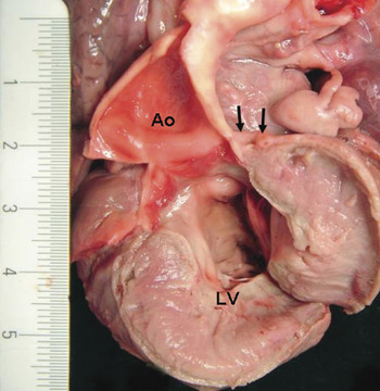
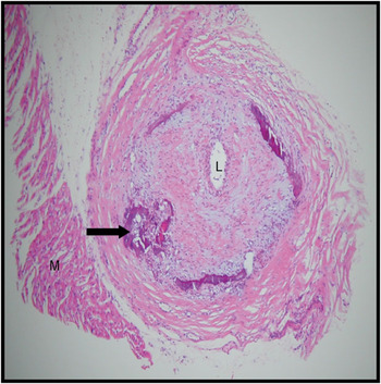
A 5 month old girl was admitted to paediatric intensive care unit with respiratory failure and hypertension. A renal ultrasonic scan was normal, with no evidence of renal arterial stenosis. Levels of renin were grossly elevated, consistent with renovascular disease. An echocardiogram showed a dilated and hypertrophied left ventricle. There was no evidence of obstruction in the left ventricular outflow tract, nor coarctation or abnormal coronary arteries. She initially responded to therapy, but suddenly collapsed 18 hours after admission with a low cardiac output state and intractable acidosis, dying shortly thereafter. The post mortem examination revealed marked left ventricular hypertrophy, with no obstruction to outflow or coarctation. The coronary arteries had a normal origin and distribution, but all of them were thick-walled, with diffusely narrowed lumen (Fig. 1). The thoracic and abdominal part of the aorta also showed diffuse mural thickening. The mesenteric and coeliac arteries showed slight diffuse thickening of the walls, but no narrowing or occlusion. Both renal arteries were thickened and completely occluded. Histology confirmed deposition of calcium in the internal elastic lamina of the coronary arteries, involving both the epicardial and intramural vessels (Fig. 2), with fibrointimal proliferation producing occlusion of the vessels. There was no inflammation of the vessel wall. Similar changes were seen in the renal vessels. All other vessels, including the pulmonary, carotids, coeliac, and mesenteric arteries, showed focal deposition of calcium, but not the marked fibrointimal proliferation noted in the coronary and renal vessels.
The heart has been opened at autopsy, revealing the left ventricular hypertrophy (LV). Arrows indicate the thickened and narrowed main stem of the left coronary artery and its interventricular branch. Ao: Aorta.

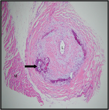
Haematoxylin and eosin stained section of the anterior interventricular coronary artery, showing the focal calcification (black arrow) and fibrointimal narrowing, with a narrowed lumen (L). Note the nearby myocardium (M).
Case 2
A two month old female presented with failure to thrive and poor feeding. On examination, she had reduced pulses in all four limbs, with a pansystolic murmur on auscultation. Echocardiography showed a large perimembranous ventricular septal defect. Her cardiac enzymes were elevated. She had an angiogram, which showed normal coronary arterial anatomy. She improved with diuretics, and an echocardiogram showed improved ventricular function. She was discharged home on digoxin to prepare for the closure of her ventricular septal defect. One month later, she was admitted to a local hospital with severe cardiac failure, and died shortly after admission. At autopsy, the orifice of the right coronary artery was found to be reduced to a pinhole, while the left coronary artery had a normal lumen. Throughout the coronary arteries, there were white patches, which were calcified and stiff on cutting. These affected the proximal right coronary artery to the acute margin of the heart, and were present throughout the left anterior interventricular and circumflex arteries. Cross sections of the affected areas revealed complete occlusion. Histologic sections showed calcification affecting the internal elastic lamina of the coronary arteries, with marked fibrointimal proliferation and occlusion. Sections of the right and left ventricles showed infarction of variable age, ranging from acute infarction with neutrophilic infiltration, to healing infarcts with granulation tissue and also widespread myocytic calcification. The subendocardial region of the left ventricle was densely fibrosed. Samples taken from the descending aorta and the left subclavian artery did not show calcification.
Case 3
An 11 year old girl with no history of cardiac disease presented with cardiac failure. Investigation revealed a dilated left ventricle with poor left ventricular function. Coronary angiography was normal. She was diagnosed as having dilated cardiomyopathy, and treated with an infusion of dobutamine. Two weeks later, she collapsed at home and died. An autopsy, limited to the heart, showed that it was heavy, weighing 249 grams, with a thin-walled left ventricle showing areas of haemorrhagic discolouration in the free wall, as well as areas of pallor indicating scarring. The coronary arteries were patent, but firm to the touch. They could only be cut with difficulty due to the hardness of the wall, indicating calcification, which was confirmed histologically. Microscopically, acute infarction was confirmed in the myocardium, as well as dense fibrosis indicating previous infarction.
Discussion
Idiopathic arterial calcification of infancy is a rare condition, and difficult to diagnose clinically because of the variable clinical features, which are confirmed in these three cases presenting with hypertension, congenital cardiac disease, and dilated cardiomyopathy, respectively. Our experience, furthermore, shows that there is a wider spectrum of age for afflicted patients than originally published. Late presentation is very rare, but the condition has been reported in a 22 year old, while our 11 year old patient has also previously been reported.Reference Marrott, Newcombe, Becroft and Friedlander6, Reference Sebire, Ramsay and Sheppard7 Clinical diagnosis is possible through radiological study of the arteries of the head, neck, abdomen and extremities,Reference Meradji, de Villeneuve, Huber, de Bruijn and Pearse2, Reference Moran3 but our experience emphasises that diagnosis prior to death is rare, and that children may often present acutely, with death following rapidly or suddenly.Reference Meurman, Somersalo and Tuuteri8 It should also be noted that 2 of our 3 cases underwent coronary angiography, albeit with reported normal coronary arterial anatomy. This could theoretically indicate rapid progress of the disease, but is more likely a consequence of widespread involvement of all coronary arteries resulting in diffuse narrowing, such that focal lesions could not be detected, and the diffuse narrowing was missed when not reviewed by a paediatric radiologist with a high index of suspicion. Thus, both clinicians and pathologists, including paediatric and forensic pathologists, must be aware of this entity when they are presented with such a clinical background. Fetal diagnosis is reported usually with sonography, but multislice detector computerised tomography is superior for detection of calcification in this condition.Reference Bellah, Zawodniak, Librizzi and Harris9, Reference Greenberg and Gibson10 To our knowledge, there have been no reports using multislice detector computerised tomography for fetal diagnosis.
A tendency of the disease to occur in siblings indicated a genetic cause,3 which was confirmed, and it is now established as a rare autosomal recessive disease caused by mutations that inactivate ectonucleotide pyrophosphatase/hosphodiesterase 1.Reference Rutsch, Ruf and Vaingankar11 This enzyme found on the surfaces of cells generates inorganic pyrophosphate, a solute that regulates cellular differentiation, and serves as an essential physiologic inhibitor of calcification. Not all cases, however, have this mutation.Reference van der Sluis, Boot, Vernooij, Meradji and Kroon12 Thus, fetal diagnosis is possible, but most patients present after birth, as reported in a review of 180 cases diagnosed prior to 2006.Reference Glatz, Pawel, Hsu, Weinberg and Chrisant13 Usually fresh blood or tissue is required for genetic analysis for the mutation. In making the diagnosis of this condition, other arterial lesions must be considered including metastatic calcification in severe renal disease, calcific arterial lesions noted in conjunction with certain cardiovascular anomalies, and hypervitaminosis D. Because of the genetic aetiology, once the diagnosis is made, the family should be informed and genetic counselling offered. Despite the same genotype and similar sonographic and radiographic features, the phenotype, prognosis, and response to therapy can vary to a great extent within one family.Reference Glatz, Pawel, Hsu, Weinberg and Chrisant13 Due to the rarity of the condition, trials of treatment are not possible, and therapy is usually based on anecdotal single case reports. Diphosphanates are usually administered, since they inhibit calcification and have been successfully given to slow calcification or reverse it, but longer-term survival has also been reported in patients receiving no therapy, like our 11 year old and the 22 year old already reported, who had reversal of calcification.