Xanthosine (XS) is a nucleoside reported to increase milk production and the stem cell population in mammary glands (Capuco et al. Reference Capuco, Evock-Clover, Minuti and Wood2009). XS overcomes p53-mediated repression of inosine monophosphate dehydrogenase (IMPDH), a rate limiting enzyme of guanine synthesis (Rambhatla et al. Reference Rambhatla, Bohn, Stadler, Boyd, Coss and Sherley2001). Suppression of IMPDH mediated repression increases the concentration of intracellular guanine ribonucleotide and favors symmetric cell division (Lee et al. Reference Lee, Crane, Merok, Tunstead, Hatch, Panchalingam, Powers, Griffith and Sherley2003). Symmetric stem/progenitor cell division produces two identical daughter cells; this type of cell division supports exponential growth. In dairy cattle, administration of XS increased the mammary stem/progenitor cell population ((Capuco et al. Reference Capuco, Evock-Clover, Minuti and Wood2009). However, not all in vivo experiments involving nucleoside administration result in increased stem/progenitor cells. Rauner & Barash (Reference Rauner and Barash2014) used a ‘bovinized’ mouse model and showed that XS decreased mammary cell proliferation by 50%. Because of these conflicting results continued research on the role of XS in lactating mammary glands is warranted. Over a two-month period of lactation, intramammary treatment of inosine (a compound similar to XS) resulted in increased milk production (26–200%) in transgenic goats (Baldassarre et al. Reference Baldassarre, Deslauriers, Neveu and Bordignon2011). Potential effect of XS on mammary gland specific gene expression and milk production is still lacking.
We hypothesized that intramammary infusion of XS alters milk synthesis genes, mammary stem/progenitor cell population and epithelial cell turn over in Beetal goat. Because milk synthesis genes expression affects milk protein synthesis and ultimately determines milk production, it was further hypothesized that XS administration increases milk yield. Therefore, the main objectives were to quantify the effects of XS on mRNA and protein expression of (1) milk synthesis genes and mammary stem/progenitor cells and (2) markers of mammary cell proliferation and evaluate milk yield and composition of TRT and CON glands in goats.
Materials and methods
Animals and experimental design
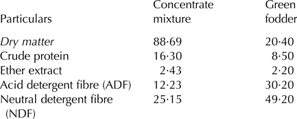
The use of goats for this study was approved by the Committee for the Purpose of Control and Supervision of Experiments on Animals (reference no. 25/20/2016-CPCSEA), Ministry of Environment, Forest and Climate Change (Animal Welfare Division), New Delhi. Beetal goats (n = 7, primiparous) were enrolled in this experiment at kidding. Goats were housed in a shed along with other farm goats and were fed a concentrate feed mixture (0·5 kg/d per lactating goat) and a mixture of oat and berseem green fodder (5·0 kg/goat per d). Composition of the concentrate feed mixture included maize (35%), soybean meal (15%), wheat bran (22%), de-oiled rice bran (25%), mineral mixture (2%) and salt (1%). Water was available at all times. Proximate composition of concentrate feed mixture and feed offer to the goats are provided in Table 1. Individual feed intake was not measured but was estimated for the group (data not shown). Kids were not allowed to nurse project goats and were bottle fed milk of other lactating goats (1/10th of kid's body weight which is around 400–500 ml/kid per day).
Proximate composition of concentrate feed mixture and fodder given to the goats during the experiment based on percentage of dry matter (DM%)

Goats were hand-milked twice daily at 0900 and 2100 h. Beginning on day 5 after kidding and ending after 3 consecutive days, each of the seven lactating goats received twice daily intramammary injections of a sterile XS solution in one gland (TRT) with the other gland serving as a control (CON). Within goat, selection of left or right gland for treatment was randomly assigned. The experimental unit was mammary gland within goat. For TRT, 20 ml of 10 mm of XS were infused through the teat canal immediately after the morning and evening milking. XS was prepared in sterile physiological saline and the resulting solution was filtered with 0·22 µm filters. The TRT glands received an additional injection of 1 ml of 10 mm XS into mammary parenchyma after each intramammary infusion for 3 d; this was to ensure delivery of XS to the peripheral regions of the glands (Capuco et al. Reference Capuco, Evock-Clover, Minuti and Wood2009). For CON, glands were left alone and no saline solution was injected; this was similar to the methodology of Capuco et al. (Reference Capuco, Evock-Clover, Minuti and Wood2009). Milk somatic cell counting was performed on both TRT and CON glands, one day after the last XS treatment. Two days after the last XS treatment (d 10 of lactation), transcutaneous mammary biopsies (7 TRT and 7 CON glands) were performed on each glands as described next.
Mammary gland biopsies
Mammary biopsies were conducted by a veterinary surgeon under anterior epidural anaesthesia as standard procedures done in veterinary clinics. Mammary parenchyma (~200 mg) was then excised with a number 22 scalpel blade. Mammary samples collected per gland were preserved and processed for RNA isolation in RNAlater (Ambion) and stored at −80 °C and fixed in 10% neutral buffered formalin for immunohistological evaluations.
RNA isolation and cDNA synthesis
Approximately 100 mg of tissue were lysed in 1 ml of Trizol (Ambion) using a handheld tissue homogenizer (Cole-Palmer, IL, USA) and proceeded for RNA isolation as per the protocol described earlier (Choudhary & Choudhary, Reference Choudhary and Choudhary2017). The purity and concentration of total RNA isolated from mammary parenchymal tissues were assessed using a NanoDrop ND-1000 Spectrophotometer (Thermo Fisher Scientific, Waltham, MA). Integrity of total RNA was evaluated by non-denaturing agarose gel electrophoresis and RNA integrity number (RIN) were evaluated using TapeStation (Agilent). First-strand cDNA was synthesized from 1000 ng of total RNA primed with oligo (dT) and random primers (3 : 1, vol/vol) using an AffinityScript One-Step RT-PCR Kit (Agilent Technology, CA, USA) according to the manufacturer's instructions.
Primer design and RT-qPCR
The cDNA was used to determine the expression of 17 selected genes of interest by reverse transcriptase-quantitative polymerase chain reaction (RT-qPCR). Genes of interest included: (1) milk synthesis related genes, (2) mammary stem/progenitor cell markers and, (3) cell proliferation markers. Genes of milk synthesis related gene whose expression was measured were beta-casein (CSN2), alpha S2- casein (CSN1S2), lactalbumin alpha (LALBA), mucin 1 (MUC1), beta-lactoglobulin (BLG), fatty acid binding protein 3 (FABP3) and CD36 molecule (CD36). Markers of basal epithelium (VIM) and mammary stem cells markers included aldehyde dehydrogenase 1 (ALDH1), hepatocyte nuclear factor 4A (HNF4A), nuclear receptor subfamily 5 group A member 2 (NR5A2), fibronectin type III domain containing 3B (FNDC3B) and notch 1 (NOTCH1). Genes related to cell proliferation including steroid receptors were marker of proliferation Ki67 (MKI67), tumour protein translationally-controlled 1 (TPT1), oestrogen receptor alpha (ESR1) and progesterone receptor beta (PRB). Endogenous control genes were two ribosomal proteins namely ribosomal protein L4 (RPL4) and ribosomal protein S23 (RPS23) (Kapila et al. Reference Kapila, Kishore, Sodhi, Sharma, Kumar, Mohanty, Jerath and Mukesh2013). mRNA sequences in the reference goat genome (NCBI), along with the NCBI Primer-BLAST tool and guidelines described previously (Thornton & Basu, Reference Thornton and Basu2011; Ye et al. Reference Ye, Coulouris, Zaretskaya, Cutcutache, Rozen and Madden2012), were used to design primers for all genes. A list of the primers and annealing temperatures are presented (Supplementary Table S1). Purchased primers (Integrated DNA Technologies, Inc., Iowa, USA) were reconstituted in nuclease-free water prior to use in qPCR. Efficiency of RT-qPCR ranged from 90–110%. PCR product specificity was confirmed using melt curve analyses and by electrophoresing representative qPCR products on 4% agarose gels to confirm singular bands of expected size (Yohe et al. Reference Yohe, Tucker, Parsons, Geiger, Akers and Daniels2016).
Each RT-qPCR assay was performed on all samples in duplicate, with each reaction mixture (10 µl) containing 0·25 µl each forward and reverse primer, 5 µl of Brilliant III Ultra-Fast SYBR® QPCR (Agilent Technologies, CA, USA), 3 µl of cDNA (1 : 10 dilution) and 1·5 µl of nuclease free water. Two-step qPCR assays were performed using a CFX96 Touch™ Real-Time PCR Detection System (BioRad Laboratories, California, USA) with the following cycling conditions: 95 °C for 3 min; 36 repeating cycles of 95 °C for 10 s (denaturation), respective annealing temperature (Supplementary Table S1) for a set of primers for 30 s (annealing); 95·0 °C for 3 min, 55·0 °C for 30 s, and then a temperature increase at 0·5 °C increment to 95·0 °C (melting curve). Each assay included a no-template control and a no reverse-transcriptase control, with the no template control receiving 1 µl of RNase/DNase free water instead of cDNA, while the no reverse-transcriptase control received a 1 µl sample of the reverse transcription product to which no reverse-transcriptase enzyme was added during cDNA synthesis.
Fluorescence immunohistochemistry and co-labelling
Formalin-fixed mammary biopsy tissue samples were dehydrated and embedded in paraffin. A microtome was used to cut 5-μm thick sections, which were then mounted onto charged microscope slides (Globe Scientific Inc. New Jersey, USA). Fluorescence immunohistochemistry was also performed for co-localization of two different markers (Supplementary Table S2) in the cellular compartment. The tissue antigens of interest were: stem cell marker (ALDH1), differentiation markers (CSN2 and MUC1), basal mammary epithelial cell markers (KRT14 and CD10) and luminal epithelial cell markers (EMA and MUC1). Following primary antibody incubation, slides were washed in PBS and incubated with Alexa conjugated secondary antibodies (Molecular Probes, Inc., OR, USA) for 1 h at room temperature at 1 : 400 dilution. Excess secondary antibodies were washed away in PBS (3 × 5 min); slides were then mounted with DAPI counterstain (4,6-diamidino-2-phenyl-indole) containing Prolong Gold anti-fade mounting medium (Molecular Probes). Slides were then coversliped and viewed by epifluorescence microscopy (Nikon Instruments Inc. NY, USA, model Eclipse 90i) at 400× magnification under FITC, TRITC and DAPI channels. Images were corrected for bright and contrast followed by merging all three channel images into one composite image using Fiji ImageJ (Schindelin et al. Reference Schindelin, Arganda-Carreras, Frise, Kaynig, Longair, Pietzsch, Preibisch, Rueden, Saalfeld, Schmid, Tinevez, White, Hartenstein, Eliceiri, Tomancak and Cardona2012).
Milk yield and composition analyses
Throughout the experiment, twice-daily milk production was measured gland-wise (TRT and CON glands milked separately) using a digital scale. Three days after finishing the XS treatment, on day 11, approximately 50 ml of milk per gland were collected from the milking bucket after completion of the morning milking for determination of milk composition. An ultrasonic milk analyser (Lactoscan; Milkotronic Ltd., Nova Zagora, Bulgaria) was utilized to measure: fat, protein, lactose and solid-not-fat (SNF).
Statistical analysis
For RT-qPCR data, Students’ t-tests were performed at the ΔCt level to evaluate significant changes in gene expression (Safayi et al. Reference Safayi, Theil, Hou, Engbaek, Nørgaard, Sejrsen and Nielsen2010). Target genes of interest were normalized to the geometric mean of RPS23 and RPL4 (target gene Ct – reference genes Ct = ΔCt) (Vandesompele et al. Reference Vandesompele, De Preter, Pattyn, Poppe, Van Roy, De Paepe and Speleman2002). For those genes in which raw data were not normally distributed (Shapiro Wilk's Test), data were transformed by logs of the ΔCt value prior to statistical analysis. For milk yield and composition data, morning and evening individual gland milk yields were summed daily. Daily yield data were then averaged on a weekly basis (Mean ± se). This resulted in 7 weekly milk yield observations per gland. Weekly data were analysed with paired t-tests using functions in Microsoft excel. Quantification of MUC1-positive area were analysed by paired t-test assuming equal variance after F-test analysis. Quantification of localization of other markers are not done and hence not presented here.
Results
Xanthosine treatment induces change in ALDH1, NR5A2, CSN2, LALBA, BLG, CD36, FABP3, MUC1 and ESR1 genes
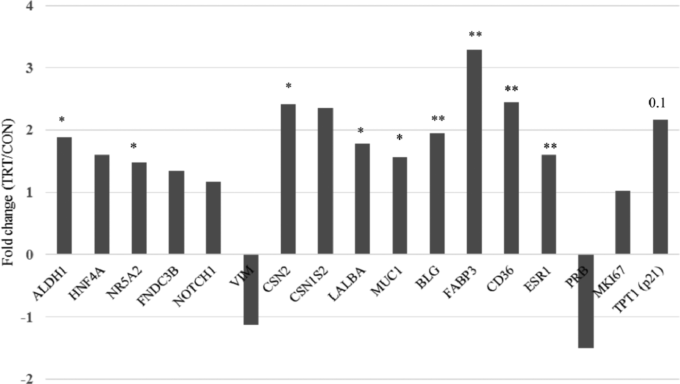
Gene expression analysis was performed to determine the effects of XS on genes relevant to key cellular mechanisms pertaining to stem cell, milk synthesis and cell proliferation. Three, twice daily intramammary injections of XS into TRT gland (either left or right half udder of the animal) caused an immediate and significant increase in RNA expression of stem cell markers (ALDH1 and NR5A2), milk protein genes (CSN2, LALBA, BLG), genes associated with fatty acid uptake and transport (CD36 and FABP3) and the oestrogen receptor-alpha (ESR1) gene (Fig. 1). Transcripts of other stem cell markers (HNF4A, FNDC3B, and NOTCH1) remained high in TRT glands in comparison to the CON glands of the same animals. Vimentin, a basal/myoepithelial cell marker and progesterone receptor (PRB) were not significantly affected although there was a tendency for downregulation. We found no decrease in mRNA expression of cell proliferation marker, Ki67 upon XS treatment. Expression of p21- a cell cycle regulator was also unaffected, although with a tendency to upregulation.
Effects of xanthosine treatment on gene expression of goat mammary glands analysed by RT-qPCR. Asterisks and figure on top of bar graphs indicate P-value (*P < 0·05, **P < 0·01) between treatment (TRT) vs. control (CON) group.

Immunohistochemical localization of protein markers
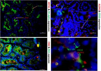
XS did not disrupt the histology of mammary tissue. Immuno-colocalization studies with various markers revealed weak abundance of cytokeratin 14 in alveolar epithelial cells with luminal presence of ESR1 (Fig. 2a). Nuclear localization of ALDH1, a mammary stem cell marker, was observed in basal mammary epithelial cells. Cytoplasmic staining of CD10 marked the basal layer of goat mammary epithelium (Fig. 2b). Immunostaining of CSN2 and EMA were abundant and present in the luminal epithelium (Fig. 2c). Individually stained CD10-positive cells (green), ALDH1-positive cells (red) and doubly labelled CD10-ALHD1-immunuopositive cells (yellow) is shown for emphasis. (Fig. 2d, not to the same scale).
Co-localization of various protein markers in goat mammary glands. Cytokeratin 14 expression (red) was limited to alveolar epithelial cells while expression of oestrogen receptor α (ESR1, green) was predominant in their cytoplasm (a). Alveolar epithelial cells showed abundant expression of casein β (CSN2, red) and epithelial membrane antigen (EMA, green) indicating secretory activity of epithelium. Colocalization of red and green channel appeared as yellow (b). Expression of goat mammary stem/progenitor cell marker ALDH1 (red) was localized in the basal layer. Basal epithelium is marked by localization of CD10 (green) (c). Enlarged morphology of ALDH1-positive mammary stem/progenitor cells (not to the scale) is shown for clarity (d). All the nuclei are stained with DAPI.

Milk yield and composition
Milk composition of TRT glands was similar to the CON glands, suggesting no adverse effects of XS treatment (Table 1). TRT glands (vs. CON glands) infused with XS in early lactation (5 to 7 d after kidding) produced more milk (average of milk produced between 2nd to 7th weeks post-biopsy; 3200·5 ± 142·7 vs. 3077·1 ± 113·5; P = 0·05) than CON glands (Table 1). Of 7 TRT and 7 CON glands, only 4 CON glands returned to normal lactation, whereas all 7 TRT gland remained lactating after post-operative care of biopsy. The effect of XS treatment varied among goats with an increase in milk production to no change in milk production by the treated gland (Table 2). Similarly, milk somatic cell count increased next day after the last day of treatment in some TRT glands, otherwise in majority of the glands showed no change in somatic cell count in comparison to CON glands.
Composition of goat milk obtained from the xanthosine treated (TRT) and control glands (CON)

ns, non-significant.
Discussion
Our first objective was to quantify the effects of XS on mRNA and protein expression of milk synthesis genes, mammary stem/progenitor cell, and proliferation markers on mammary glands of goats. Given past research (Capuco et al. Reference Capuco, Evock-Clover, Minuti and Wood2009; Choudhary & Capuco, Reference Choudhary and Capuco2012), we hypothesized that XS increases mammary stem cell and progenitor cell population which ultimately differentiates into progenitor cells to mammary epithelial cells and affects milk production. Our data showed that the XS administration for 3 d into the half udder of the goat mammary gland was able to induce changes in gene expression of milk protein genes (CSN2, LALBA, BLG, FABP3, CD36), stem/progenitor cell markers (ALDH1 and NR5A2) and differentiation markers (MUC1 and ESR1). Relative expression of other stem/prognitor cell related markers (HNF4A, FNDC3B and NOTCH1) was higher in XS treated glands, but due to high variation in their expression among animals, this change was not significant. Vimentin (VIM) and progesterone receptor (PRB) expression tended to be downregulated in TRT glands. A decrease in VIM expression indicated increased differentiation of basal epithelial cells into luminal cells. A decreased PRB and increased ESR1 expression might indicate role of XS in inducing mammary growth and lactation. The role of increasing estradiol and decreasing progesterone levels has been proposed for mammary growth and lactation in dairy goats (Hart & Morant, Reference Hart and Morant1980). Aldehyde dehydrogenase 1 (ALDH1) and nuclear receptor subfamily 5 group A member 2 (NR5A2; also called liver receptor homologue 1 or LRH1) are adult stem/progenitor cell markers. ALDH1 is expressed in a variety of cells, including cancer stem cells (Ginestier et al. Reference Ginestier, Hur, Charafe-Jauffret, Monville, Dutcher, Brown, Jacquemier, Viens, Kleer, Liu, Schott, Hayes, Birnbaum, Wicha and Dontu2007). NR5A2 is a pluripotency transcription factor of adult stem cell like that of OCT4 in embryonic stem cells (Heng et al. Reference Heng, Feng, Han, Jiang, Kraus, Ng, Orlov, Huss, Yang, Lufkin, Lim and Ng2010), and HNF4A, FNDC3B and NOTCH1 are potential candidate markers of mammary stem/progenitor cells (Dontu et al. Reference Dontu, Jackson, McNicholas, Kawamura, Abdallah and Wicha2004; Choudhary et al. Reference Choudhary, Li, Evock-Clover and Capuco2013, Reference Choudhary, Choudhary, Kaur and Pathak2016; Osińska et al. Reference Osińska, Wicik, Godlewski, Pawłowski, Majewska, Mucha, Gajewska and Motyl2014). Fatty acid transporter genes, CD36 and FABP3, are co-regulatory genes and have been associated with mammary cell differentiation (Spitsberg et al. Reference Spitsberg, Matitashvili and Gorewit1995). Fully differentiated mammary epithelial cells secrete more milk in comparison to intermediately and poorly differentiated cells. Increased expression of stem/progenitor cell marker genes in XS treated glands indicates role of the XS in increasing stem cell population. Despite the marginal increase in milk production in TRT glands, XS was able to induce significant changes in fatty acid transporter genes, indicating its role in functional differentiation of milk producing epithelial cells. A comparable increase in expression of milk protein genes suggests a positive effects of XS on milk volume and milk production. Contrary to the report of Rauner & Barash (Reference Rauner and Barash2014), we were unable to see negative effects of XS on cell proliferation marker Ki67, possibly due to a lack of immediate response of XS to Ki67 expression. Interestingly, p21 (TPT1) expression tended to be higher in TRT glands. The role of p53-mediated TPT1 has been indicated in reducing oxidative cell damage, minimizing apoptosis and promoting cell survival (Chen et al. Reference Chen, Wang, Tao, Zheng, Wu, Lian, Jaramillo, Fang and Zhang2013).
Our second objective was to quantify the effects of XS on protein expression of milk synthesis genes, mammary stem/progenitor cells and proliferation in mammary glands of goats. Protein expression results have shown higher expression of MUC1 in TRT glands. Increased expression of MUC1 in apical surface of alveolar epithelium of TRT glands are likely due to enhanced cell differentiation. Transmembrane protein MUC1 is expressed on apical surface of luminal epithelial cells and its expression is primarily in the cytoplasm. A recent study demonstrated the role of the cytoplasmic domain of MUC1 (MUC1-CD) by inducing hyperplasia of mammary epithelium (Li et al. Reference Li, Yi, Yao, Liao, Xie, Yang, Yan, Wang, Lu, Kuang, Gu, Fei, Wang and Huang2011). Aberrant expression of MUC1 is seen in breast cancer (Bouillez et al. Reference Bouillez, Gnemmi, Gaudelot, Hemon, Ringot, Pottier, Glowacki, Butruille, Cauffiez, Hamdane, Sergeant, Van Seuningen, Leroy, Aubert and Perrais2014), including goat mammary carcinoma (Gonçalves et al. Reference Gonçalves, Morais, de CG Alencar, Mota, Silva, Batista and Mendonça2011). In our study, we did not find overexpression or aberrant expression of MUC1 in TRT glands suggesting the XS do not promote tumorogenesis. Interestingly, cytoplasmic expression of ESR1 was seen in both the TRT and CON glands. Immunoreactivity of ESR1 was detected in nuclei of epithelial cells and fat cells of prepubertal bovine mammary gland (Meyer et al. Reference Meyer, Capuco, Boisclair and Van Amburgh2006), as well as stromal and epithelial cells in sheep mammary gland (Colitti & Parillo, Reference Colitti and Parillo2013) and, in the present study, cytoplasmic compartment of epithelial cells in lactating goats. Localization of ESR1 in the nuclei of cells is its normal expression, whereas, localization of ESR1 outside the nucleus is observed in cancer cells (De Stefano et al. Reference De Stefano, Zannoni, Prisco, Fagotti, Tortorella, Vizzielli, Mencaglia, Scambia and Gallo2011). In our case, expression of ESR1 was in the cytoplasm of mammary epithelial cells which could be the transient expression, and might be associated with growth and survival of cells (Welsh et al. Reference Welsh, Lannin, Young, Sherman, Figueroa, Henry, Ryden, Kim, Love, Schiff and Rimm2012). A weak non-nuclear expression of ESR1 has been reported in breast cancer (Welsh et al. Reference Welsh, Lannin, Young, Sherman, Figueroa, Henry, Ryden, Kim, Love, Schiff and Rimm2012).
Our final objective was to quantify the effects of XS on milk yield and composition in goats. our We showed only a marginal increase in milk production by the TRT glands. The possible reasons for this include variable responses amongst animals to XS treatment, which indicates goats are either XS-responders or non-responders. Response of XS treatment on global gene expression of mammary tissue (analysed by two sets of RNA sequencing mammary tissue and milk fat layer mammary specific RNA) showed similar observations indicated by the varied numbers of differentially expressed genes between TRT and CON glands (data not shown). Three days of consecutive infusion of XS in morning and evening (6 doses in total) could be suboptimal and milk yield responses might require best possible treatment strategy in terms of dose, frequency and duration. We hypothesize that XS should be administered for 5–7 d consecutively for the pronounced effects on milk production. However, increasing the days of XS administration beyond 14 d may have negative effects on cell proliferation and have no effects on stem cell population as suggested in the literature (Rauner & Barash, Reference Rauner and Barash2014). In earlier experiments with inosine, milk production of transgenic goats were compared with non-transgenic goats, whereas in our case, glands of the same goat were used as TRT and CON. Of seven goats under the study, only four goats were considered for milking after the gland biopsy. All seven TRT gland returned into normal lactation one week after the biopsy, whereas only four CON glands remained functional after biopsy. This observation suggests that XS may play a role in wound healing and resuming lactation after the surgical wound. This result supports the hypothesis that XS promotes mammary specific gene expression and stem cell transcripts along with a small increase in milk production.
Conclusion
In summary, this study demonstrated that XS increases expression of milk synthesis genes and mammary stem/progenitor cells of goat mammary glands without deleterious effects on mammary cyto-architecture. Our short XS treatment only marginally increased milk production up to 7 wk when administered during first wk of lactation, and XS had no effect on milk composition. We were unable to observe an increase in cell proliferation of mammary epithelial cells with 3 d of XS infusion. Increased duration of XS treatment for 5–7 d could be merited, alternatively, a comparative study using inosine treatment should be planned to evaluate if inosine is a better treatment than XS in increasing milk yield and cell proliferation of milk secreting epithelial cells.
Supplementary material
The supplementary material for this article can be found at https://doi.org/10.1017/S0022029918000493
Authors would like to thank Dr A. L. Saini, former Head of the Department of Livestock Production and Management, GADVASU, Ludhiana for providing and keeping goats in the farm. This study was supported by grant received from the Department of Biotechnology, Govt. of India, New Delhi, grant # BT/PR8352/AAQ/1/549/2014. Authors also would like to acknowledge Dr Vikas Vohra, Senior Scientist at ICAR- National Bureau of Animal Genetic Resources, Karnal, India for scientific inputs pertaining to the milk yield data. We duly acknowledge Paulnisha Granger and her M.S. advisor Dr Kristy Daniels (Assistant Professor, Department of Dairy Science, Virginia Tech, Blacksburg, VA) for English-language and technical editing.