Introduction
Hairy polyp was first described by Arnold in 1870.Reference Arnold1 It is a rare congenital skin-covered mass that is most commonly found in the oropharynx. Infants with hairy polyps may present with breathing and feeding difficulties, depending on the size, shape and location of the polyps. We present a four-month-old girl with such a polyp arising from the left posterior tonsillar pillar. The mass periodically disappeared when ‘swallowed’ and initially went unappreciated on physical examination. However, it was captured on the parent's smartphone video, illustrating the increasingly broad integration of smartphone use into medical diagnostics.
Case report
A concerned mother came in worried about the occasional appearance of a finger-like lesion protruding from the mouth and intermittent inspiratory stridor, which was particularly prominent during the feeding of her four-month-old daughter. The infant would periodically ‘swallow it’, thereby allowing the lesion to fall behind the tongue out of sight. The lesion was not seen on initial inspection; however, the mother had taken a video of it on her smartphone. This showed a mass emanating from the left side of the mouth.
The patient was born by normal spontaneous, full-term vaginal delivery, with normal Apgar scores. She was seen previously in clinic for intermittent inspiratory stridor, which was particularly noticeable during feeding. On occasions, she would relieve the stridor by extending her neck. She was otherwise healthy, with no other medical problems.
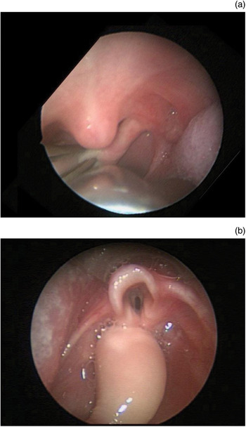
The patient underwent direct laryngoscopy and bronchoscopy, and removal of the lesion. Under general anaesthesia, a Parson's laryngoscope was used to inspect the oropharynx and larynx. The larynx and trachea were normal; however, a pedunculated mass emanated from the superior portion of the left posterior pillar on the palatal side (Figure 1a). The mass would lie in the oropharynx with the potential to occlude both the oesophagus and larynx (Figure 1b). The mass was removed transorally using forceps and a Bovie electrocautery. There were no post-operative complications and the patient was discharged the day after surgery. Follow up at four weeks post-surgery was unremarkable.
Endoscopic views of (a) the polypoid mass attached to left posterior tonsillar pillar, and (b) the mass residing over the oesophagus and potentially the larynx.

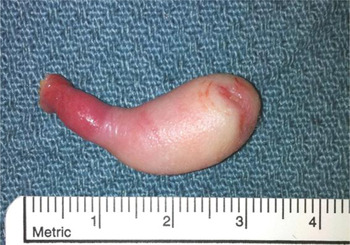
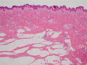
Gross examination showed a skin-covered polypoid mass that was 3.5 × 1.2 × 1.0 cm in size (Figure 2). Microscopic examination showed a hairy polyp, consisting of a core of fibroadipose tissue and cartilage covered by skin (Figure 3).
The resected specimen: a 3.5 cm pedunculated polyp covered in skin bearing fine vellus hair.

Microscopic view of the epidermal surface of the polyp showing numerous pilosebaceous units. Underlying lobules of adipose tissue with fibrous septa were seen. At a deeper level an Island of cartilage was present. (H&E; ×40)

Discussion
Whilst the oropharynx is the commonest site for hairy polyps, the posterior tonsillar pillar is an unusual subsite. The one previously reported case of a hairy polyp in this location occurred in a five-week-old female with breathing difficulties.Reference Shvidler, Cable and Sheridan2
Hairy polyps are rare, benign congenital malformations. Their outer layer is epidermal and thus may have hairs, which is why they are known as ‘hairy’. A recent study has shown that they are eight times more common in females than males and, as in the patient presented herein, they appear mainly on the left side.Reference Kalcioglu, Can and Aydin3 The term hairy polyp has occasionally been used to describe similar polyps occurring anywhere in the body. However, it is usually reserved for those occurring in the nasopharynx and oropharynx, and more rarely, in the middle ear, mastoid cavity, eustachian tube and tongue. They can occasionally occur in association with other congenital malformations of the head and neck, such as hard palate clefts, low set ears, microtia, absence of outer ears and lateral facial dysplasia.Reference Burns, Axon and Pahade4 There have been no reports of malignant transformation of a hairy polyp. They usually occur singly but there have been rare cases of bilaterality.Reference Franco, Florena, Lombardo and Restivo5
Macroscopically, the hairy polyp is typically pedunculated, fleshy or rubbery, and covered with tan hair-bearing skin. Microscopically, it is made of a core of mesodermal structures such as fat, muscle, cartilage and fibroconnective tissue. The core is covered in stratified squamous keratinising epithelium, with normal skin appendages such as pilosebaceous glands (ectodermal). Cases of hairy polyps usually present early in life with breathing and feeding difficulties. The age of presentation depends on the size and shape of the polyp, and it can remain asymptomatic until later life; the oldest case in the literature is a 71-year-old male.Reference Resta, Santangelo and Bastilla6
When Arnold first described the hairy polyp in 1870, he termed it a type of ‘dermoid’.Reference Arnold1 Hairy polyps have also been described as teratomas or teratoid lesions;Reference New and Erich7 these terms are not suitably applied to hairy polyps because they imply a neoplastic process. Extragonadal teratomas are tumours containing tissue or organ components resembling normal derivatives of all three embryonic layers (ectoderm, mesoderm and endoderm).Reference Tapper and Ernest8 They are commonly congenital and, whilst they are mostly seen in the sacrococcygeal region, a substantial proportion of these teratomas are located in the cervicofacial region. They can contain tissue such as brain, gut, skin, cartilage and respiratory tract. When they contain primitive neuroepithelial tissue, they are said to be an ‘immature teratoma’. They may contain malignant germ cell elements such as yolk sac tumour, and they can develop malignancy such as Ewing's sarcoma or other ‘somatic’ tumours.
• Hairy polyps are rare congenital polypoid growths comprising two germ cell layers: the ectoderm and mesoderm
• Cases usually present at or shortly after birth with breathing and feeding difficulties; excision is usually curative
• They are most likely due to a malformation of the first and second branchial arches, and should be distinguished from teratomas
• This is the second paper to report a case of hairy polyp located on the left posterior tonsillar pillar
• The diagnosis was made using the parent's smartphone, highlighting the growing use of patients' electronic devices in aiding diagnosis and management
However, hairy polyp is clearly not a neoplastic tumour. It is instead a type of congenital malformation that resembles accessory tissue from the external ear, much like an accessory tragus. Heffner et al. noted that the histological appearance of the hairy polyp and the fetal auricle (which is derived from first and second branchial arches) were very similar. He subsequently proposed that hairy polyps were accessory pharyngeal auricles.Reference Heffner, Thompson, Schall and Anderson9 Hairy polyps can be considered a form of heterotopia or, more specifically, a choristoma, which is a benign growth that contains normal tissues but in an abnormal location. Its increased prevalence in females is unexplained.
The diagnosis of a hairy polyp is usually made clinically by visualising the characteristic polypoid mass in the oral cavity. Symptoms of breathing and feeding difficulties are also present. Radiological investigation, whilst helpful in delineating the size and location of the mass, cannot differentiate between other masses. Management of a hairy polyp is resection of the mass, usually transorally. Excision is curative; recurrence has been noted only once in the literature.Reference Frech and McAlister10
We believe that the use of a parent's smartphone video was, in this case, an integral part of the initial diagnosis. This illustrated the noteworthy trend in modern diagnosis of the integration of patient-derived videos into the diagnostic armamentarium. Smartphones and hand-held electronic devices have been integrated into medical care primarily with physicians and caretakers as the users. The use of patient-operated devices in diagnosis is rare, but has been very successful in the past. A study by Rich et al. showed that video intervention assessment, wherein patients were given video camcorders to record their daily lives, yielded a more complete and accurate understanding of exacerbating environmental exposures and inappropriate medication usage in children and adolescents with asthma.Reference Rich, Lamola, Amory and Schneider11 Other examples include the use of a smartphone as a Holter monitor to record electrocardiograms,12 which is potentially very useful in diagnosing intermittent heart disease. We believe this trend of patient-operated devices in diagnosing will continue, and will undoubtedly help physicians in guiding the management of their patients.
In conclusion, this paper described the case of a hairy polyp. This rare congenital malformation, which resembles heterotopic external ear tissue, arose from the posterior tonsillar pillar. This site has been documented only once previously in the literature. We also highlighted the noteworthy trend of patient-operated devices such as smartphones being used to help aid the modern diagnosis of disease. There is also the potential for the use of such devices to aid in the gathering of clinical information, which may help the physician to formulate a more effective management plan.
Acknowledgements
The primary author (SS) wrote this as a medical student at Imperial College London during his elective at Harvard Medical School. He would like to thank the paediatric otolaryngology team at Children's Hospital Boston for delivering an enjoyable and educational experience.