




No CrossRef data available.
Article contents
Characterization of a GABAergic neurotransmission in adult Schistosoma mansoni
Published online by Cambridge University Press: 06 August 2004
Abstract
The neuromuscular systems of parasitic helminths are targets that are particularly amenable for anthelmintics. In this study, we describe a GABAergic neurotransmission in adult Schistosoma mansoni, the trematode responsible for high levels of morbidity in people living in developing countries. GABA immunoreactivity (GABA-IR) was detected in nerve cells and fibres of the cerebral ganglia and longitudinal nerve cords and the nerve plexuses ramifying throughout the parenchyma of male adult worms. In addition, strong GABA-IR was also found associated with the oral and ventral suckers as well as in testes indicating a role for GABA in fixation to the host vascular wall and spermatogenesis. The capacity to synthesize GABA from glutamate was confirmed by measurement of a glutamate decarboxylase (GAD) activity. Supporting these data, a single band with an apparent molecular weight of about 67 kDa was detected using an antibody raised against mammalian GAD. In vivo studies revealed that picrotoxin, a non-competitive antagonist of the GABAA receptor, produced a modification of the motility and locomotory behaviour of adult worms, suggesting that GABAergic signalling pathway may play a physiological role in the motonervous system of S. mansoni and could be considered as a potential target for the development of new drugs.
Keywords
Information
- Type
- Research Article
- Information
- Copyright
- 2004 Cambridge University Press
INTRODUCTION
Many authors have suggested that the neuromuscular system of helminths is a target that is particularly amenable for anthelmintics. A better understanding of the neuromuscular physiology of the platyhelminth Schistosoma mansoni could thus provide valuable information for the discovery of new drugs active against that particular parasite, responsible for 130000 deaths per year, and possibly for the other species of this genus. Thus 200 million people infected with schistosomiasis worldwide could be concerned (Fenwick et al. 2003). Furthermore, the World Health Organization recently identified schistosomiasis among its top five disease priorities and identified research of new drugs as a major need (Remme et al. 2002).
Gamma-aminobutyric acid (GABA), the major inhibitory neurotransmitter in vertebrates, has been reported to be present in the nervous system of several invertebrate phyla (Walker & Holden-Dye, 1991). In helminths, such as the nematodes Ascaris suum and Caenorhabditis elegans (Johnson & Stretton, 1987; McIntire et al. 1993), GABA could act as an inhibitory interneuronal and neuromuscular transmitter. Indeed, the ablation of motor neurons containing GABA receptors induced defects in physiological processes such as locomotion and defecation in C. elegans (McIntire et al. 1993). Moreover, GABA receptors present on somatic muscle cells of A. suum mediated an increase in chloride conductance as observed with mammalian GABAA receptors. However, these Ascaris receptors were insensitive to classical mammalian GABAA antagonists such as bicuculline and picrotoxin, as well as to GABAA potentiators such as barbiturates, benzodiazepines and neurosteroids (Holden-Dye et al. 1989). Such differences between the pharmacological profiles of mammalian and helminthic GABAA receptors became attractive targets for the development of selective drugs. For instance, the anthelminthic piperazine acts as a GABA agonist at muscular receptors inducing a reversible flaccid paralysis of Ascaris (Martin, 1987).
Despite these numerous studies on GABA activities in nematodes, the physiological actions of GABA in platyhelminths has remained relatively poorly explored. This is due, in part, to the small size and inaccessibility of their individual nerve and muscle cells, which make difficult the study of nerve or muscle systems in the absence of interfering tissues. Furthermore, since the majority of adult platyhelminths are endoparasites, the impact of in vitro isolation on their behaviour and physiology is largely undefined (Pax & Bennett, 1992). Nevertheless, some results support a putative role for GABA in the neurotransmission of platyhelminths, in part because GABA-immunoreactivity (IR) has been demonstrated in the central and peripheral nervous system of several species, including the cestode, Moniezia expansa, the turbellarian, Girardia tigrina and the trematode, Faciola hepatica (Eriksson & Panula, 1994; Eriksson et al. 1995). The planarian, G. tigrina, contains a large number of neurons that exhibit GABA-IR and is also able to synthesize GABA through glutamate decarboxylation (Eriksson & Panula, 1994). In F. hepatica, a GABA-IR was detected in the nerve cords and in subtegumental fibres (Eriksson et al. 1995). In S. mansoni, there is no direct evidence for a GABA transmission pathway. Furthermore, direct application of GABA to whole worms did not alter their basal motor activity (Mellin et al. 1983).
The aim of the present work was to identify the presence of a GABA signalling pathway in male adult S. mansoni and to investigate its involvement in the control of worm motility. We initially applied immunohistochemical techniques to localize GABA, present mainly in nerves, but also in suckers and testes. We also measured a glutamate decarboxylase (GAD) activity and confirmed its presence by Western blot, demonstrating that the worm is able to synthesize GABA from glutamate. Finally, we performed some in vivo measurements indicating that the GABAergic signalling pathway described here for the first time may play a physiological role in the motonervous system of S. mansoni.
MATERIALS AND METHODS
Animals
Adult male S. mansoni (BH strain) were recovered from the portal and mesenteric veins of mice infected 45 days earlier, each with approximately 150 male cercariae, as previously described by Cunha et al. (1992).
Immunohistochemical localization of GABA
The worms were washed with cold sodium phosphate-buffered saline (PBS), pH 7·2, and fixed in 4% paraformaldehyde in 0·16 M PBS for 4 h. After several washes in PBS, the worms were cryoprotected in 10% and 30% sucrose in PBS until cryosectioned. The worms were then embedded in OCT medium (Sakura Finetek, Torrance, CA, USA), quickly frozen in liquid nitrogen and sliced in 15–20 μm thick sections. These sections were collected on gelatinized slides and stored at −20 °C until immunostained. The sections were washed in PBS plus 0·25% Triton X-100 and incubated with 5% goat normal serum for 20 min. Then, the sections were incubated overnight in a guinea-pig polyclonal antibody against GABA (Vector Labs, Burlingame, CA, USA) diluted 1[ratio ]1000 or 1[ratio ]2000 at room temperature. After several washes in PBS containing 0·25% Triton X-100, the sections were incubated for 1 h with goat anti-guinea pig IgG biotinylated antiserum (Protos Biotech Corp, New York, NY, USA) diluted 1[ratio ]200. After several washes in PBS, the sections were incubated for 30 min in a mixture of biotin and avidin-peroxidase (ABC Elite Kit, Vector Labs, Burlingame, CA, USA) diluted 1[ratio ]50. After that, the sections were incubated with 3,3′-diaminobenzidine tetrahydrochloride in 0·1 M Tris buffer, pH 7·2, plus 0·01% hydrogen peroxide.
All slides were examined and photographed using a light microscope (Zeiss Axioscope).
Determination of GAD activity
The GAD activity was determined using the method described previously (de Mello, Bachrach & Nirenber, 1976). Briefly, about 200 worms were homogenized at 4 °C in 50 mM potassium phosphate buffer, pH 6·8, and 1 mM EDTA using 3 sequences of 10 passes of the pestle in a Dounce homogenizer. The homogenates were incubated in a reaction mixture containing the following components in a final volume of 100 μl: 50 mM potassium phosphate buffer, pH 6·8, 1 mM EDTA, 0·5% Triton X-100, 1 μCi L-[U-14C]-glutamate, 0·5 mM pyridoxal phosphate, 1 mM GABA and 1 mg of protein. Each reaction mixture was incubated at 37 °C for 30 min in a 16×25 mm plastic tube equipped with a rubber stopper supporting a polyethylene centre well; then 0·1 ml of 10% trichloroacetic acid and 0·3 ml of hyamine hydroxide were injected through the stopper into the reaction mixture and centre well, respectively, and the tubes were incubated for an additional 30 min to collect 14CO2. The centre wells were transferred to vials containing scintillation mixture [1,4-bis-[2-(5-phenyloxazolyl)]-benzene (4 mg/l) and 2,5-diphenyloxazole (0·1 mg/l) in toluene], and the radioactivity was measured in a Tri-Carb Packard liquid scintillation counter. Protein concentration was determined by the method of Bradford (1976), using bovine serum albumin as standard.
Immunoblot detection of GAD
Sodium dodecyl sulfate–polyacrylamide gel electrophoresis (SDS–PAGE) was carried out with worm homogenates according to the method described by Laemmli (1970). After their transfer to nitrocellulose membranes, the proteins were identified with rouge de Ponceau, washed with an appropriate buffer and incubated for 2 h with 5% casein and 1% bovine serum albumin at room temperature. The membranes were incubated overnight with diluted (1[ratio ]5000) sheep polyclonal antibody against GAD (kindly provided by W. H. Oertel, see Oertel et al. 1981) at 4 °C. Blots were rinsed and incubated for 2 h with anti-sheep horseradish peroxidase-conjugated antibody (Sigma–Aldrich Chemicals, Milwaukee, WI, USA). Immunoreactivity was detected by enhanced chemiluminescence and exposure to Hyperfilm ECL (Pharmacia-Amersham Biotech Ltd, Little Chalfont, UK). The molecular weight of the immunoreactive bands was estimated by comparison with molecular weight standards run with each experimental sample.
In vivo studies
About 12–15 adult male worms were recovered from portal veins of mice 45 days after infection. The worms were then washed rapidly and placed in a glass dish with 2 wells containing 250 μl of a buffered saline solution, consisting of 82·5 mM Na+, 4·1 mM K+, 3·6 mM Ca2+, 3·3 mM Mg2+, 100·4 mM Cl−, 79·9 mM glucose, 10 μM 5-hydroxytryptamine (5-HT), and 15 mM HEPES (pH 7·4). In each well, 4 worms were pre-incubated for 10 min at 37 °C. Then, the solution was replaced by a fresh one containing 100 μM picrotoxin or 1 mM bicuculline. After 10 min, the latter solution was exchanged and worms further incubated for 10 min. Body area measurements were made by analysis of the images captured with a CDC camera using the Image-Pro® Plus program (Media Cybernetics, Silver Spring, MD, USA) according to previously published procedures (Mendonça-Silva, Pessôa & Noël, 2002). Data represent the means (±S.E.M.) of at least 3 different dishes with 3–4 worms each (n=11). One-way ANOVA analysis was used for determination of statistical significance.
Drugs
Picrotoxin, bicuculline, GABA and 5-HT were obtained from Sigma–Aldrich Chemicals (Milwaukee, WI, USA). L-[U-14C]-glutamate was from New England Nuclear (Boston, MA, USA).
RESULTS
Immunohistochemical studies
According to Pax & Bennett (1992), the nervous system of platyhelminths consists of a pair of cerebral ganglia that are situated on both sides of the pharynx, just posterior to the oral sucker, and are connected by a dorsal commissure. From the cerebral ganglia arise a variable number of nerve cords running anteriorly and posteriorly. In addition, from the longitudinal cords, numerous branches run in a lateral direction forming a peripheral nerve plexus. Based on these morphological characteristics, the present study identified an intense and widespread GABA-IR throughout the nervous system of S. mansoni (Fig. 1). In the central nervous system (CNS), GABA-IR was detected in the paired cerebral ganglia, whereas it could not be identified with confidence in the dorsal commissure (Fig. 4A). Strong GABA-IR was also detected in the longitudinal nerve cords throughout the entire length of the animal, identifying large amounts of nerve cells and nerve fibres (Fig. 2A–C). GABA-IR was also detected in numerous fibres extending from the main nerve cords both dorsally and ventrally, providing an extensive nerve plexus of fine fibres and abundant nerve cells (Figs 1 and 2A–C). In the peripheral nerve plexuses, structures that appeared to be cell somata with processes running in the direction of the tegument were observed. The putative cell bodies are usually bipolar, with a neural process extending to the tegument and the other one reaching the main nerve cord (Fig. 2D–F). Although GABA-IR was observed in some nerve endings at the outer surface of the worm, almost no GABA-IR nerve endings were identified along the gynaecophoral canal (Fig. 1). In addition, strong and diffuse GABA-IR was also seen along with muscle tissue of both oral and ventral suckers (Fig. 3A–B). This immunoreactivity was closely associated to the surface of the individual muscle fibres (Fig. 3B).

Fig. 1. Digitized image of transverse section (20 μm) of midbody of adult male Schistosoma mansoni showing GABA-IR in main nerve cords (long arrows) and peripheral nerve net (P). GABA-IR in peripheral nerve net (P) was formed by neuronal cell bodies (arrowheads) and nerve fibres. Note numerous GABA-immunoreactive nerve endings (small arrows) in protusion of tegument along the outer surface. Gut (G).

Fig. 2. Digitized image of longitudinal section (20 μm) of adult male Schistosoma mansoni showing GABA-IR in the nervous system. (A) GABA-IR in the longitudinal nerve cords (MC) with numerous cell bodies (arrows) and branching nerves (arrowheads). (B) Detail of longitudinal nerve cords showing GABA-immunoreactive nerve fibres (arrowheads). (C) Typical appearance of longitudinal nerve cords, with GABA-immunoreactive nerve fibres running in bundles or alone towards the entire length of the body. Note lateral nerves that run to outer surface and several bundles of fibres running to the centre of the animal. (D–F) Detail of GABA-immunoreactive neuronal endings (arrowheads) in protusions of tegument. Note bipolar GABA-IR in neuronal cells (arrows) with some processes extending to the tegument and other ones running to the centre of the body.

Fig. 3. Digitized image of longitudinal section (20 μm) of Schistosoma mansoni showing GABA-IR in the attachment structures. (A) Strong and diffuse GABA-IR in both oral and ventral suckers. (B) Detail of oral sucker GABA-IR. Oral sucker (OS) and ventral sucker (VS).
The reproductive system of male S. mansoni is composed of a single testis with 6–12 combining testicular lobes ventrally intercommunicated between them, as recently characterized by confocal microscopy (Machado-Silva et al. 1998). Interestingly, an intense GABA-IR was found restricted to the testicular lobes that exhibited an amorphous appearance (Fig. 4C–E).

Fig. 4. Digitized images of longitudinal sections (20 μm) of adult male Schistosoma mansoni showing GABA-IR in the reproductive system (A–B). Intense GABA-IR with an amorphous aspect is shown in testicular lobes (TL). (C–E) Detail of testicular lobes GABA-IR. In (A), cerebral ganglia-like structures (arrows) can be seen inside the anterior part of the animal, but neither cell somata nor cerebral commissures can be clearly identified. From the cerebral ganglia, nerves extend in anterior and posterior directions (arrowheads). (B) GABA-IR in the longitudinal nerve cords (arrows) on each side of the gut (G) (arrow). Main nerve cords (MC) and gut (G).
Note that control sections were incubated with sheep normal serum in the absence of primary antibody against GABA and no immunoreactivity was detected. Control sections were also pre-incubated with glutamate in order to exclude the possibility of a cross-reaction with this amino acid since the presence of glutamate-like IR has been described in cercariae of S. mansoni (Solis-Soto & Brink, 1994). Pre-incubation with 10 μg/ml glutamate caused no detectable decrease in the GABA-IR (data not shown).
GAD determination
The conversion of glutamate to GABA was measured in homogenates prepared from whole adult S. mansoni. The glutamate decarboxylase specific activity obtained in these worm homogenates was relatively high (5·36±1·00 nmol CO2/h/mg protein, n=4). In order to check whether the GAD present in S. mansoni was similar to that found in mammalian tissues, immunoblot detection of this enzyme was performed with a mammalian polyclonal antibody, which recognizes both GAD65 and GAD67 isoforms. As shown in Fig. 5, a single band corresponding to an apparent molecular weight of 67 kDa was detected in worm homogenates.

Fig. 5. Immunoblot analysis of glutamate decarboxylase (GAD) in adult Schistosoma mansoni homogenates.
In vivo studies
As we mentioned before (Mendonça-Silva et al. 2002), adult male worms in saline solution supplied with 5-HT exhibited a variety of body movements including small and fast generalized shortening and increasing of the body length, and undulatory propagating body waves along the anterior/posterior axis, similar to peristaltic waves. In the presence of 100 μM picrotoxin, the worm bodies bent rapidly in ring- or corkscrew-like coils leading to an alteration of their activity behaviour (Fig. 6). In order to quantify the picrotoxin effect, we measured the body area of the worms before and after drug addition. We observed that 100 μM picrotoxin produced a time-dependent reduction of the body area (Fig. 7). On the other hand, addition of 1 mM bicuculline to saline solution did not affect the worm activity mentioned above (data not shown).

Fig. 6. Digitized images showing the body postures of individual adult male Schistosoma mansoni in the absence (control, A,B) and presence of 100 μM picrotoxin (C,D). Control worms presented a variety of postures, including body waves (B). Note that 100 μM picrotoxin produced corkscrew-like coilings of parasite bodies, modifying the motor activity in a time-dependent manner (C,D). t=time after addition of picrotoxin (C,D) or vehicle (A,B).

Fig. 7. Time-course of picrotoxin effect on body area of adult male Schistosoma mansoni. Each point represents the mean±S.E.M. of 11 worms in the absence (○) or presence ([squf ]) of 100 μM picrotoxin. Note that these experiments were performed on 3 different days with 3–4 worms each. The effect of picrotoxin was statistically significant (P<0·05) as determined by one-way ANOVA analysis.
DISCUSSION
GABA-IR has been demonstrated in the nervous system of members of every invertebrate phylum thus far investigated, including some platyhelminths. However, the present work is the first report of GABA-IR in S. mansoni. As in the planarian G. tigrina and trematode F. hepatica (Eriksson & Panula, 1994; Eriksson et al. 1995), widespread GABA-IR was demonstrated in central and peripheral nerve structures. Strong GABA-IR was observed in the longitudinal nerve cords and in their lateral nerves. GABA-IR was also identified in the peripheral nerve plexus, where the cell bodies are frequent. However, in contrast with F. hepatica, where the GABA-IR occurs in the longitudinal nerve cords and lateral nerves in the posterior half of the worm (Eriksson et al. 1995), the GABA-IR was seen in the main nerve cords throughout the entire length of the S. mansoni. Furthermore, no difference was seen in GABA-IR detected in the ventral and dorsal subepidermal nerve plexus of S. mansoni whereas the planarian G. tigrina and the flatworm F. hepatica exhibit more abundant GABA-IR in the ventral part of the subtegumental plexus (Eriksson & Panula, 1994; Eriksson et al. 1995). Although some differences were noticeable in the patterns of GABA-IR observed in these platyhelminths, GABA is extensively distributed in the nervous system of all these worms. The distribution pattern of GABA-IR described here in S. mansoni was similar to the immunoreactivity against neuropeptides that are important neurotransmitters in this worm (Gustafsson, 1987).
Most of the GABA-IR neurons located laterally to the main nerve cords have neural processes extending out to the surface of the worm, suggesting that GABA is associated with sensory receptors in S. mansoni. In fact, ciliated and non-ciliated sensory receptors have been localized in the tegument of platyhelminths. They consist essentially of a bulb-like expansion of the dendrite from which a process extends to the tegument (Silk & Spence, 1969). Sensory endings have been stained by other neuronal signalling substances like neuropeptides and 5-HT (Gustafsson, 1987).
In the endoparasitic trematodes, extensive cholinergic, aminergic, peptidergic and serotoninergic nerve fibres innervate the oral and ventral suckers (Halton & Gustafsson, 1996). In S. mansoni, serotoninergic fibres provide innervations to both oral and ventral suckers through extensive nerve nets that are localized beneath the body wall musculature, where they anastomose between the muscle fibres (Mair et al. 2000). GABA-IR followed the muscle fibres so closely that it was impossible to identify whether the staining resides inside the muscle fibres or in very thin nerve fibres between the muscles, since nerve tissue is closely associated with the musculature of the suckers (Silk & Spence, 1969). As a consequence of this high level of GABA in the sucker, we could hypothesize that drugs acting on GABA transmission could interfere with worm fixation to the vascular wall.
The localization of GABA-IR in the testicular lobes of adult S. mansoni shows that GABA is not limited to its nervous system and that GABA may have functions other than neurotransmission in this worm. This hypothesis is supported by reports describing the presence of GABA and GABA receptors in mammalian testes and spermatozoa (Ritta, Calamera & Bas, 1998). In addition, the existence of a pathway for GABA synthesis was also reported in mammalian reproductive tissues. Indeed, according to Tillakaratne et al. (1992), GAD67 is expressed in rat testis, whereas GAD65 is expressed in rat oviduct. Under these circumstances, numerous studies have shown that GABA at relatively low concentrations may mimic the effects of progesterone, by promoting the acrosome reaction of mammalian spermatozoa (Shi, Yuan & Roldan, 1997). Moreover, GABA increases the percentage of spermatozoa that move with a hyperactivated motility (Calogero et al. 1996). These findings indicate that GABA plays an important role in the mammalian fertilization process by acting on GABA receptors present in the testis. Our data indicate that such a role for GABA could be present in adult S. mansoni and so opens new perspectives for drug development since inhibition of spermatogenesis would be beneficial.
The presence of a GABA pathway in S. mansoni is further supported by our description of a glutamate decarboxylase (GAD) activity in worm homogenate, indicating that GABA can be synthesized from glutamate, as is the case in mammals. Note that the specific GAD activity measured in schistosome homogenate (5·36±1·00 nmol CO2/h/mg protein) may be considered relatively high if compared with the activities measured in chick retina (50 nmol CO2/h/mg protein (De Mello et al. 1976) and porcine brain synaptosomes (24·4±0·8 nmol CO2/h/mg protein (Bao, Cheung & Wu, 1995). Indeed, the schistosome activity was measured in a crude homogenate of the whole worm and not in preparations enriched in nervous tissue as was the case for porcine synaptosomes and avian retina homogenates. The existence of a GAD in S. mansoni was reinforced by immunoblot analysis that indicated the presence of a GAD-IR molecule in worm homogenate. Although we observed only a single band corresponding to an apparent molecular weight of 67 kDa, the possible presence of the GAD65 isoform cannot be discarded, as two isoforms of GAD with different molecular weights and subcellular localizations are detected in the mammalian central nervous system (Erlander et al. 1991). Since the molecular weight of GAD from other invertebrates (C. elegans and Drosophila melanogaster) is considerably smaller than the mammalian enzyme (Jin et al. 1999; Phillips, Salkoff & Kelly, 1993), we have to consider whether the immunoreactive band detected in S. mansoni homogenate is of host origin. Contamination of our preparation by host material could be disregarded since we washed the worms several times with buffered saline solution to remove the blood cells before the homogenization procedure. In addition, the quantity of GAD in blood cells is very low so that the contamination with few cells could not produce either a dense immunoreactive band or a relatively high GAD specific activity as obtained in our experiments. On the other hand, we cannot rule out that the GAD present in S. mansoni is of host origin since we did not find evidence for a GAD homologue in schistosome EST databases (Verjovski-Almeida et al. 2003) and this parasite is known to be able to acquire different kinds of macromolecules from the host. Indeed, S. mansoni is able to incorporate host antigens into their surface membrane as a way for reducing its overall immunogenicity and favouring its survival in the host environment (Smithers & Doenhoff, 1982). For instance, the class I of major histocompatibility complex antigens expressed on S. mansoni are not synthesized by the parasite but acquired from the host as intact glycoproteins (Simpson et al. 1983). Regardless of the GAD origin, our data suggest that a GAD isoform similar to the major mammalian isoform (67 kDa) is present, and active, in adult S. mansoni and probably of relevance for its physiology.
Most authors have suggested that GABA is an inhibitory transmitter in the neuromuscular system of many helminths. In the nematodes A. suum and C. elegans, GABA-gated chloride channels are present in inhibitory motoneurons which are implicated in locomotory behaviour (Brownlee & Fairweather, 1999). In the marine polyclad flatworm, Notoplana acticola, treatment with GABA or glycine decreases the electrical activity in the ventral nerve cords. This inhibitory effect is reversible and can be prevented with picrotoxin, bicuculline or strychnine (Keenan, Koopowitz & Bernardo, 1979). In S. mansoni, our present data provide preliminary insights into the physiological role of GABA showing that the addition of picrotoxin to the culture medium induced an important alteration of the worm motility pattern. Similar to kainic acid (Mendonça-Silva et al. 2002), an agonist of non-NMDA receptors, picrotoxin produces a corkscrew-like coiling of the worm bodies. In contrast with present data, Mellin et al. (1983) found that 100 μM picrotoxin had no effect on schistosome motor activity. This apparent discrepancy could be due to different experimental conditions. In our experiments, the worms were tested in a buffered saline solution containing 10 μM serotonin, which is required for maintenance of the worm motor activity in vitro as described for dissociated muscle fibers of S. mansoni (Day, Bennett & Pax, 1994). The presence of serotonin, which has an excitatory effect on motor activity, could be essential to observe the inhibitory effect of 100 μM picrotoxin in such experiments. The picrotoxin effect might reflect an indirect excitatory stimulus due to blocking of a GABA inhibitory tonus. On the other hand, since picrotoxin inhibits the glutamate-gated chloride channels (GluCl) present in C. elegans (Raymond & Sattelle, 2002), a possible interaction with such inhibitory receptor activated by glutamate can not be discounted in S. mansoni, although there is no evidence for the presence of such GluCl in trematodes. In contrast, another classical antagonist of mammalian GABAA, bicuculline, had no effect on the motor activity of S. mansoni, even when tested at a high concentration (1 mM). A priori, a difference in pharmacological modulation between mammalian and helminth receptors is expected as exemplified by Holden-Dye et al. (1989) who reported that several GABAA antagonists, including bicuculline, are inactive in GABA receptors present in A. suum. With respect to S. mansoni, particularly, we have already reported the presence of such differences in pharmacological modulation when studying two important transport ATPases (Noël et al. 2001). The inability of GABA to alter schistosome motility (data not shown, and Mellin et al. 1983) might be due to the inability of this hydrophilic molecule to cross the tegument, a diffusion barrier for very polar substances, and to reach its site of action.
In conclusion, the present work indicates the presence of a GABAergic signalling pathway in adult S. mansoni that is supported by the very recent report of transcripts for GABA transporter and GABA receptor-associated protein (Verjovski-Almeida et al. 2003), and opens new perspectives for drug development. Indeed, drugs interfering with GABA transmission could not only alter worm motility but could also modify two important physiological functions related to worm pathogenicity, namely attachment to the host vascular wall and spermatogenesis. The description of a new putative target for schistosomicidal drugs is relevant since the WHO has recently recognized the need for the identification of new compounds to complement praziquantel, the only commercially available treatment against schistosomiasis (Fenwick et al. 2003).
The authors thank Dr Lygia Paraense (FIOCRUZ, Rio de Janeiro, Brazil) for kindly providing infected mice. This work was supported by CNPq. F. Noël, and F. G. de Mello, are Fellows of CNPq. D. L. Mendonça-Silva is recipient of a CAPES Fellowship.
References
REFERENCES
BAO, J.,
CHEUNG, W. Y. &
WU, J-Y.
(1995).
Brain L-glutamate decarboxylase inhibition by phosphorylation and activation by dephosphorylation.
Journal of Biological Chemistry 270, 6464–6467.CrossRefGoogle Scholar
BRADFORD, M. M.
(1976).
A rapid and sensitive method for the quantitation of microgram quantities of protein utilizing the principle of protein-dye binding.
Analytical Biochemistry 72, 248–254.CrossRefGoogle Scholar
BROWNLEE, D. J. A. &
FAIRWEATHER, I.
(1999).
Exploring the neurotransmitter labyrinth in nematodes.
Trends in Neurosciences 22, 16–24.CrossRefGoogle Scholar
CALOGERO, A. E.,
HALL, J.,
FISHEL, S.,
GREEN, S.,
HUNTER, A. &
D'AGATA, R.
(1996).
Effects of gamma-aminobutyric acid on human sperm motility and hyperactivation.
Molecular Human Reproduction 2, 733–738.CrossRefGoogle Scholar
CUNHA, V. M. N.,
MEVER-FERNANDES, J. R. &
NOËL, F.
(1992).
A (Ca2+–Mg2+) ATPase from Schistosoma mansoni is coupled to an active transport of calcium.
Molecular and Biochemical Parasitology 52, 167–174.CrossRefGoogle Scholar
DAY, T. A.,
BENNETT, J. L. &
PAX, R. A.
(1994).
Serotonin and its requirement for maintenance of contractility in muscle fibres isolated from Schistosoma mansoni.
Parasitology 108, 425–432.CrossRefGoogle Scholar
DE MELLO, F. G.,
BACHRACH, U. &
NIRENBER, G. M. (1976).
Ornithine and glutamic acid decarboxylase activities in the developing chick retina.
Journal of Neurochemistry 27, 847–851.CrossRefGoogle Scholar
ERIKSSON, K. S. &
PANULA, P.
(1994).
Gamma-aminobutyric acid in the nervous system of a planarian.
The Journal of Comparative Neurology 345, 528–536.CrossRefGoogle Scholar
ERIKSSON, K. S.,
MAULE, A. G.,
HALTON, D. W.,
PANULA, P. A. J. &
SHAW, C.
(1995).
GABA in the nervous system of parasitic flatworms.
Parasitology 110, 339–346.CrossRefGoogle Scholar
ERLANDER, M. G.,
TILLAKARATNE, N. J. K.,
FELDBLUM, S.,
PATEL, N. &
TOBIN, A. J.
(1991).
Two genes encode distinct glutamate decarboxylases.
Neuron 7,
91–100.CrossRefGoogle Scholar
FENWICK, A.,
SAVIOLI, L.,
ENGELS, D.,
BERGQUIST, N. R. &
TODD, M. H.
(2003).
Drugs for the control of parasitic diseases: current status and development in schistosomiasis.
Trends in Parasitology 19, 509–515.CrossRefGoogle Scholar
GUSTAFSSON, M. K. S.
(1987).
Immunocytochemical demonstration of neuropeptides and serotonin in the nervous system of adult Schistosoma mansoni.
Parasitology Research 74, 168–174.CrossRefGoogle Scholar
KEENAN, L.,
KOOPOWITS, H. &
BERNARDO, K.
(1979).
Primitive nervous system. Action of aminergic drugs and blocking agents on activity in the ventral nerve cord of the flatworm Notoplana acticola.
Journal of Neurobiology 10, 397–408.Google Scholar
HALTON, D. W. &
GUSTAFSSON, M. K. S.
(1996).
Functional morphology of the platyhelminth nervous system.
Parasitology 113, S47–S72.CrossRefGoogle Scholar
HOLDEN-DYE, L.,
KROGSGAARD-LARSEN, P.,
NIELSEN, L. &
WALKER, R. J.
(1989).
GABA receptors on the somatic muscle cells of the parasitic nematode Ascaris suum: stereoselectivity indicates similarity to a GABAA-type agonist recognition site.
British Journal of Pharmacology 98, 841–850.CrossRefGoogle Scholar
JIN, Y.,
JORGENSEN, E.,
HARTWIEG, E. &
HORVITZ, H. R.
(1999).
The Caenorhabditis elegans gene unc-25 encodes glutamic acid decarboxylase and is required for synaptic transmission but not synaptic development.
The Journal of Neuroscience 19, 539–548.Google Scholar
JOHNSON, C. D. &
STRETTON, A. O. W.
(1987).
GABA-like immunoreactivity in inhibitory motor neurons of the nematode Ascaris.
Journal of Neuroscience 7, 223–235.CrossRefGoogle Scholar
LAEMMLI, U. K.
(1970).
Cleavage of structural proteins during the assembly of the head of bacteriophage T4.
Nature, London 227, 680–685.CrossRefGoogle Scholar
MACHADO-SILVA, J. R.,
PELAJO-MACHADO, M.,
LENZI, H. L. &
GOMES, D. C.
(1998).
Morphological study of adult male worms of Schistosoma mansoni sambon, 1907 by confocal laser scanning microscopy.
Memórias do Instituto Oswaldo Cruz 93, 303–307.CrossRefGoogle Scholar
MAIR, G. R.,
MAULE, A. G.,
DAY, T. A. &
HALTON, D. W.
(2000).
A confocal microscopical study of the musculature of adult Schistosoma mansoni.
Parasitology 121, 163–170.CrossRefGoogle Scholar
MARTIN, R. J.
(1987).
The γ-aminobutyric acid of Ascaris as a target for anthelmintics.
Biochemical Society Transactions 17 61–65.CrossRefGoogle Scholar
McINTIRE, S. L.,
JORGENSEN, E.,
KAPLAN, J. &
HORVITZ, H. R.
(1993).
The GABAergic nervous system of Caenorhabditis elegans.
Nature, London 364, 337–341.CrossRefGoogle Scholar
MELLIN, T.,
BUSCH, R.,
WANG, C. &
KATH, G.
(1983).
Neuropharmacology of the parasitic trematode Schistosoma mansoni.
American Journal of Tropical Medicine and Hygiene 32, 83–93.CrossRefGoogle Scholar
MENDONÇA-SILVA, D. L.,
PESSÔA, R. F. &
NOËL, F.
(2002).
Evidence for the presence of glutamatergic receptors in adult Schistosoma mansoni.
Biochemical Pharmacology 64, 1337–1344.CrossRefGoogle Scholar
NOËL, F.,
CUNHA, V. M. N.,
SILVA, C. L. M. &
MENDONÇA-SILVA, D. L.
(2001).
Control of calcium homeostasis in Schistosoma mansoni.
Memórias do Instituto Oswaldo Cruz 96 (Supp.), 85–88.CrossRefGoogle Scholar
OERTEL, W. H.,
SCHMECHEL, D. E.,
TAPPAZ, M. L. &
KOPIN, I. J.
(1981).
Production of a specific antiserum to rat brain glutamic acid decarboxylase by injection of an antigen-antibody complex.
Neuroscience 6, 2689–2700.CrossRefGoogle Scholar
PAX, R. A. &
BENNETT, J. L.
(1992).
Neurobiology of parasitic flatworms: How much “neuro” in the biology?
Journal of Parasitology 78, 194–205.Google Scholar
PHILLIPS, A. M.,
SALKOFF, L. B. &
KELLY, L. E.
(1993).
A neural gene from Drosophila melanogaster with homology to vertebrate and invertebrate glutamate decarboxylases.
Journal of Neurochemistry 61, 1291–1301.CrossRefGoogle Scholar
RAYMOND, V. &
SATTELLE, D. B.
(2002).
Novel animal-health drug targets from ligand-gated chloride channels.
Nature Reviews Drug Discovery 1, 427–436.CrossRefGoogle Scholar
REMME, J. H.,
BLAS, E.,
CHITSULO, L.,
DESJEUX, P. M.,
ENGERS, H. D.,
KANYOK, T. P.,
KENGEYA KAYONDO, J. F.,
KIOY, D. W.,
KUMARASWAMI, V.,
LAZDINS, J. K.,
NUNN, P. P.,
ODUOLA, A.,
RIDLEY, R. G.,
TOURE, Y. T.,
ZICKER, F. &
MOREL, C. M.
(2002).
Strategic emphases for tropical diseases research: a TDR perspective.
Trends in Parasitoligy 18, 421–426.CrossRefGoogle Scholar
RITTA, M. N.,
CALAMERA, J. C. &
BAS, D. E.
(1998).
Occurrence of GABA and GABA receptors in human spermatozoa.
Molecular Human Reproduction 4, 769–773.CrossRefGoogle Scholar
SHI, Q. X.,
YUAN, Y. Y. &
ROLDAN, E. R.
(1997).
γ-aminobutyric acid (GABA) induces the acrosome reaction in human spermatozoa.
Molecular Human Reproduction 3, 677–683.CrossRefGoogle Scholar
SILK, M. H. &
SPENCE, I. M.
(1969).
Ultrastructural studies of the blood fluke – Schistosoma mansoni. II. The musculature.
South African Journal of Medical Science 34, 11–20.Google Scholar
SIMPSON, A. J. G.,
SINGER, D.,
McCUTCHAN, T. F.,
SACKS, D. L. &
SHER, A.
(1983).
Evidence that schistosome MHC antigens are not synthesized by the parasite but are acquired from the host in intact glycoproteins.
Journal of Immunology 131, 962–965.Google Scholar
SMITHERS, S. R. &
DOENHOFF, M. J.
(1982).
Schistosomiasis. In
Immunology of Parasitic Diseases
(
ed. Cohen, S. &
Warren, K. S.), pp.
527–607. Blackwell Scientific, Oxford, UK.
SOLIS-SOTO, J. M. &
BRINK, M. D. J.
(1994).
Immunocytochemical study on biologically active neurosubstances in daughter sporocysts and cercariae of Trichobilharzia ocellata and Schistosoma mansoni.
Parasitology 108, 301–311.CrossRefGoogle Scholar
TILLAKARATNE, N. J.,
ERLANDER, M. G.,
COLLARD, M. W.,
GREIF, K. F. &
TOBIN, A. J.
(1992).
Glutamate decarboxylases in nonneural cells of rat testis and oviduct: differential expression of GAD65 and GAD 67.
Journal of Neurochemistry 58, 618–627.CrossRefGoogle Scholar
VERJOVSKI-ALMEIDA, S.,
DEMARCO, R.,
MARTINS, E. A. L.,
GUIMARÃES, P. E. M.,
OJOPI, E. P. B.,
PAQUOLA, A. C. M.,
PIAZZA, J. P.,
NISHIYAMA Jr., M. Y.,
KITAJIMA, J. P.,
ADAMSON, R. E.,
ASHTON, P. D.,
BONALDO, M. F.,
COULSON, P. S.,
DILLON, G. P.,
FARIAS, L. P.,
GREGORIO, S. P.,
HO, P. L.,
LEITE, R. A.,
MALAQUIAS, L. C. C.,
MARQUES, R. C. P.,
MIYASATO, P. A.,
NASCIMENTO, A. L. T. O.,
OHLWEILER, F. P.,
REIS, E. M.,
RIBEIRO, M. A.,
SÁ, R. G.,
STUKART, G. C.,
SOARES, M. B.,
GARGIONI, C.,
KAWANO, T.,
RODRIGUES, V.,
MADEIRA, A. M. B. N.,
WILSON, R. A.,
MENCK, C. F. M.,
SETUBAL, J. C.,
LEITE, L. C. C. &
DIAS-NETO, E. (2003). Transcriptome analysis of the acoelomate human parasite Schistosoma mansoni.
Nature Genetics 35, 148–157.CrossRefGoogle Scholar
WALKER, R. J. &
HOLDEN-DYE, L.
(1991).
Evolutionary aspects of transmitter molecules, their receptors and channels.
Parasitology 102, S27–S29.CrossRefGoogle Scholar

Fig. 1. Digitized image of transverse section (20 μm) of midbody of adult male Schistosoma mansoni showing GABA-IR in main nerve cords (long arrows) and peripheral nerve net (P). GABA-IR in peripheral nerve net (P) was formed by neuronal cell bodies (arrowheads) and nerve fibres. Note numerous GABA-immunoreactive nerve endings (small arrows) in protusion of tegument along the outer surface. Gut (G).
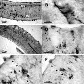
Fig. 2. Digitized image of longitudinal section (20 μm) of adult male Schistosoma mansoni showing GABA-IR in the nervous system. (A) GABA-IR in the longitudinal nerve cords (MC) with numerous cell bodies (arrows) and branching nerves (arrowheads). (B) Detail of longitudinal nerve cords showing GABA-immunoreactive nerve fibres (arrowheads). (C) Typical appearance of longitudinal nerve cords, with GABA-immunoreactive nerve fibres running in bundles or alone towards the entire length of the body. Note lateral nerves that run to outer surface and several bundles of fibres running to the centre of the animal. (D–F) Detail of GABA-immunoreactive neuronal endings (arrowheads) in protusions of tegument. Note bipolar GABA-IR in neuronal cells (arrows) with some processes extending to the tegument and other ones running to the centre of the body.

Fig. 3. Digitized image of longitudinal section (20 μm) of Schistosoma mansoni showing GABA-IR in the attachment structures. (A) Strong and diffuse GABA-IR in both oral and ventral suckers. (B) Detail of oral sucker GABA-IR. Oral sucker (OS) and ventral sucker (VS).
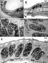
Fig. 4. Digitized images of longitudinal sections (20 μm) of adult male Schistosoma mansoni showing GABA-IR in the reproductive system (A–B). Intense GABA-IR with an amorphous aspect is shown in testicular lobes (TL). (C–E) Detail of testicular lobes GABA-IR. In (A), cerebral ganglia-like structures (arrows) can be seen inside the anterior part of the animal, but neither cell somata nor cerebral commissures can be clearly identified. From the cerebral ganglia, nerves extend in anterior and posterior directions (arrowheads). (B) GABA-IR in the longitudinal nerve cords (arrows) on each side of the gut (G) (arrow). Main nerve cords (MC) and gut (G).
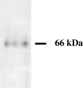
Fig. 5. Immunoblot analysis of glutamate decarboxylase (GAD) in adult Schistosoma mansoni homogenates.
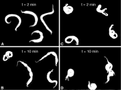
Fig. 6. Digitized images showing the body postures of individual adult male Schistosoma mansoni in the absence (control, A,B) and presence of 100 μM picrotoxin (C,D). Control worms presented a variety of postures, including body waves (B). Note that 100 μM picrotoxin produced corkscrew-like coilings of parasite bodies, modifying the motor activity in a time-dependent manner (C,D). t=time after addition of picrotoxin (C,D) or vehicle (A,B).
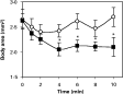
Fig. 7. Time-course of picrotoxin effect on body area of adult male Schistosoma mansoni. Each point represents the mean±S.E.M. of 11 worms in the absence (○) or presence ([squf ]) of 100 μM picrotoxin. Note that these experiments were performed on 3 different days with 3–4 worms each. The effect of picrotoxin was statistically significant (P<0·05) as determined by one-way ANOVA analysis.
 You have
Access
- 15
- Cited by