Introduction
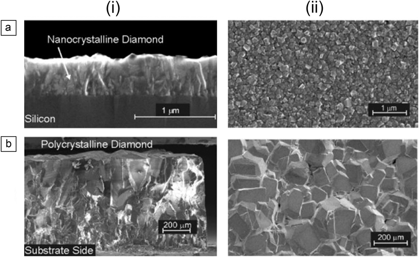
There is a great deal of current interest in modifying solid substrate surfaces with biomolecules due to their utility as biosensors and for implantable biomedical devices. Physical and chemical properties such as wear resistance, chemical inertness, and biocompatibility are the most important parameters guiding the choice of the substrate. As a wide-bandgap semiconductor, diamond combines ultimate electronic properties with exceptional physicochemical properties and bio-inertness (Table I).Reference Nebel1 Applications of diamond ultimately depend on the commercial availability of diamond films. Recent advances made in the controlled deposition of diamond thin films with varying morphologies (Figure 1) have pushed researchers to consider diamond as an alternative carbon-based material.Reference Narayan2
Properties of diamond.

Comparison of different diamond films: (a) nanocrystalline diamond: (i) grain structures and (ii) surface morphology where the typical surface roughness is 30–50 nm; (b) polycrystalline diamond: (i) grain structures and (ii) surface roughness of several tens of micrometers.Reference Nebel1
Ultranano-, nano-, and microcrystalline diamond layers are dominated by grain boundaries that are decorated with sp 2-bonded and amorphous carbon.Reference Nebel1 The volume fraction of sp Reference Narayan2 carbon and grain boundaries depends on the growth parameters used and varies from layer to layer. While nanocrystalline diamond typically has a surface roughness in the range of 30–50 nm, single-crystalline CVD diamond has been optimized to electronic grade quality with atomically smooth surfaces and defect densities of less than 102 cm–2.
To make diamond thin films valuable for biosensing applications, stable and site-specific immobilization of enzymes, proteins, and peptides is required. Covalent grafting of biological molecules is considered the most viable approach for the formation of extremely robust and stable biointerfaces even at elevated temperatures. The high chemical stability and inertness of diamond for a long time have been considered to be incompatible with surface functionalization strategies developed on other semiconductor surfaces, in particular silicon. Modification of hydrogen-terminated silicon surfaces through silicon-carbon (Si–C) bonds or chemical functionalization of the oxide layer through Si–O–Si bonds using silane chemistry are two of many means for providing new functionalities to the semiconductor surface.Reference Boukherroub3 The chemical stability of the resulting semiconductor/organic interface and its biocompatibility were found to be limited, as surface-active electronic defects are easily formed. The rather rapid formation of an insulating oxide layer on silicon also means that surface modification needs to be performed under well-controlled conditions. These disadvantages have been shown to be overcome using diamond interfaces. In the last 15 years, progress has been made in the development of easy, controllable, and specific surface modification methods for the introduction of different functional groups on the diamond surface. These myriad approaches are based on chemical, photochemical, and, in the case of doped diamond, electrochemical concepts.Reference Szunerits, Boukherroub and Narayan4 Thus, despite the high chemical stability of diamond even under harsh environmental conditions, the formation of C–C and C–N bonds is now available. Our discussion here focuses on some of the most common surface functionalization strategies employed on diamond surfaces in order to control the resulting interactions with proteins, peptides, and biological cells.
Approaches for the surface functionalization of thin-film diamond
Although diamond is composed of only carbon atoms, bonding to elements other than carbon (H, O, N) stabilizes the diamond surface. Reproducible diamond surfaces are obtained by exposing freshly grown diamond films to atomic hydrogen, which removes oxidized sites and leaves surface atoms terminated with hydrogen atoms.Reference Thoms, Owens, Butler and Spiro5 H-terminated surfaces can be modified with different organic ligands using two approaches: electrochemical reduction of aryldiazonium salts, and grafting of organic alkenes under UV light illumination at 254 nm.Reference Shin, Rezek, Tokuda, Takeuchi, Watanabe, Nakamura, Yamamoto and Nebel6
Reduction of aryldiazonium salts
The functionalization of diamond by electrochemical reduction of aryldiazonium salts was first reported in 1999 by Kuo, McCreery, and Swain,Reference Kuo, McCreery and Swain7 which generated much interest.Reference Uetsuka, Shin, Tokuda, Saeki and Nebel8,Reference Zhong, Ng, Yang and Loh9 The success of this approach is based on the fact that diazonium salts with a wide range of functional groups (-COOH, -Br, -NO2) are commercially available and can be introduced onto the surface in one step. Diazonium salts can also be easily prepared from aromatic amines, widening their application scope. The unique characteristics of aryldiazonium cations are the very strong electron-withdrawing effect of the diazonium moiety and the high stability of the dinitrogen leaving group (evolution of N2 gas). Both factors account for the relative ease of reducing aryldiazonium salts. The electro-reduction of aryldiazonium salts proceeds through a concerted mechanism, in which electron transfer and dinitrogen loss are concomitant (Figure 2a).Reference Andrieux and Pinson10
(a) Electrochemical linking of organic layers through reduction of aryldiazonium salts; (b) cyclic voltammograms of 4-nitrobenzene diazonium tetrafluoroborate (1 mM in 0.1 M NBu4BF4/acetonitrile, scan rate 0.2 V/s); and (c) schematic illustration of covalent linking of thiolated DNA on diamond modified with 4-nitrobenzene diazonium tetrafluoroborate and using 4-(N-maleimidomethyl)cyclohexane-1-carboxylate (SSMCC) as a bifunctional linker.Reference Shin, Rezek, Tokuda, Takeuchi, Watanabe, Nakamura, Yamamoto and Nebel6,Reference Yang, Baker, Butler, Lee, Russell, Shang, Sun and Hamers11
Electrochemical reduction of the 4-nitrobenzene diazonium salt to 4-nitrophenyl radicals occurs at –0.17 V (versus Ag/Ag+) on boron-doped diamond electrodes, as seen in Figure 2b. The reduction peak decreases rapidly due to surface passivation of grafted aromatic moieties. The interest of nitro aryl-modified diamond for the formation of a biological interface lies in the ability to selectively reduce nitro groups to primary amines,Reference Shin, Rezek, Tokuda, Takeuchi, Watanabe, Nakamura, Yamamoto and Nebel6 followed by a reaction with bifunctional linkers to covalently link DNA and other biomolecules of interest (Figure 2c).Reference Yang, Baker, Butler, Lee, Russell, Shang, Sun and Hamers11–Reference Wenmackers, Vermeeren, vandeVen, Ameloot, Bijnens, Haenen, Michiels and Wagner13 The significance of using electrochemical reduction is that it is a simple pathway for the fabrication of high-density, highly stable bimolecular arrays for applications such as electronic sensing.
While the electrochemical aryldiazonium reduction method is a convenient way for the derivatization of diamond surfaces, precise control of the density of grafted molecules, however, is not a straightforward task. Phenyl radical species, formed next to nitrogen during the grafting process, react directly with the diamond surface, but also with the phenyl ring grafted on the surface to form multilayers.Reference Pinson and Podvorica14 Monolayer formation is only achieved through careful control of the grafting parameters. One way to overcome the problems of multilayer formation using the electrochemical grafting process is the use of chemical grafting, as shown by Lud et al. using 4-nitro-1,1-biphenyl-4-diazonium tetrafluoroborate.Reference Lud, Steenackers, Jordan, Bruno, Gruen, Feulner, Garrido and Stutzmann15 The advantage of the chemical grafting step is its simplicity and that it can be applied to intrinsic undoped hydrogenated diamond. The electrons necessary for the reduction of the diazonium salt are most likely provided by the diamond itself. Other recent examples of the electroless chemical grafting approach are based on the use of “chemical initiators” for the reduction of diazonium salts, such as sodium dodecyl sulfate,Reference Yang, Baker, Butler, Lee, Russell, Shang, Sun and Hamers11,Reference Shul, Actis, Marcus, Opallo, Boukherroub and Szunerits16 hydrochloric acid (0.1 M),Reference Zhong, Loh, Midya and Chen17 or ionic liquids.Reference Shul, Actis, Marcus, Opallo, Boukherroub and Szunerits16
Photochemical grafting
Modification of H-terminated diamond surfaces by photochemical-initiated chlorination/amination/carboxylation processes was one of the first methods for the introduction of different functional groups onto diamond.Reference Takahashi, Tanga, Takai and Okamura18–Reference Ando, Nishitani-Gamo, Rawles, Yamamoto, Kamo and Sato21 Based on this concept, Hamers and co-workers proposed ultraviolet irradiation (λ = 254 nm) of H-terminated diamond covered with liquid films of an appropriate alkene for the covalent modification of H-terminated diamond.Reference Yang, Auciello, Butler, Cai, Carlisle, Gerbi, Gruen, Knickerbocker, Lasseter, Russell, Smith and Harmers22 As depicted in Figure 3a, organic alkenes will graft to a H-terminated surface when illuminated with UV light. This chemistry has been largely used for the covalent bonding of DNA to diamond using, in most cases, amine- or carboxylic acid-protected alkynes (e.g., 10-aminodec-1-ene protected with a trifluoroacetamide functional group (TFAAD); 2,2,2-trifluoroehtyl undecenoate).Reference Yang, Auciello, Butler, Cai, Carlisle, Gerbi, Gruen, Knickerbocker, Lasseter, Russell, Smith and Harmers22–Reference Knickerbocker, Strother, Schwartz, Russell, Butler, Smith and Hamers25 One of the potential benefits of covalent attachment of DNA to diamond is increased long-term stability (Figure 3b). No significant loss of DNA occurred on diamond over 30 hybridization and dehybridization cycles, while DNA-modified silicon, gold, and glassy carbon interfaces showed a significant loss in fluorescence intensity over the 30 hybridization cycles.
(a) Photochemical grafting of alkenes to a H-terminated surface of diamond; (b) comparison of hybridization stability of DNA on different surfaces; and (c) cyclic voltammograms of untreated diamond (black) and an enzyme-modified diamond electrode (red). Reprinted with permission from Reference 28. © 2004 Nature Publishing Group.
Mixed monolayers can be easily formed with this strategy by using two alkenes. Using triethylene glycol undec-1-ene (EG3-ene) in addition to TFAAD, the hydrophilic ethylene glycol groups accessible at the exposed surface allowed for minimization of nonspecific binding of proteins.Reference Lasseter, Clare, Abbott and Hamers26,Reference Stavis, Clare, Butler, Radadia, Carr, Zeng, King, Carlisle, Aksimentiev, Bashir and Hamers27 1-dodecene can be used to control the spacing between functional groups through the formation of mixed monolayers. Härtl and colleagues showed the very challenging task of the covalent linking of proteins to diamond using the photochemical grafting approach.Reference Härtl, Schmich, Garrido, Hernando, Catharino, Walter, Feulber, Kromka, Steinmuller and Stutzmann28 The authors have gone further and demonstrated the ability of the diamond surface to electrochemically drive and detect redox reactions of immobilized enzymes (Figure 3c).
Amine-terminated diamond surfaces
The use of amine functions is one of the most widely employed strategies for linking biomolecules to surfaces through amide bond formation. Amination of diamond represents an attractive strategy for building complex and functional surfaces for biomedical applications. The first report on the possibility of forming NH2-terminated diamond dates back to 1996,Reference Miller and Brown20 where amine functions were incorporated onto diamond by reaction of chlorinated diamond with ammonia gas under UV irradiation. The group of Kawarada reported on the possibility of the formation of DNA micropatterns on diamond via direct amination of H-terminated diamond surfaces (Figure 4a).Reference Zhang, Song, Nakamura, Ueno, Funatsu, Ohdomari and Kawarada29 More recently, plasma rather than gas phase amination processes have been the focus of attention.Reference Szunerits, Jama, Coffinier, Marcus, Delabouglise and Boukherroub30–Reference Wang, Kromka, Houdkova, Babchenko, Rezek, Li, Boukherroub and Szunerits32 Based on the diamond structure, it is expected that the sp Reference Boukherroub3 C–H bonds on the (111) facets will be terminated with -NH2 groups, while the CH2 bonds on the (100) facets will be transformed to imine (C=N) and/or (C–N–C) functional groups. From high-resolution x-ray photoelectron spectroscopy analysis, this behavior was confirmed next to where primary amines (-NH2), secondary amines (C–N–C), and imides (C=N) are found (Figure 4b).Reference Wang, Kromka, Houdkova, Babchenko, Rezek, Li, Boukherroub and Szunerits32 Such surfaces allowed the decoration of diamond with horseradish peroxidase enzymeReference Wang, Kromka, Houdkova, Babchenko, Rezek, Li, Boukherroub and Szunerits32 or with peptides (Figure 4b).Reference Coffinier, Szunerits, Jama, Desmet, Melnyk, Marcus, Gengembre, Payen, Delabouglise and Boukherroub31 The fluorescent images of peptide-modified diamond obtained after incubation with fluorescent-labeled antibody anti-peptide showed that the fluorescence observed from peptide 2 is more important than with peptide 1. This is consistent with specific covalent ligation of peptide 2 with the semicarbazide surface and the non-covalent interaction of peptide 2 with the semicarbazide surface.
(a) Micropatterning DNA on diamond via direct amination in ammonia gas under 254 nm irradiation, surface passivation by fluorination, and DNA immobilization. Fluorescence image after hybridization with Cy5-labeled complementary and noncomplementary DNA. Reprinted with permission from Reference 29. © 2006. American Chemical Society. (b) High-resolution N1s x-ray photoelectron spectroscopy spectrum of diamond aminated by a linear-antenna microwave plasma chemical vapor deposition process.Reference Wang, Kromka, Houdkova, Babchenko, Rezek, Li, Boukherroub and Szunerits32 Formation of peptide array through site-specific α-oxo semicarbazone ligation on aminated diamond and corresponding fluorescence image, top (peptide 1: FLAG-NH2) and bottom (peptide 2: FLAG-COCHO).Reference Wang, Kromka, Houdkova, Babchenko, Rezek, Li, Boukherroub and Szunerits32
Oxygen-terminated diamond surfaces
In spite of the high stability of H-terminated diamond, oxidation is relatively easy to carry out, and diverse processes, including thermal, singlet oxygen, ozone treatment, and electrochemical approaches, have been reported.Reference Szunerits and Boukherroub33 The resulting oxidized surface is so stable that it can be recovered only by hydrogen plasma treatment at elevated temperatures. Since oxygen atoms are divalent, they can simultaneously saturate two surface dangling bonds. Consequently, all oxidation techniques result in a combination of surface oxygen moieties, including hydroxyl (-OH), ketone (C=O), and ether (C–O–C) groups (Figure 5a). The detailed surface composition following oxidation depends sensitively on the specific processing conditions, surface orientation, and surface morphology. Characterization of these oxidized diamond surfaces remains a subject of active research.Reference Klausner, Ghodbane, Boukherroub, Szunerits, Steinmueller-Nethel, Bertel and Memmel34–Reference Wang, Simon, Decorse-Pascanut, Bouttemy, Etcheberry, Li, Boukherroub and Szunerits36 For example, it has been concluded that high concentrations of C–O functions can be obtained on nanocrystalline diamond when oxidized by radio frequency (RF) oxygen plasma, whereas the highest C=O/C–O ratio has been achieved by electrochemical oxidation. Indeed, a diamond surface oxidized by RF plasma was completely free of C=O functions.Reference Ghodbane, Haensel, Coffinier, Szunerits, Steinmüller-Nethl, Boukherroub, Imad-Uddin Ahmed and Schaefer35 The two primary means of functionalization of oxygen-terminated diamond are silanizationReference Boukherroub, Wallart, Szunerits, Marcus, Bouvier and Mermoux37–Reference Coffinier, Galopin, Szunerits and Boukherroub40 and esterification,Reference Szunerits, Shirahata, Actis, Nakanishi and Boukherroub41,Reference Marcon, Wang, Coffinier, Le Normand, Melnyk, Boukherroub and Szunerits42 both using surface hydroxyl groups (Figure 5b).
(a) Oxidation of H-terminated diamond with formation of different oxygen-containing functions and further reaction of surface OH groups with functional silanes or with acids such as 4-benzoyl benzoic acid benzophenone via surface esterification. Photoactivation of benzophenone unit results in H-abstraction reaction to covalently link biomolecules. (b) Fluorescence images after photoimmobilization (i) of streptavidin and revelation with biotin-conjugated Qdots, (ii) of green fluorescence protein, and (iii) of DNA followed by hybridization with Cy5-labeled complementary DNA.Reference Szunerits, Shirahata, Actis, Nakanishi and Boukherroub41,Reference Marcon, Wang, Coffinier, Le Normand, Melnyk, Boukherroub and Szunerits42
Recent developments
One class of reaction that is very attractive for the quick, simple, and efficient grafting of organic moieties onto diamond is the copper (I) catalyzed cycloaddition of azides to alkynes (Figure 6a). The underlying cycloaddition reaction fulfils many of the crucial requirements for mild surface immobilization, including excellent coupling yields, versatility, regioselectivity, excellent functional group tolerance, and stability of the catalytic process in different solvents and pH. Szunerits and co-workers have used azide-terminated mono- and disaccharides (Figure 6a) for the construction of a glycan-modified diamond, where electrochemical impedance spectroscopy allowed for the measurement of the affinity of the sugars to different lectins.Reference Szunerits, Niedziółka-Jönsson, Boukherroub, Woisel, Baumann and Siriwardena43 In addition, the use of the proposed “click” reaction (describes a stereospecific reaction with high yield that has a wide scope and creates only byproducts that can be easily removed along with the solvent) allows for varying the density of the surface-immobilized carbohydrate by performing the “click” reaction of the carbohydrate analog in the presence of a diluting agent such as 4-azidopropanol. Mixed monolayers can be formed in an easy and well-controlled fashion.
“Click” chemistries on diamond: (a) Huisgen 1,3 dipolar cycloaddition between azide and alkyne forming a triazole ring for the formation of glycan-modified diamond interfaces.Reference Szunerits, Niedziółka-Jönsson, Boukherroub, Woisel, Baumann and Siriwardena43 (b) Photo-addition of thiols to alkenes and alkynes; thiol-yne reaction on amine-terminated diamond for the formation a DNA-modified diamond interface DNA hybridization kinetics followed by electrochemical impedance spectroscopy with fully complementary (●) and mismatched DNA (●).Reference Mezian, Barras, Kromka, Houdkova, Boukherroub and Szunerits48
Catalyst-free “click” scenarios are thiol-ene and thiol-yne-based “click” reactions (Figure 6b).Reference Hoyle, Lowe and Bowman44–Reference Caipa Campos, Paulusse and Zuilhof46 While the “click” chemistry characteristics are preserved, the versatility of the reaction is enhanced as it is initiated thermally or photochemically. Alkanes are used in the case of the thiol-ene reaction for the radical addition of one thiolated molecule, while in the case of the thiol-yne-based “click,” two thiol molecules react with one alkyne function, resulting in the formation of a double addition product.Reference Hensarling, Doughty, Chan and Patton47 This reaction was developed on diamond interfaces to immobilize thiolated single-stranded oligonucleotides.Reference Mezian, Barras, Kromka, Houdkova, Boukherroub and Szunerits48 The sensitivity of the DNA-modified diamond interface was assessed through the change in the electrochemical impedance during hybridization with the fully complementary target at different concentrations, and a detection limit of ∼1 nM was recorded.
Conclusion and future trends
A large effort has been expended in understanding and controlling the surface chemistry of diamond over the last decade. To date, diamond can be functionalized with a vast array of biological and therapeutic molecules. Simple surface treatments allow the adjustment of boron-doped diamond to specific biochemical requirements of proteins.Reference Hoffmann, Kriele, Obloh, Tokuda, Smirnov, Yang and Nebel49 The knowledge gained on diamond interfaces can be easily transposed to other types of diamond-based materials such as diamond nanowiresReference Yang, Uetsuka and Nebel50,Reference Szunerits, Coffinier, Galopin, Brenner and Boukherroub51 ,Reference Nebel, Yang, Uetsuka, Osawa, Tokuda and Williams54–Reference Yang, Smirnov and Nebel56 and diamond nanoparticles.Reference Barras, Martin, Bande, Baumann, Ghigo, Boukherroub, Beloin, Siriwardena and Szunerits52,Reference Barras, Szunerits, Marcon, Monfilliette-Dupont and Boukherroub53 ,Reference Krueger57–Reference Rojas, Gispert, Martín, Abad, Menchón, Pareto, Víctor, Álvaro, García and Herance62
Diamond nanowire electrodes (Figure 7a), formed by oxygen plasma etching of diamond, are currently considered as a competitive alternative to carbon nanotubes, as they possess all the advantages of diamond together with that of a nanomaterial. Reported surface functionalization schemes of such wires are currently limited to electrochemical grafting of diazonium salts,Reference Yang, Uetsuka and Nebel50 silanization reactions,Reference Coffinier, Galopin, Szunerits and Boukherroub40 and decoration with nickel nanoparticles.Reference Subramanian, Mortorina, Yeap, Haenen, Coffinier, Zaitsev, Niedziolka-Jonsson, Boukherroub and Szunerits55,Reference Yang, Smirnov and Nebel56 Nebel showed that the enhanced electrical field at the very end of the diamond tips results in a preferential modification of the tip. This gave rise to a detection limit of 2 pM toward complementary DNA (Figure 7b).Reference Yang, Uetsuka and Nebel50 It has recently been shown that nanostructured boron-doped diamond modified with nickel nanoparticles are sensitive interfaces for the investigation of antibody-antigen interactions (Figure 7c).Reference Subramanian, Mortorina, Yeap, Haenen, Coffinier, Zaitsev, Niedziolka-Jonsson, Boukherroub and Szunerits55
(a) SEM image of vertically aligned diamond nanowires (NWs). (b) Sensitivity of DNA-modified diamoond NWs to complementary DNA targets determined by using differential pulse voltammetry. (c) Fabrication of an immunosensor based on a diamond NWs electrode modified with nickel nanoparticles (NPs) and chelated with biotin-tagged proteins.Reference Subramanian, Mortorina, Yeap, Haenen, Coffinier, Zaitsev, Niedziolka-Jonsson, Boukherroub and Szunerits55
Interest in the use of diamond nanoparticles (mostly referred to as nanodiamonds [NDs]) has emerged recently, as these particles are considered particularly well-suited for biological applications that require optical transparency, chemical inertness, hardness, and high specific area (Figure 8). Surface functionalization is applied to customize the particles for different applications, such as increased solubility in biological media, bioconjugation to antibodies, or receptors for drug delivery and diagnostics.Reference Krueger57,Reference Khanal, Vausellin, Barras, Bande, Turcheniuk, Benazza, Zaitsev, Teodurescu, Boukherroub, Siriwardena, Dubuisson and Szunerits58
Interest in and the use of diamond particles as therapeutics and as a contrast agent. (a) Positively charged particles to integrate DNA for gene delivery studies. (Reprinted with permission from Reference 60. © 2010 American Chemical Society.) (b) Phenylboronic acid-modified nanodiamonds (NDs) for HCV treatment (Reprinted with permission from Reference 58. © 2013 American Chemical Society); (c) mannose-modified NDs as antibacterial agent (Reprinted with permission from Reference 52. © 2013 Royal Society of Chemistry); and (d) radiolabeling of organs with 18F-modified ND (1) lung, (2) liver, (3) spleen, (4) kidney, and (5) bladder (Reprinted with permission from Reference 62. © 2011 American Chemical Society).
As the surface chemistry of NDs is more complex than that of diamond, direct surface functionalization often leads to inhomogeneous and often uncontrolled surface chemistry. Intense research efforts have been dedicated to the surface homogenization of the particles.Reference Markovic, Adzic, Cahan and Yeager59–Reference Girard, Petit, Perruchas, Gacoin, Gesset, Arnault and Bergonzo61 In analogy to diamond interfaces, “click” chemistry has been used for the development of glycol-NDs. These newly developed “glyco-NDs” have proven to be of interest for anti-bacterial applications and are currently setting new anti-adhesive strategies against E. coli-derived infection.Reference Barras, Martin, Bande, Baumann, Ghigo, Boukherroub, Beloin, Siriwardena and Szunerits52 However, many challenges remain when working with nanostructured diamond, such as the production of ND particles with sizes smaller than 4 nm, while preventing their aggregation. The future of the development of highly flexible and versatile surface chemistry on NDs is thus promising.
Acknowledgments
This work is funded, in part, by the European Union through a FP7-PEOPLE-IRSES (PHOTORELEASE) and FP7-PEOPLE-ITN (MATCON) grant, the Centre National de Recherche Scientifique (CNRS), the Université Lille 1, and the Nord Pas de Calais region. S.S. especially wants to thank colleagues, students, and post-docs involved in the work discussed, in particular Rabah Boukherroub, Yannick Coffinier, Alexander Barras, Wang Qi, Palaniappan Subramanian, Manakamana Khanal, Paolo Actis, Ken Haenen, Alexander Kromka, Nathalie Simon, Aloysius Siriwardena, and Jean Dubuission.