Introduction
Researchers have investigated the potential of mammalian gonadotrophin releasing hormone (GnRH-I) immunoneutralization for the control of steroidogenesis and spermatogenesis (Khan et al., Reference Khan, Ferro, Koyama, Kinugasa, Song, Ogita, Tsutsui, Murata and Kimura2007). For the past 2 decades, GnRH-I sterilization vaccines have been produced by coupling GnRH-I to an antigenic carrier protein using chemical conjugation techniques. However, such methods generally create some reaction products that require extensive purification in order to provide a homogenous protein (Ulker et al., Reference Ulker, Kanter, Gokdal, Aygun, Karakus, Sakarya, deAvila and Reeves2005). In an attempt to overcome this problem, a recombinant fusion protein was developed to serve as a sterilization vaccine in our laboratory (Fang et al., Reference Fang, Yang, Liu, Zhang, Tao, Wang, Pu and Zhang2009, Reference Fang, Liu, Pu, Wang, Wang and Zhang2010b). The recombinant fusion protein consists of six copies of GnRH-I in a linear sequence (GnRH-I6) and maltose-binding protein (MBP) (MBP-GnRH-I6) (Fang et al., Reference Fang, Yang, Liu, Zhang, Tao, Wang, Pu and Zhang2009, Reference Fang, Liu, Pu, Wang, Wang and Zhang2010b). Its effectiveness in suppressing reproductive function has been demonstrated previously in mice (Fang et al., Reference Fang, Yang, Liu, Zhang, Tao, Wang, Pu and Zhang2009) and in boars (Fang et al., Reference Fang, Li, Liu, Zhang, Tao, Li, Cao, Wang, Wang and Zhang2010a). Initial immunocastration experiments in boars, using MBP-GnRH-I6 in AL(OH)3 administered at 9 and 13 weeks of age, were completely successful in reducing testis size and weight (Fang et al., Reference Fang, Li, Liu, Zhang, Tao, Li, Cao, Wang, Wang and Zhang2010a). Within our research framework of developing an effective vaccine against GnRH-I to immunocastrate boars, several experiments were planned, in which different immunization procedures, including the initial immunization age of the animal and the best dose of immunogen, would be further tested.
The ultrastructure is the detailed structure of a biological specimen, such as a cell, tissue or organ, that can be observed by electron microscopy. It refers in general to the study of cellular structures that are too small to be seen with an optical microscope. Previous studies on testes obtained from immunocastration animals at the light microscopy level have showed marked atrophy of the seminiferous tubules (Brown et al., Reference Brown, Mattner, Carroll, Hoskinson and Rigby1995; D'Occhio et al., Reference D'Occhio, Aspden and Trigg2001; Hilbe et al., Reference Hilbe, Jaros, Ehrensperger, Zlinszky, Janett, Hassig and Thun2006; Einarsson et al., Reference Einarsson, Andersson, Wallgren, Lundstrom and Rodriguez-Martinez2009). Nevertheless, to the best of our knowledge, there is no report to date on ultrastructural testicular changes in GnRH-I immunized animals. Thus, the objectives of the present study were as follows: (1) to evaluate the effectiveness of MBP-GnRH-I6 administered at 12 and 18 weeks of age in suppressing testicular development and testosterone concentrations; and (2) to investigate the ultrastructural appearance of the testis in boars immunized against MBP-GnRH-I6.
Materials and methods
Animals
Eighteen Chinese boars, reared at the DaDun Animal Farm, Shucheng, China, were used in the study. The study has been approved by Animal Care and Use Committee of Anhui Agricultural University. Animals were randomly assigned to two groups that received the following treatments: MBP-GnRH-I6 immunization (n = 9) and MBP immunization (n = 9). All boars had access to food and water ad libitum.
Preparation of antigens and immunization
MBP-GnRH-I6 was prepared using recombinant DNA techniques as have been described previously (Fang et al., Reference Fang, Liu, Pu, Wang, Wang and Zhang2010b). Briefly, GnRH-I6 cDNA was cloned into a pMAL-c4x vector (New England Biolabs, Beijing, China) using standard techniques. MBP-GnRH-I6 fusion protein was over-expressed in Escherichia coli and purified using a maltose affinity column. Nine milligrams of MBP-GnRH-I6 or MBP were dissolved in 9 ml phosphate-buffered saline (PBS) and 9 ml Al(OH)3 adjuvant (Tianbang, Nanjing, China). The first immunization was administered at 12 weeks of age by intramuscular injection of 2 ml of emulsion. The booster injection was given by the same route and at the same dose 6 weeks later. The pigs were slaughtered 6 weeks after the booster immunization.
Collection of blood samples
Blood samples were taken via the jugular vein when animals were 12, 18, 21 and 24 weeks old and centrifuged at 2000 g for 15 min at 4 °C. Serum was harvested and stored at −80 °C until assayed.
Determination of anti-GnRH-I antibody titres
The percentage of [125I]GnRH-I bound by antibodies in each serum sample (1:10, 1:100, 1:1 000 dilution) was quantified in duplicate using methods described by Johnson et al., (Reference Johnson, DeAvila, Chang and Reeves1988). The anti-GnRH-I titre is expressed as a per cent of total [125I]GnRH-I bound to antibody. An anti-GnRH-I titre was not evident (<2% [125I] GnRH-I bound by a 1:100 serum dilution) in controlled pigs. Antibody response to immunization was considered to be significant when a 1:100 dilution of serum bound more than 10% of [125I]GnRH-I added. A ‘good’ response to immunization was arbitrarily defined as antibody in a 1:1000 dilution of serum sufficient to bind more than 10% of [125I] GnRH-I added (Adams et al., Reference Adams, Daley, Adams and Sakurai1996).
Determination of testosterone
Serum testosterone concentrations were measured using a commercial radioimmunoassay (RIA) kit according to the manufacturer's instructions (Beifang Biotechnology Research Institute, Beijing China). The limit of detection was 0.002 ng/ml. The intra- and inter-assay CV was less than 5.42% and 9.75%, respectively. The sensitivity was 2.03 pg/ml. The correlation coefficient of the standard curve was 0.9998.
Measurement of testicular size
The testes were analyzed and the epididymides were removed. Testicular weight was recorded and the external size of the testis was measured using a caliper.
Light microscopy of paraffin sections
Testicular tissue samples were fixed in 4% formalin, and were paraffin-embedded, and then sectioned at 5 μm thickness. The slices were stained with hematoxylin and eosin (H&E). Morphological changes in the testes were evaluated under a microscope.
Transmission electron microscopy of thin sections
After slaughter, a small amount of testicular tissue was fixed in 2.5% glutaraldehyde for 4 to 6 h, and post-fixation was accomplished in 1% osmium tetroxide for 1 h. The samples were subjected to an alcohol dehydration series (30% 15 min, 50% 15 min, 70% 6–12 h, 80% 15 min, 95% 15 min, 100% 40 min). The tissues were immersed in 1,2-epoxypropane (Lingfeng Chemical Co. Ltd) for 30 min, and then transferred to 1,2-epoxypropane and resin Epon812 (1:1) for 2 h. Samples were individually embedded in Epon812 (Serva) for 2 h. Resin blocks were solidified at 45°C for 12 h and 65°C for at least 48 h. Ultrathin sections (70 nm thick) were prepared from each tissue with an ultrathin section machine (LKBNUBA, NOVA) and blade (LKB2178, knife maker II, BROMMA). Sections were stained with 1% (w/v) methanolic uranyl acetate (Lanzhou State-owned Factory 404) for 30 min, and then washed three times in deionized water for a total of 15 min, and stained with lead citrate for 30 min.
The sections were rinsed in a stream of distilled water and dried prior to examination. Sections were visualized on a transmission electron microscopy (JEM-1230, Japan).
Acquisition and analysis of data
Eight to 10 random sections were taken to represent each tissue. The diameter of the seminiferous tubule was measured, and the number of germ cells at different developmental stages in the tubules was counted using morphology analysis software (Images Advanced 3.2, MOTIC, Hong Kong, China). The thickness of basement membranes was measured with the specific software (Image-Pro Plus 6.0) and data obtained were finally transported to Excel spreadsheets for calculation of average values. Final data were expressed as mean ± standard deviation (SD).
Results
Anti-GnRH-I antibody titre
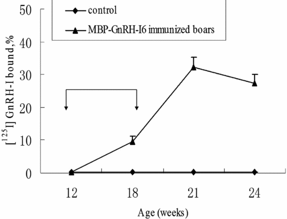
Results revealed that mean antibody binding of [125I] GnRH-I in vaccinated boars increased steadily from 12 weeks of age, and reached a peak value at 21 weeks (Fig. 1). Antibody activity differed significantly between MBP-GnRH-I6 and control pigs from week 18 through to week 24 (P < 0.05) (Fig. 1), suggesting that MBP-GnRH-I6 immunization induced a strong anti-GnRH-I immune response.
Mean percentage of [125I]GnRH-I bound in a 1:1000 dilution of serum from pigs immunized against MBP-GnRH-I6 or MBP (control). Arrows (↓) indicate immunization schedule. Antibody activity differed (P < 0.05) between MBP-GnRH-I6 and control pigs from week 18 through to week 24.

Testosterone concentrations
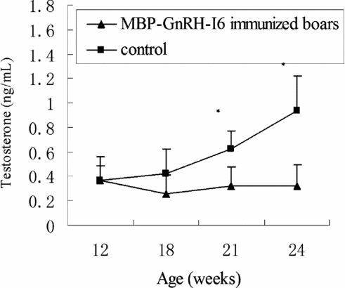
Serum testosterone levels in control animals increased steadily from 12 weeks of age, but serum testosterone levels in MBP-GnRH-I6 immunized boars remained low throughout the study (Fig. 2), serum testosterone levels were lower than control animals, although not statistically significant (P > 0.05) at 18 weeks of age. Except at 12 and 18 weeks of age, serum testosterone concentrations in immunized animals were lower than those of the control boars (P < 0.05) (Fig. 2).
Changes of serum testosterone concentrations in MBP-GnRH-I6 immunized boars and control animals from 12 to 24 weeks of age. The first immunization was delivered at 12 weeks of age and the booster at 18 weeks. Values presented are mean ± standard deviation (SD) (n = 9). *Significant difference (P < 0.05) between groups.
Testicle parameters and light microscopy
Table 1 summarizes the mean testicular weight and size, and the diameter of the seminiferous tubule. Mean ± standard deviation (SD) testicular weight and size were significantly (P < 0.01) reduced in all MBP-GnRH-I6 immunized boars compared with control animals. Histologically, a significant (P < 0.01) diminution of the mean (±SD) diameter of the seminiferous tubules was observed in treated compared with control pigs.
Testes weights and sizes, and the diameter of seminiferous tubules of boars immunized with MBP-GnRH-I6 as compared with control
Note: The values are mean ± standard deviation (SD). aSignificant difference (P < 0.01).
In normal testicular tissue, spermatogenesis was fully developed from spermatogonia up to spermatozoa (Fig. 3a,c). Testes of all MBP-GnRH-I6 immunized animals demonstrated severe hypoplasia, with small seminiferous tubules that contained degenerated spermatogonia, spermatocytes or spermatids (Fig. 3b,d). Active spermatozoa were not observed (Fig. 3d). Furthermore, the number of germ cells at every developmental stage in the seminiferous tubules of the MBP-GnRH-I6 immunized boars was significantly less (P < 0.01) than that of the control group (Table 2).
The number of germ cells at various developmental stages in seminiferous tubules of boars (unit: number)
Note: The values are mean ± standard deviation (SD). aSignificant difference (P < 0.01).
Testicular tissue of boars immunized with control (a, c) and MBP-GnRH-I6 (b, d). (a, b) Overall structure of testicular tissue. (c, d) Cells in spermatogenesis are shown as spermatogonia (short arrows), primary spermatocytes (long arrows), spermatids (ellipse area), and spermatozoa (square area). Testicular tissue was stained with hematoxylin and eosin stain. (a, b) Scale bar = 100 μm. (c, d) Scale bar = 25 μm. S, seminiferous tubules.
Testicle electron microscopy
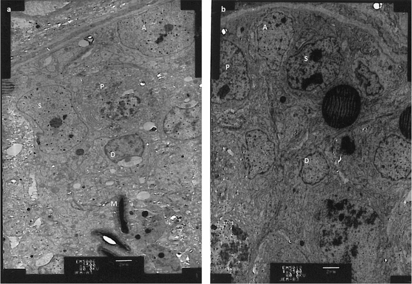
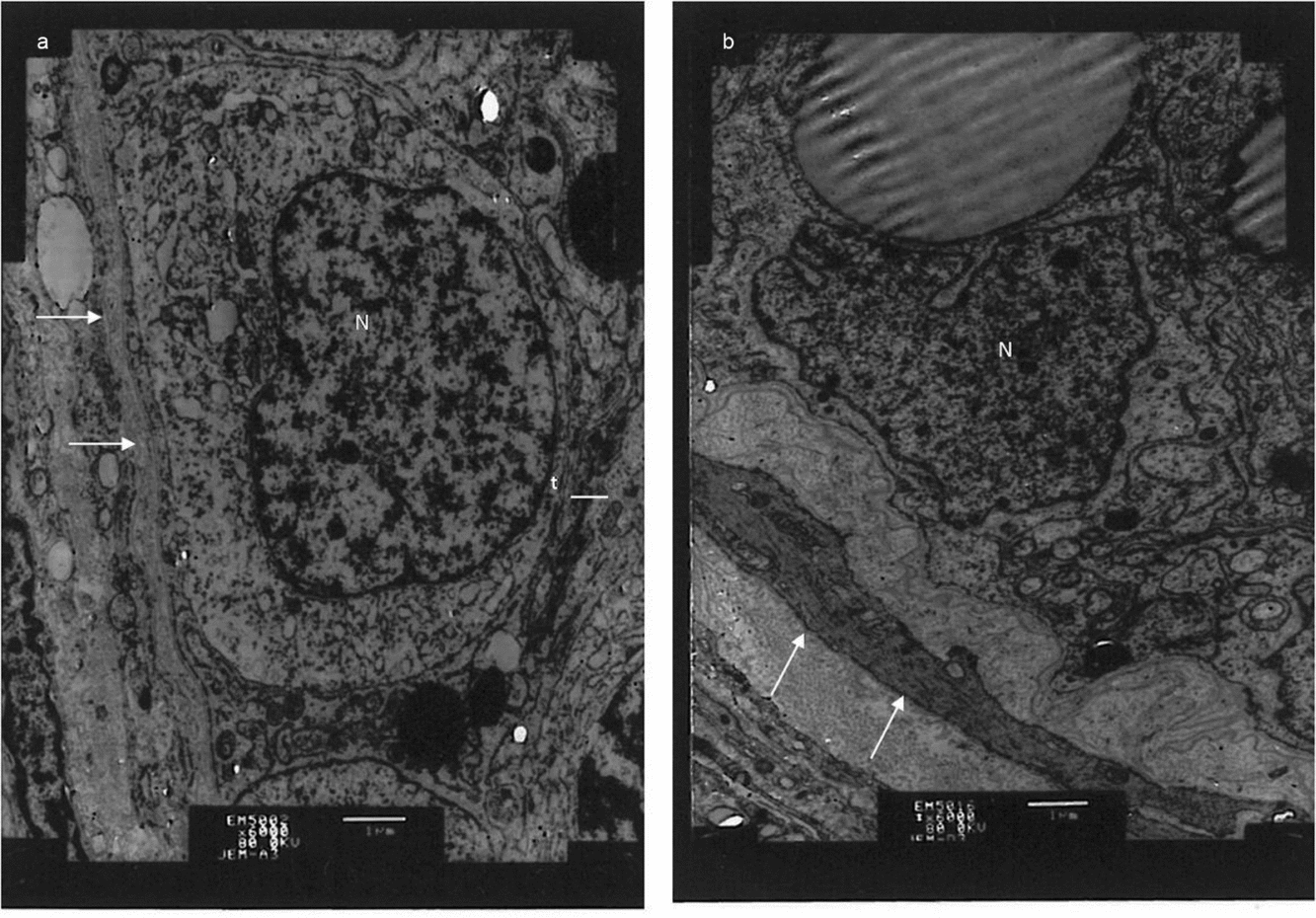
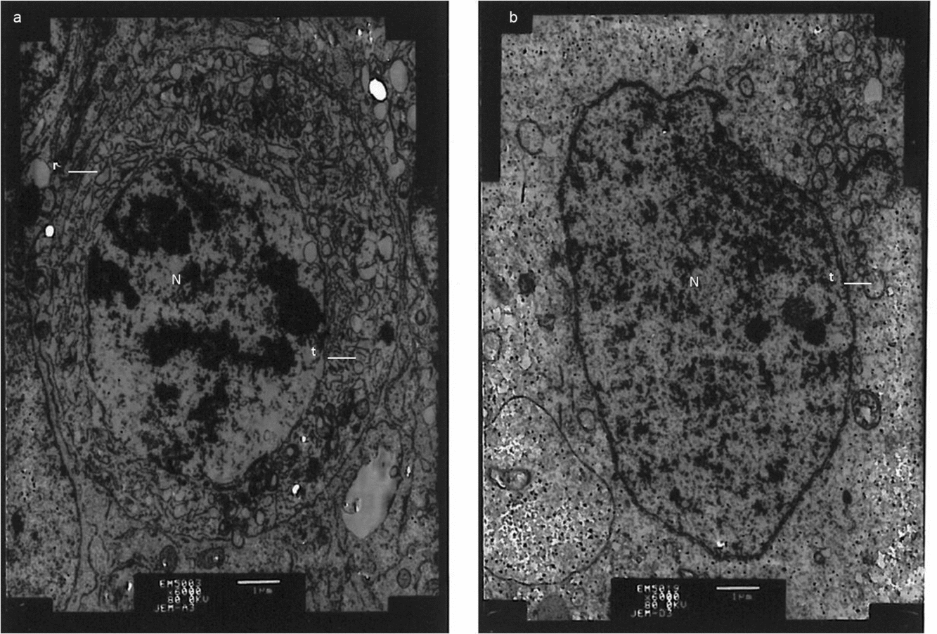
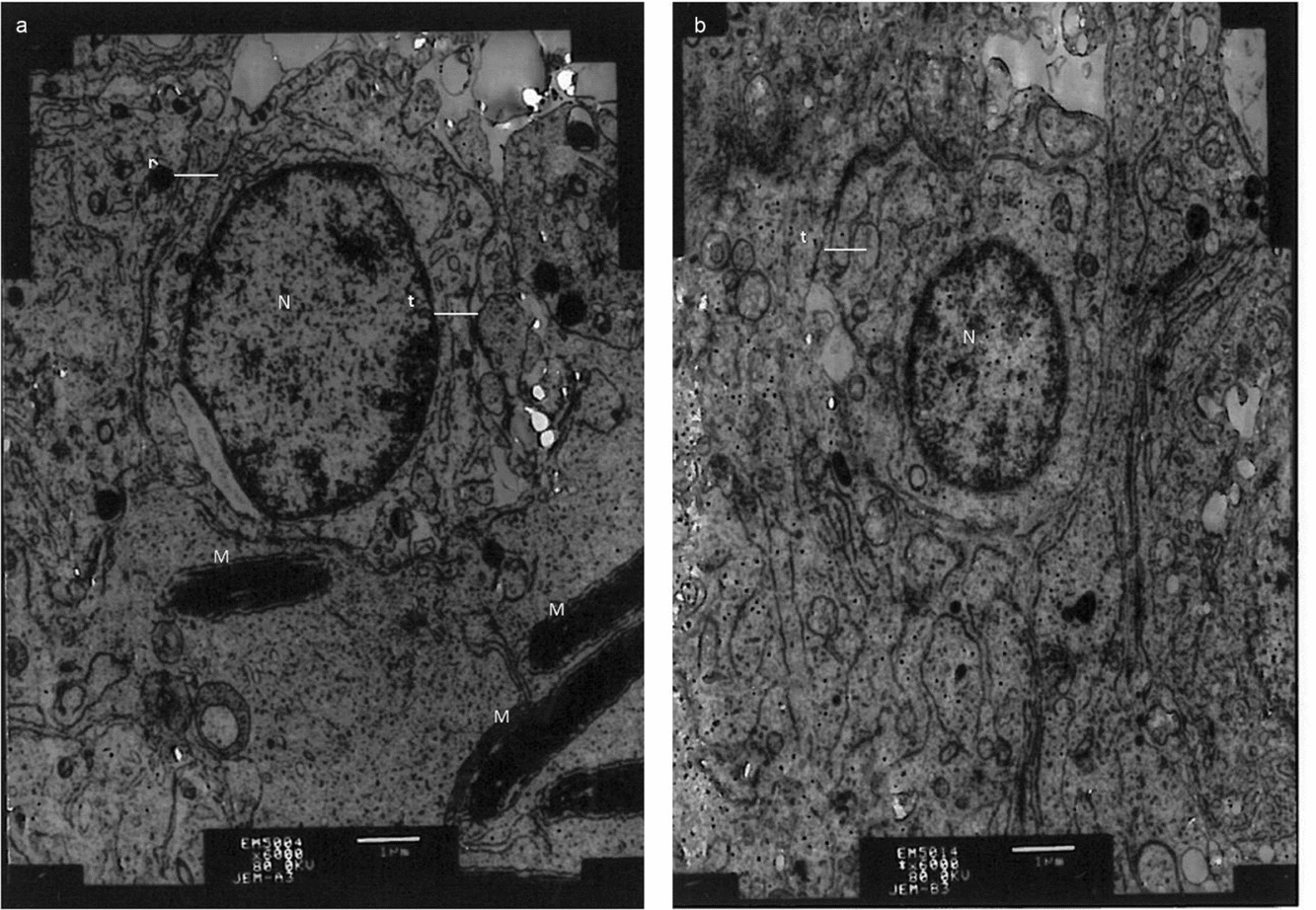
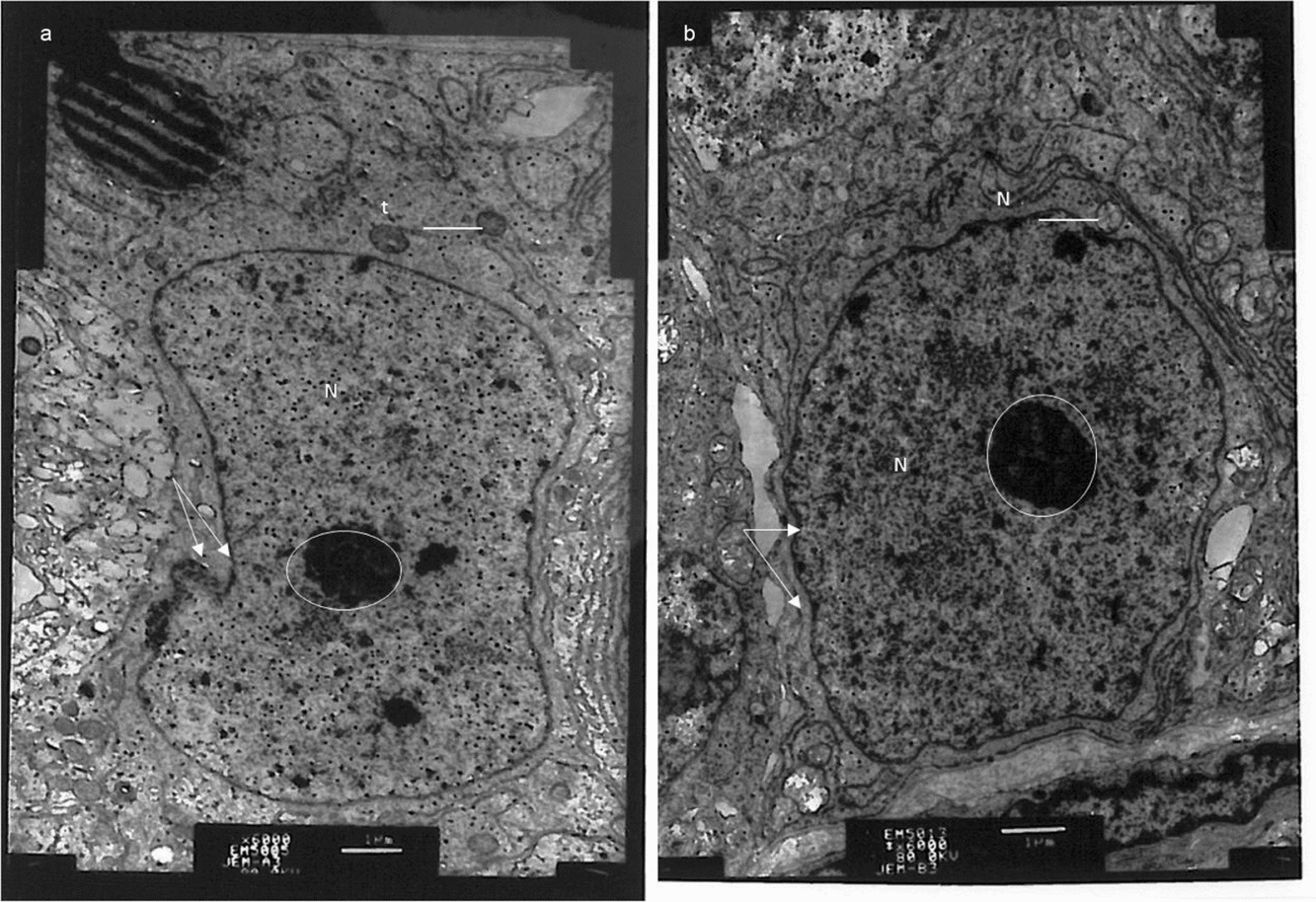
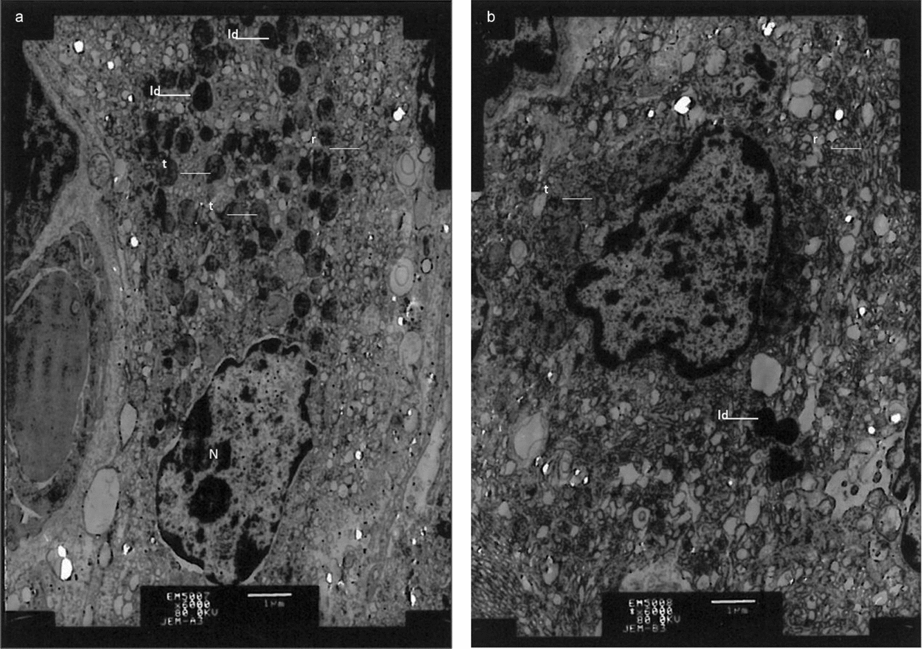
Low power electron microscopic observations confirmed and extended the information obtained from light microscopic images. Seminiferous tubules of the testes of control animals possessed epithelia that contained Sertoli cells and germ cells at various stages, covering the complete process of spermatogenesis (Fig. 4a). The spermatogonia, oval in shape, were closely associated with the basal lamina, and the nuclei contained distinct chromatin networks in the process of condensation and with well defined nuclear membranes (Fig. 5a). The spermatocytes presented as round configurations with prominent nuclei. The nuclei contained distinct chromatin networks in the process of condensation and with well defined nuclear membranes. The cytoplasm contained dispersed mitochondria and loose networks of rough endoplasmic reticulum (Fig. 6a). Round spermatids were characterized by well defined nuclei with distinct nuclear membranes and chromatin networks, and the cytoplasm was occupied by mitochondria (Fig. 7a). The Sertoli cells exhibited typical columnar nuclei with a well defined nucleolus, and the nuclear membrane appeared as a deep hollow and a few shallow hollows (Fig. 8a). The Leydig cells showed distinct nuclear characteristics. The cytoplasm showed prominent lipid bodies, well defined vesicles of smooth endoplasmic reticulum, and mitochondria (Fig. 9a).
Ultrastructure of the testis of animals showing seminiferous tubules. (a) Control animals. (b) MBP-GnRH-I6 immunized boars. Cells or cell organs are shown as Sertoli cells (S), spermatogonia (A), primary spermatocytes (P); spermatids (D), spermatozoa (M). Scale bar = 2 μm.
Ultrastructure of the testis of animals showing spermatogonia and the basement membrane. (a) Control animals. (b) MBP-GnRH-I6 immunized boars. Cell organs are shown as the basement membrane (arrows), mitochondrion (t) and nucleus (N). Scale bar = 1 μm.
Ultrastructure of the testis of animals showing primary spermatocyte. (a) control animals. (b) MBP-GnRH-I6 immunized boars. Cell organs are shown as mitochondrion (t) and endoplasmic reticulum (r), nucleus (N). Scale bar = 1 μm.
Ultrastructure of the testis of animals showing spermatids. (a) Control animals. (b) MBP-GnRH-I6 immunized boars. Cells or cell organs are shown as a mitochondrion (t) and endoplasmic reticulum (r), nucleus (N), spermatozoa (M). Scale bar = 1 μm.
Ultrastructure of the testis of animals showing Sertoli cells. (a) Control animals. (b) MBP-GnRH-I6 immunized boars. Cells or cell organs are shown as the nucleolus (ellipses); nuclear membrane hollow (double arrows); mitochondrion (t) and nucleus (N). Scale bar = 1 μm.
Ultrastructure of the testis of animals showing Leydig cells. (a) Control animals. (b) MBP-GnRH-I6 immunized boars. Cells or cell organs are shown as nucleus (N); mitochondrion (t) and endoplasmic reticulum (r), lipid droplet (ld). Scale bar = 1 μm.
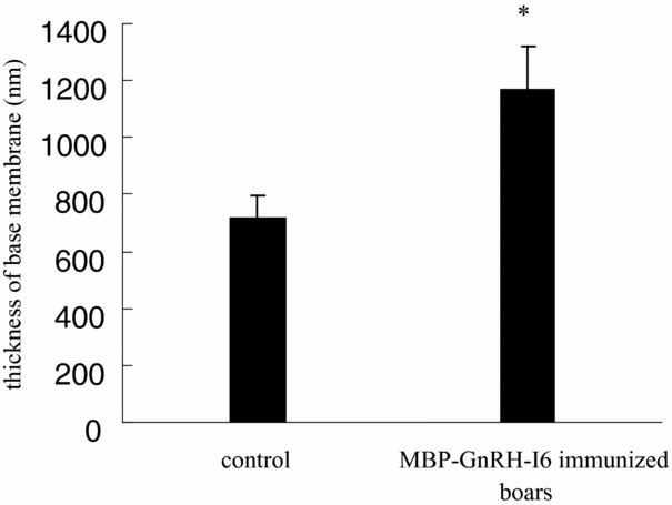
The testes of animals treated with MBP-GnRH-I6 displayed germ cells with abnormal structure (Fig. 4b) and seminiferous tubules with a thick basement membrane (Figs. 5b and 10). The ultrastructural characteristics of the Sertoli cells and the germ cells were indicative of disturbed spermatogenesis (Fig. 4b). The nuclear and cytoplasmic volumes of the spermatogonia were reduced (Fig. 5b). The cytoplasmic organelles were sparse, and mitochondria were few and vacuolated in the spermatocytes (Fig. 6b) and spermatids (Fig. 7b). The Sertoli cells showed evidence of severe changes in the nuclei, the shape was oval and the nuclear membrane appeared as many shallow hollows and no deep hollow (Fig. 8b). The Leydig cells showed fewer lipid droplets and mitochondrial vacuolization (Fig. 9b).
The thickness of the basement membrane in seminiferous tubule of the boar testis. *Significant difference (P < 0.05) between groups.
Discussion
The results of the present study demonstrate that administration of recombinant MBP-GnRH-I6 to boars elicits clear inhibitory changes in serum testosterone levels and testicular development, in particular thickening of the basement membrane of the seminiferous tubule, and aberrant cell organelles of germ cells, Sertoli cells and Leydig cells.
Effective immunization against GnRH-I is a practically feasible alternative to surgical castration in pigs. Previously, the authors have shown that boars immunized against recombinant MBP-GnRH-I6 at the age of 9 weeks demonstrated a reduction in testosterone levels and testicular size (Fang et al., Reference Fang, Li, Liu, Zhang, Tao, Li, Cao, Wang, Wang and Zhang2010a). In the current study, we change the immunization schedule, that is, the first immunization took place at the age of 12 weeks, and the very effective second (booster) immunization at the age of 18 weeks, with the result that the pigs could profit from the presence of testosterone until such time that an effective amount of anti-GnRH-I antibody levels had been produced. Antibody binding of [125I] GnRH-I appeared to be maximal at the age of 21 weeks. The findings of the present study are consistent with other similar reports (Hilbe et al., Reference Hilbe, Jaros, Ehrensperger, Zlinszky, Janett, Hassig and Thun2006; Einarsson et al., Reference Einarsson, Andersson, Wallgren, Lundstrom and Rodriguez-Martinez2009).
Although testicular development was suppressed in all mentioned animal studies, including those involving the boar (Ulker et al., Reference Ulker, Kanter, Gokdal, De Avila and Reeves2001), the rat (Giri et al., Reference Giri, Chaudhuri, Jayashankar, Neelaram, Jayaraman and Talwar1990), the dog (Jung et al., Reference Jung, Moon, Cho, Yeh, Kim, Chang, Park, Song, Kim, Park, McOrist, Choi and Lee2005), the bull (Adams et al., Reference Adams, Daley, Adams and Sakurai1996; Huxsoll et al., Reference Huxsoll, Price and Adams1998; Cook et al., Reference Cook, Popp, Kastelic, Robbins and Harland2000), the ram (Kiyma et al., Reference Kiyma, Adams, Hess, Riley, Murdoch and Moss2000) and the monkey (Chappel et al., Reference Chappel, Ellinwood, Huckins, Herbert and Spies1980), the timing of the first immunization had a significant effect on testicular development. Brown et al. (Reference Brown, Mattner, Carroll, Holland, Paull, Hoskinson and Rigby1994) obtained better testicular suppression in ram lambs immunized at 20–21 weeks of age than those immunized at 3–4 weeks. In contrast, in the present study, immunization was performed at 12 weeks of age, and testicular size and weight were reduced approximately 29.96% and 65.78%, respectively, when compared with the control boars. However, in the previous study, immunization was performed at 9 weeks, and testicular size and weight were reduced approximately 21% and 46%, respectively (Fang et al., Reference Fang, Li, Liu, Zhang, Tao, Li, Cao, Wang, Wang and Zhang2010a). In addition, anti-GnRH-I antibody levels in pigs immunized at 12 weeks of age was higher than those at 9 weeks of age (27.19% versus 15.89%). These results suggest that the optimal age for immunization is near prepuberty. Alternatively, the reduced response to immunization at 9 weeks of age may be due, at least in part, to the extended interval between immunization and slaughter in this group, to account for the declining anti-GnRH-I titre at the slaughter. Similar studies reported that nearly half of the bulls immunized at 1.5 months of age failed to reach the anti-GnRH titre level characteristic of a ‘good’ response. In contrast, all bulls immunized at 7 or 12 months of age reached the ‘good’ antibody response threshold (Adams et al., Reference Adams, Daley, Adams and Sakurai1996). Enhanced suppression was thus obtained using the current immunization schedule. As the anti-GnRH-I vaccine, whether administered at 9 weeks or 12 weeks, is effective in suppressing testicular development for considerable periods, commercial producers have a wide scope in terms of the timing of immunization.
Active immunization against GnRH-I in this study clearly demonstrates that boars are capable of generating high GnRH-I antibody titres. When chronically sustained, these antibody titres can result in depletion of luteinizing hormone (LH) and testosterone, with concomitant changes in testicular structure and function (Awoniyi et al., Reference Awoniyi, Chandrashekar, Arthur, Schanbacher and Falvo1988). Having succeeded in reducing serum testosterone levels to those observed following hypophysectomy, there was a striking reduction in testicular weight at autopsy, and histological alterations in both the seminiferous epithelium and the Leydig cells (Morat Reference Morat1977).
Immunological castration is achieved by immunizing boars against GnRH-I to disrupt the hypothalamic–pituitary–gonad axis, thereby effectively inhibiting pituitary LH and follicle-stimulating hormone (FSH) secretion. This action leads to a reduced production of testosterone, as LH is necessary for testosterone production. In our study, the observed reduction in serum testosterone levels and in testicular weights in GnRH-I immunized boars correlated well with the morphologic appearance of the Leydig cells and the seminiferous tubules. As the rate of testosterone synthesis and secretion by Leydig cells is primarily dependent upon LH secretion, depletion of LH as a result of GnRH-I immunization disrupted the cell organelles, and resulted in testosterone depletion.
The reduction in the seminiferous tubule diameter, the absence of spermatids, vacuolization and disruption of Sertoli cells, Leydig cells and germ cells in GnRH-I immunized boars, all support the curtailment of spermatogenesis. Normal Sertoli cells were observed in rabbits after immunization against GnRH-I (Arimura et al., Reference Arimura, Sato, Kumasaka, Worobec, Debeljuk, Dunn and Schally1973). In contrast with this finding, Sertoli cells appear to have affected to a greater extent in our study, suggesting that these cells are principal targets for the action of testosterone in the testis. This finding also indicates that Sertoli cells in boars are more sensitive to testosterone deprivation. In addition, a thickened basement membrane was observed in the current study. A thickened basement membrane is often a feature of the diseased human testes (Jarow et al., Reference Jarow, Budin, Dym, Zirkin, Noren and Marshall1985; Haider et al., Reference Haider, Talati and Servos1999). The thickened membrane may interfere with blood-borne substances reaching the seminiferous epithelium, thus interfering with normal germ cell development. The ultrastructural defects in spermatocytes and spermatids, particularly in those that interfere with the spermiation process, could be a result of disturbances in the microenvironment of the Sertoli cells and of the basement membrane.
In conclusion, active immunization against GnRH-I affects the structure of Leydig cells, Sertoli cells and the basement membrane of seminiferous tubules, with a corresponding depletion of testosterone and germinal cells, and curtailment of spermatogenesis.
Acknowledgements
This work was supported by a grant from the Natural Science Foundation of Anhui Education Department (Grant KJ2011A117), and Anhui Provincial Natural Science Foundation (Grant 1208085MC39), and Specialized Research Fund for the Doctoral Program of Higher Education from Ministry of Education Science and Technology Development Center, Beijing, P. R. China (Grant 20123418110004). The authors report no conflicts of interest. The authors alone are responsible for the content and writing of the paper.