Introduction
Sutra neti is an ancient Indian practice of passing a thread or similar material like a rubber catheter (rubber neti) through the nose and out of the mouth.Reference Ramalingam and Smith1 These practices are believed to be relatively safe and are widely practised with or without prescription around the world. Acquired velopharyngeal stenosis is a rare entity usually resulting as a complication of uvulopalatopharyngoplasty (UPPP), adenoidectomy and in post-radiotherapy cases of carcinoma nasopharynx.Reference Hussein, Tan, Chong, Narayanan and Omar2,Reference Krespi and Kacker3
To our knowledge, this is the first case report correlating velopharyngeal stenosis to the practice of sutra neti. We also report successful treatment of velopharyngeal stenosis by coblation.
Case report
A 67-year-old male patient presented to the out-patient department of a tertiary care hospital with complaints of bilateral nasal obstruction, mouth breathing, anosmia and a change in voice in the last two years. The patient had a history of practising sutra neti and rubber neti on a regular basis. While practising sutra neti, the patient had multiple separate events of oral bleeding and bleeding from both nostrils. Despite this, the patient continued his practice of sutra neti.
In 2015, the patient sought medical attention at another hospital because of his progressing symptoms. A diagnosis of velopharyngeal stenosis was made. In June 2015, the patient underwent adhesiolysis by laser. The symptoms improved transiently but came back within 18 months. In 2017, the patient underwent palatoplasty and removal of adhesions using cold steel instruments. Because of the persistence of symptoms even after the procedure, the patient sought an opinion at our hospital.
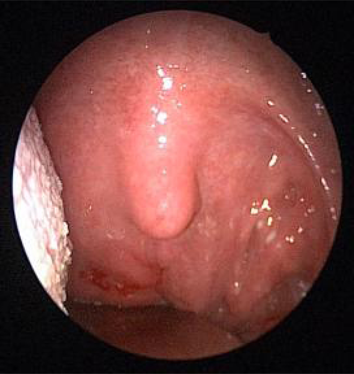
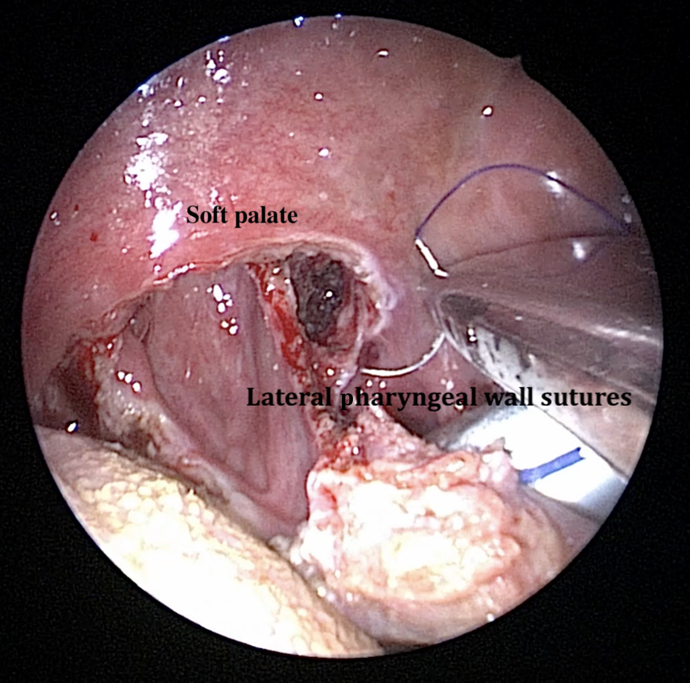
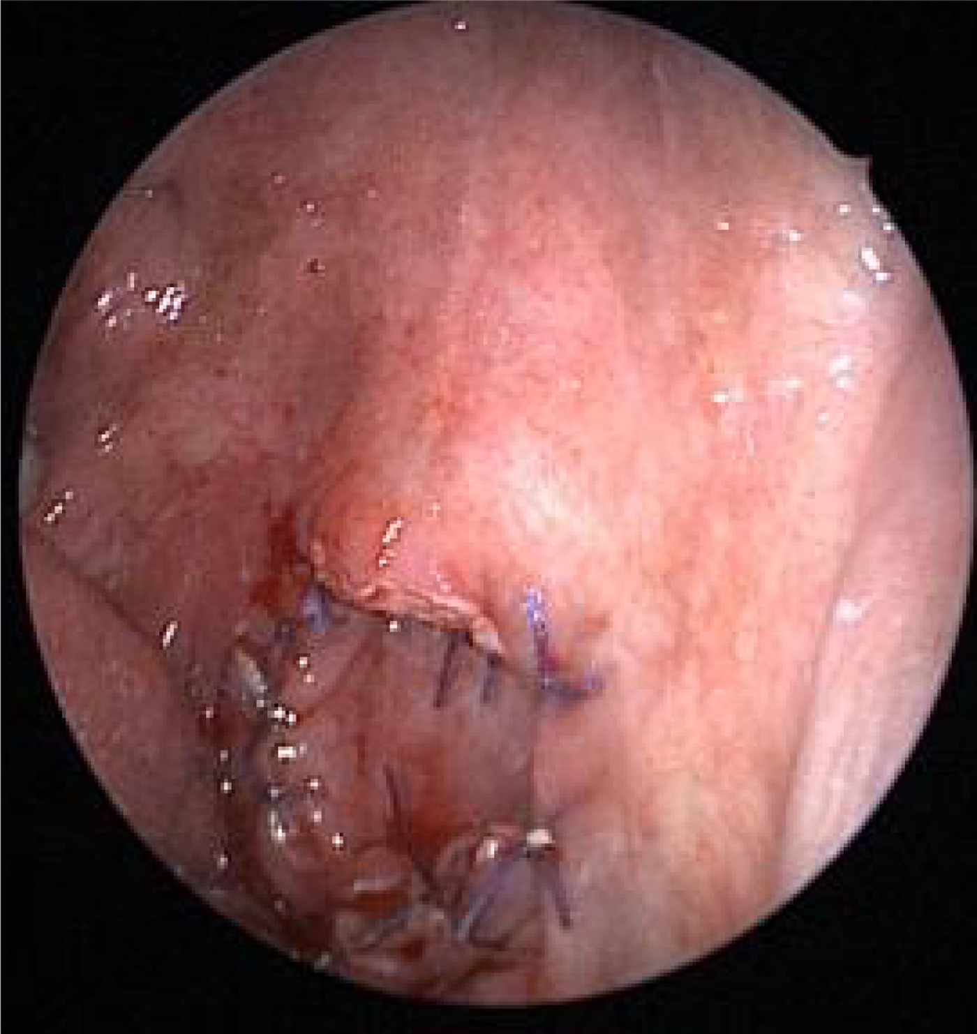
Nasal endoscopy examination showed that the soft palate was completely adherent to the posterior pharyngeal wall causing complete closure of the velopharyngeal inlet (Figure 1). The adhesions were released using the EVAC 70 Coblation Wand (Smith & Nephew, London, UK), and the raw areas on the palate were sutured using 3-0 Vicryl (Ethicon, Cincinnati, Ohio, USA; Figures 2 and 3). No stent was placed. Post-operatively, the patient was relieved of his symptoms, and the velopharyngeal inlet was patent at 12 months’ follow up.
Image showing complete stenosis between the uvula and posterior pharyngeal wall.

Image showing the intra-operative appearance after excision of the stenosed segment.

Image showing the immediate post-operative appearance after excision of the adhesions.

Discussion
Historically, acquired velopharyngeal stenosis was associated with maxillofacial trauma and infective conditions like syphilis.Reference Krespi and Kacker3 Conditions such as lupus, rhinoscleroma, acid burns, diphtheria and scarlet fever are also known to cause this entity.Reference Giannoni, Sulek, Friedman and Duncan4 Currently, surgical trauma, due to procedures like UPPP and adenoidectomy, is the most common cause.Reference Stevenson5 The non-judicious use of electrocautery, infection, creation of raw areas due to improper tissue handling and trauma caused by posterior nasal packing are the predisposing factors.Reference Fairbanks6
In the present case study, repeated use of a thread and rubber catheter led to trauma to the posterior pharyngeal wall and the velum, as evidenced by multiple episodes of bleeding after the procedure. The continued practice led to the persistence of raw areas, resulting in synechiae formation between the soft palate and posterior pharyngeal wall leading to velopharyngeal stenosis.
The management of velopharyngeal stenosis is challenging because of the high chance of recurrence despite complex surgical procedures.Reference Lee, Tang, Singh, Kumar and Singh7 The different management options described in the literature include: palatal eversion, buccal mucosal graft repair, bilateral Z-pharyngoplasty, adhesiolysis by carbon dioxide laser, nasopharyngeal stenting and mitomycin C application.Reference Abdel-Fattah8–Reference Eppley, Sadove, Hennon and van Aalst12 Relatively newer techniques include robot-assisted bivalved palatal flap technique and plasma radiofrequency-based ablation (coblation).Reference Vicini, D'Agostino, Meccariello, De Virgilio and Montevecchi13,Reference Karakoc, Polat, Akcam and Gerek14 Coblation is a relatively simple method and is known to cause significantly less mucosal damage, tissue oedema and earlier re-epithelisation when compared with electrocautery.Reference Madgy, Belenky, Dunkley and Shinhar15 Karakoc et al. described successful treatment of velopharyngeal stenosis in two patients using a coblator-assisted transnasal endoscopic approach.Reference Karakoc, Polat, Akcam and Gerek14 Madgy et al. reported three cases in which transoral adhesiolysis was done by coblation.Reference Madgy, Belenky, Dunkley and Shinhar15 No procedure-related complication was noted in any of the patients.
• Sutra neti is an ancient Indian yoga technique for keeping the nasal passages healthy
• This technique can cause velopharyngeal stenosis
• The general population and physicians need to know that this complication can occur
• Treatment requires surgical intervention
Conclusion
Any procedure or disease condition that results in raw areas over the posterior nasopharynx and velum can lead to velopharyngeal stenosis. Vigorous sutra neti can lead to the creation of raw areas at these locations.
With the ancient practice of yoga becoming popular in the west, the general population and physicians need to be informed of the side effects of this ancient Indian practice, even if the incidence is minimal. The use of coblation adhesiolysis is a relatively safe, physician and patient friendly option for management of velopharyngeal stenosis.
Competing interests
None declared