Introduction
Atrophy and cerebral small-vessel changes, characterized by white-matter lesions (WMLs) and lacunar infarcts, are common findings on magnetic resonance imaging (MRI) in the elderly (Brickman et al. Reference Brickman, Schupf, Manly, Luchsinger, Andrews, Tang, Reitz, Small, Mayeux, DeCarli and Brown2008; Geerlings et al. Reference Geerlings, Appelman, Vincken, Algra, Witkamp, Mali and van der Graaf2010). These changes result from loss of the neuronal and dendritic architecture and chronic or acute ischaemia, leading to incomplete infarction, demyelination and concomitant necrosis (Pantoni, Reference Pantoni2002; Freeman et al. Reference Freeman, Kandel, Cruz, Rozkalne, Newell, Frosch, Hedley-Whyte, Locascio, Lipsitz and Hyman2008). Although atrophy and small-vessel changes are often asymptomatic, they have been associated with an increased risk of cognitive impairment (van der Flier et al. Reference van der Flier, van Straaten, Barkhof, Verdelho, Madureira, Pantoni, Inzitari, Erkinjuntti, Crisby, Waldemar, Schmidt, Fazekas and Scheltens2005) and late-life depression (Steffens et al. Reference Steffens, Krishnan, Crump and Burke2002; Ikram et al. Reference Ikram, Luijendijk, Vernooij, Hofman, Niessen, van der Lugt, Tiemeier and Breteler2010).
The underlying mechanisms contributing to the increased risk of late-life depression are not yet fully understood. The ‘vascular depression’ hypothesis postulates that small-vessel lesions predispose to late-life depression by disrupting emotion-regulating prefrontal structures or their modulating pathways (Alexopoulos et al. Reference Alexopoulos, Meyers, Young, Campbell, Silbersweig and Charlson1997a; Krishnan et al. Reference Krishnan, Hays and Blazer1997). In addition, age-related degenerative changes could also play a role in the disruption of emotion-regulating pathways (Alexopoulos, Reference Alexopoulos2001). Motivational symptoms are considered as a dominant feature of vascular depression, including psychomotor retardation and loss of interest (Alexopoulos et al. Reference Alexopoulos, Kiosses, Klimstra, Kalayam and Bruce2002; Taylor et al. Reference Taylor, Steffens and Krishnan2006).
Direct evidence supporting the hypothesis that disruption of frontal-subcortical pathways by small-vessel or degenerative changes is associated with a motivational symptom profile is scarce, because few studies examined whether specific locations of small-vessel and degenerative changes were associated with characteristic depressive symptoms. Studies examining anatomic locations of cerebral changes on MRI often did not assess individual depressive symptoms, but an overall depressive symptom score (de Groot et al. Reference de Groot, de Leeuw, Oudkerk, Hofman, Jolles and Breteler2000; Steffens et al. Reference Steffens, Krishnan, Crump and Burke2002). One study that did, observed that deep WML severity was associated with symptoms of impaired motivation, concentration and decision making, but associations with lacunar infarcts and atrophy were not assessed (Nebes et al. Reference Nebes, Vora, Meltzer, Fukui, Williams, Kamboh, Saxton, Houck, DeKosky and Reynolds2001). Others reported that subcortical small-vessel lesion severity was associated with an increased risk of apathy in subjects with major depression, but lesion location was not distinguished (Krishnan et al. Reference Krishnan, Taylor, McQuoid, MacFall, Payne, Provenzale and Steffens2004). In addition, a direct relation between cerebral changes on MRI and depressive symptoms is more likely if greater lesion progression increases the risk of motivational or mood symptom profiles, but no studies have examined this.
We examined whether location and progression of WMLs, lacunar infarcts and atrophy were associated with an increased risk of mood or motivational symptom profiles in patients with symptomatic atherosclerotic disease. In addition, we excluded patients with major depressive disorder to investigate whether associations were independent of depressive state. We expect that as a result of their vascular burden, these patients are more vulnerable for the presence of cerebrovascular and degenerative changes than the general population.
Method
Subjects
Data were used from the Second Manifestations of ARTerial disease-Magnetic Resonance (SMART-MR) study, a prospective cohort study intended to investigate brain changes on MRI in 1309 independently living patients with symptomatic atherosclerotic disease. Details of the design and participants have been described elsewhere (Muller et al. Reference Muller, Appelman, van der Graaf, Vincken, Mali and Geerlings2011). In brief, between May 2001 and December 2005, all patients newly referred to the University Medical Center Utrecht with manifest coronary artery disease, cerebrovascular disease, peripheral arterial disease, or an abdominal aortic aneurysm, and without MR contraindications, were invited to participate. During a 1-day visit to our medical centre, an MRI of the brain, a physical examination, and blood and urine sampling were performed. Risk factors, medical history and functioning were assessed with questionnaires.
Between January 2006 and May 2009, all participants still alive were invited for follow-up measurements, including MRI of the brain, neuropsychological testing, a physical examination, blood and urine sampling, risk factors, medical history and functioning. In addition, as part of the SMART-Medea (Memory, depression and aging) study, an ancillary study to the SMART-MR study, intended to investigate brain changes associated with psychosocial vulnerability and stress factors, measurements of depression were added from March 2006. The SMART-MR and SMART-Medea study were approved by the ethics committee of our institution, and after complete description of the study to the subjects, written informed consent was obtained from all participants.
In total, 754 of the surviving cohort (61% of 1238 participants) gave written informed consent; 466 (38%) persons refused, and 18 (1%) were lost to follow-up.
MRI protocol
MR investigations were performed at baseline and follow-up on a 1.5 T whole-body system (Gyroscan ACS-NT; Philips Medical Systems, The Netherlands). The protocol consisted of a transversal T1-weighted gradient-echo sequence [repetition time (TR)/echo time (TE)=235/2 ms; flip angle=80°], a transversal T2-weighted turbo spin-echo sequence (TR/TE=2200/11 ms and 2200/100 ms, turbo factor=12), a transversal T2-weighted fluid attenuating inverse recovery (FLAIR) sequence [TR/TE/inversion time (TI)=6000/100/2000 ms] and a transversal inversion recovery (IR) sequence (TR/TE/TI=2900/22/410 ms) (field of view=230×230 mm, matrix size=180×256, slice thickness=4.0 mm, slice gap=0.0 mm, 38 slices).
Brain segmentation
We used the T1-weighted gradient-echo, IR sequence and FLAIR sequence for brain segmentation. The probabilistic segmentation technique has been described elsewhere (Anbeek et al. Reference Anbeek, Vincken, van Osch, Bisschops and van der Grond2004, Reference Anbeek, Vincken, van Bochove, van Osch and van der Grond2005). The segmentation program distinguishes cortical grey matter, white matter, sulcal and ventricular cerebrospinal fluid (CSF), and lesions. The results of the segmentation analysis were visually checked for the presence of infarcts and adapted if necessary to make a distinction between WML and infarct volumes. Total brain volume was calculated by summing the volumes of grey and white matter and, if present, the volumes of WMLs and infarcts. All volumes cranial to the foramen magnum were included. As a result, the total brain volume includes the cerebrum, brainstem and cerebellum. Total intracranial volume (ICV) was calculated by summing the total brain volume and the volumes of the sulcal and ventricular CSF. At baseline, the IR and T1-weighted sequence were missing in 188 patients due to a temporary change in MRI protocol, and the brain segmentation was based on the FLAIR sequence. Intra-class correlation coefficients between the segmentation using all three sequences and FLAIR only based on a subset of 740 patients were 0.995, 0.996, 0.961, 0.996 and 0.985 for ICV, total brain volume, CSF, ventricular volume and WML volume, respectively.
Infarcts and WMLs
The whole brain was visually searched for infarcts by a trained investigator and a neuroradiologist. Raters were blinded to the history and diagnosis of the patient. Discrepancies in rating were re-evaluated in a consensus meeting. Infarcts were defined as focal hyperintensities on T2-weighted images of at least 3 mm in diameter. Hyperintensities located in the white matter also had to be hypointense on T1-weighted and FLAIR images in order to distinguish them from WMLs. Peri-lesional hyperintensity surrounding a lacunar infarct was considered as infarct volume and not as WML volume. Dilated perivascular spaces were distinguished from infarcts on the basis of their location (along perforating or medullary arteries, often symmetric bilaterally, usually in the lower third of the basal ganglia or centrum semiovale), form (round/oval), and the absence of gliosis. The location, affected flow territory and type were scored for every infarct. Brain infarcts were categorized as cortical infarcts, lacunar infarcts, large subcortical infarcts and infratentorial infarcts. Large subcortical infarcts were sized >15 mm and were not confluent with cortical infarcts. We defined lacunar infarcts as infarcts of 3–15 mm in diameter and located in the frontal, parietal, temporal and occipital lobes, corona radiata, internal capsule, semioval centre, thalamus or basal ganglia. Infratentorial infarcts were located in the brainstem or cerebellum.
Periventricular lesions were defined as WMLs adjacent to or within 1 cm of the lateral ventricles in both hemispheres. Deep lesions were located in the deep white-matter tracts and may or may not have adjoined periventricular lesions. Volumes of periventricular and deep WMLs were summed to obtain the total volume of WMLs. Volumes of WMLs were normalized for regular subject variations by expressing WML volumes as percentage of ICV.
Brain volumes
All brain volumes (total brain volume, ventricular volume and cortical grey-matter volume) were expressed relative to ICV. Brain parenchymal fraction was used as an indicator of global brain atrophy, ventricular volume as an indicator of subcortical brain atrophy, and cortical grey-matter volume as an indicator of cortical brain atrophy.
Depressive symptom profiles
At the follow-up examination of SMART-MR, as part of SMART-Medea, depressive symptoms were measured with the Patient Health Questionnaire-9 (PHQ-9) (Kroenke et al. Reference Kroenke, Spitzer and Williams2001; Thombs et al. Reference Thombs, Ziegelstein and Whooley2008). It assesses the presence of the nine DSM-IV symptoms for major depressive disorder in the past 2 weeks. Responses are scored on a four-point Likert scale of 0 to 3, indicating that the participant experienced the symptom ‘not at all’, ‘on several days’, ‘on more than half the days’ or ‘nearly every day’, with higher scores indicating more severe symptoms. According to previously reported classifications, we distinguished motivational (anhedonia, energy loss, concentration problems and psychomotor retardation) and mood profiles (depressed mood, appetite disturbance, feelings of guilt and suicidal thoughts) by counting the positively rated criteria (score range 0–12) (Forsell et al. Reference Forsell, Jorm, Fratiglioni, Grut and Winblad1993; Janzing et al. Reference Janzing, Teunisse, Bouwens, van ‘t Hof and Zitman1999). Sleep disturbance was not included in either profile (Forsell et al. Reference Forsell, Jorm, Fratiglioni, Grut and Winblad1993; Janzing et al. Reference Janzing, Teunisse, Bouwens, van ‘t Hof and Zitman1999). In addition, we dichotomized the nine individual items into presence (‘several days’ to ‘nearly every day’) or absence (‘not at all’) of symptoms in line with recently recommended cut-off scores (Zuithoff et al. Reference Zuithoff, Vergouwe, King, Nazareth, van Wezep, Moons and Geerlings2010).
The presence of major depressive disorder in the preceding 12 months was assessed in all participants according to DSM-IV criteria (APA, 1994) using the Composite International Depression Interview (version 2.1; Robins et al. Reference Robins, Wing, Wittchen, Helzer, Babor, Burke, Farmer, Jablenski, Pickens and Regier1988).
Other variables
Educational level was divided into eight categories, graded from primary school to academic degree, according to the Dutch educational system. During the visit to the medical centre, an overnight fasting venous blood sample was taken to determine lipid and glucose levels. Height and weight were measured without shoes and heavy clothing, and body mass index (BMI) was calculated (kg/m2). Systolic and diastolic blood pressures (mmHg) were measured twice with a sphygmomanometer and averaged. Hypertension was defined as mean systolic blood pressure ⩾160 mmHg, mean diastolic blood pressure ⩾95 mmHg or self-reported antihypertensive drug use. Hyperlipidaemia was defined as total cholesterol >5.0 mmol/l, low-density lipoprotein cholesterol >3.2 mmol/l or self-reported use of lipid-lowering drugs. Diabetes mellitus was defined as a history of diabetes mellitus, glucose ⩾7.0 mmol/l or self-reported use of oral antidiabetic drugs or insulin. Type and dose of antidepressant medication, smoking habits and alcohol intake were assessed with questionnaires. Pack-years of smoking was calculated, and alcohol use was categorized into <1 drink per week, 1–20 drinks per week, and >20 drinks per week. Physical functioning was assessed with the Physical Component Summary scale of the Short Form-12, a shortened version of the Short Form-36 Medical Outcomes Study Health Survey (Ware et al. Reference Yoshita, Fletcher and DeCarli1996).
Global cognitive functioning was measured with the Mini-Mental State Examination (MMSE; Folstein et al. Reference Folstein, Folstein and McHugh1975). Executive functioning was assessed with three tests. The visual elevator test (subtest of the Test of Everyday Attention; Robertson et al. Reference Robertson, Ward, Ridgeway and Nimmo-Smith1996) is a timed test of 10 trials that measures mental flexibility and shifting of attention. The Brixton Spatial Anticipation test (Burgess & Shallice, Reference Burgess and Shallice1996) was used to assess the capacity to discover logical rules and mental inhibition and flexibility. The Verbal Fluency test was used to assess mental flexibility and employment of strategies. Before calculating z scores, scores of the Visual Elevator test and Brixton Spatial Anticipation test were multiplied by –1, so that lower scores represented poorer performance. A composite z score was computed by averaging z scores of the three subtests.
Study sample
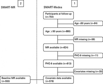
Of the 754 patients participating at follow-up, all patients aged 50 years or older (n=680) were included in the current study. Of these, data on MRI variables were missing in 56 patients (no MRI, 40; motion or artefacts, 16). Of the remaining 624 patients, the PHQ-9 questionnaire was missing in 11 patients, and data on covariates in 35 patients. This resulted in a cross-sectional analytical sample of 578 patients (Fig. 1).
Flowchart illustrating the composition of the study sample for cross-sectional analyses on the Second Manifestations of ARTerial disease (SMART)-Medea data (1), and for longitudinal analyses including the baseline data on magnetic resonance imaging (MRI) variables (2). PHQ-9, Patient Health Questionnaire-9.

For analyses on lesion progression we used data of 553 patients, because data on baseline MRI variables were missing in 25 patients (no MRI, 4; motion or artefacts, 21).
No significant differences in patient characteristics were found between patients with missing data and our cross-sectional sample, except for lower MMSE score [median, 28.5 (10th–90th percentile 26–30) versus 29.0 (10th–90th percentile 27–30), p=0.01].
Data analysis
Because of overdispersion of the positively skewed component scores, we used negative binomial regression analysis to investigate the cross-sectional relationships between location of WML volume, lacunar infarcts and atrophy as independent variables, and motivational and mood component scores as dependent variables. Using negative binomial regression analysis, incidence rate ratios can be calculated. In addition, we investigated relationships between location of WML volume, lacunar infarcts and atrophy with individual depressive symptoms using log binomial models and Poisson regression models with robust standard errors. We estimated relative risks (RRs) and accompanying confidence intervals (CIs) (Robbins et al. Reference Robbins, Chao and Fonseca2002; Blizzard & Hosmer, Reference Blizzard and Hosmer2006) rather than odds ratios, which overestimate the relative risk, particularly for outcomes that are common (>10%) (Barros & Hirakata, Reference Barros and Hirakata2003; McNutt et al. Reference McNutt, Wu, Xue and Hafner2003). WML volume (% of ICV) and atrophy were first entered in quartiles, with the lowest quartile serving as the reference category to check for non-linear relationships, and then entered on a continuous scale (per s.d. increase) if the association was linear to increase precision. Locations of lacunar infarcts were divided into frontal, deep white matter (corona radiata, semioval centre or internal capsule), other white matter (parietal, temporal or occipital lobe), thalamus and basal ganglia. In model I, associations were adjusted for age, gender, education and MMSE score. We additionally adjusted for physical functioning, antidepressant use, smoking, alcohol use, BMI, hyperlipidaemia, hypertension and diabetes mellitus in a separate model (model II), because it is not clear to what extent these vascular risk factors are confounders or preceding factors in the pathway between small-vessel lesions or atrophy and depressive symptoms, or both.
Next, we investigated the relationship between progression of cerebral changes during 3.9 (s.d.=0.4) years of follow-up and depressive symptoms at follow-up. Progression of WML volume and atrophy was defined as difference in WML volume (% of ICV) and brain parenchymal fraction between baseline and follow-up, and was dichotomized into the highest quartile versus the lower three quartiles. Progression of lacunar infarcts was defined as any new lacunar infarct between baseline and follow-up. All three progression measures were entered in the same model; adjustment for covariates was performed as described above.
As a final step, all the above-described analyses were additionally adjusted for the diagnosis of atherosclerotic disease at inclusion (model III).
In additional analyses, we examined associations of executive functioning with motivational and mood profile scores and individual symptoms. To investigate whether poorer executive functioning could be a confounder or intermediate factor in the pathway between cerebral changes and depressive symptoms, we additionally adjusted model II for z score of executive functioning.
To investigate whether associations of degenerative and small-vessel changes in frontal-subcortical regions with symptom profiles were independent of depressive status, we repeated the analyses of location and progression of cerebral changes with profile scores and individual symptoms, after excluding patients with major depressive disorder (n=35).
Finally, we repeated the analyses of location and progression of cerebral changes with profile scores and individual symptoms after exclusion of patients with a cortical infarct (n=84) or large subcortical infarct (n=3), to examine whether associations were influenced by the presence of these infarcts.
Results
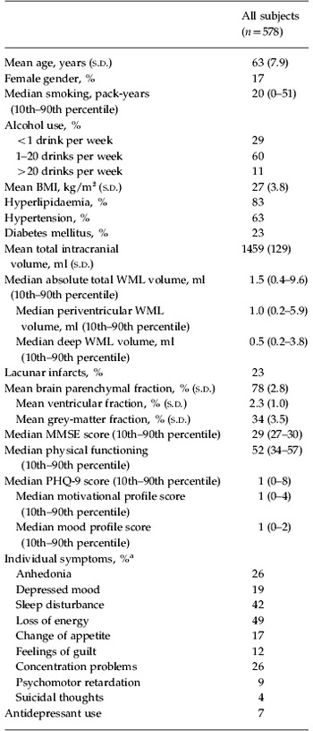
Of the study sample, 136 patients (24%) were included at baseline with cerebrovascular disease; 362 (63%) were included with coronary artery disease; 104 (18%) were included with peripheral arterial disease; and 40 (7%) were included with abdominal aortic aneurysm. Patient characteristics at follow-up are summarized in Table 1. Mean age was 63 (s.d. 8) years and 17% of the sample was female. Median PHQ-9 score was low, but most individual symptoms were frequently reported.
Patient characteristics (n=578)

s.d., Standard deviation; BMI, body mass index; WML, white-matter lesion; MMSE, Mini-Mental State Examination; PHQ-9, Patient Health Questionnaire-9.
a Presence of individual symptoms was defined as a score of ‘several days’ to ‘nearly every day’.
Location of cerebral changes
WML volume
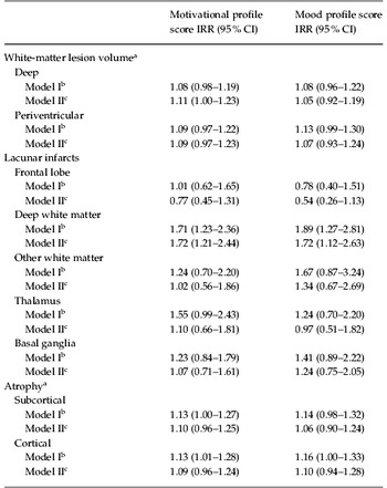
Increased periventricular and deep WML volumes were not significantly associated with greater motivational and mood profile scores after adjustment for age, gender, education and MMSE (Table 2). When individual symptoms were distinguished, larger periventricular WML volume was associated with an increased risk of anhedonia (RR 1.19, 95% CI 1.04–1.35) and appetite disturbance (RR 1.19, 95% CI 1.01–1.40), and larger deep WML volume with anhedonia (RR 1.18, 95% CI 1.09–1.27), energy loss (RR 1.08, 95% CI 1.02–1.15), appetite disturbance (RR 1.20, 95% CI 1.09–1.32) and concentration problems (RR 1.15, 95% CI 1.05–1.26) in model II.
Results of regression analyses with locations of white-matter lesion volume, lacunar infarcts and atrophy as independent, and depressive symptom profiles as dependent variables (n=578)

IRR, Incidence rate ratio; CI, confidence interval; s.d., standard deviation; MMSE, Mini-Mental State Examination.
a Per 1 s.d. increase (white-matter lesions: deep, 0.27%; periventricular, 0.23%; atrophy: subcortical, 0.96%; cortical, 3.48%).
b Adjusted for age, gender, education and MMSE.
c Additionally adjusted for physical functioning, antidepressant use, smoking, alcohol intake, body mass index, hyperlipidaemia, hypertension and diabetes mellitus.
Lacunar infarcts
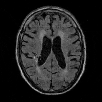
Patients with lacunar infarcts in the deep white matter (Fig. 2) compared to those without, while holding all other variables constant in the model, had a rate 1.7 times greater for mood profile score, and a rate 1.7 times greater for motivational profile score, while lacunar infarcts in other locations were not associated with increased rates (Table 2). When we distinguished individual depressive symptoms, presence of lacunar infarcts in the deep white matter increased the risk of energy loss (RR 1.34, 95% CI 1.07–1.68), psychomotor retardation (RR 2.68, 95% CI 1.48–4.86) and depressed mood (RR 1.69, 95% CI 1.08–2.64) in model II. In addition, lacunar infarcts in the thalamus were associated with psychomotor retardation (RR 2.67, 95% CI 1.25–5.74, model II).
T2-weighted fluid attenuating inverse recovery (FLAIR) image illustrating a lacunar infarct in the corona radiata in the right hemisphere.

Atrophy
Associations of greater subcortical or cortical atrophy with motivational and mood profile scores were not significant (Table 2). When individual symptoms were distinguished, subcortical atrophy increased the risk of energy loss (RR 1.12, 95% CI 1.03–1.22) and feelings of guilt (RR 1.26, 95% CI 1.03–1.55) in model I; associations attenuated and became non-significant (NS) in model II (data not shown). Cortical atrophy was significantly associated with anhedonia (RR 1.20, 95% CI 1.03–1.39) and appetite disturbance (RR 1.24, 95% CI 1.01–1.51) in model II.
Progression of cerebral changes
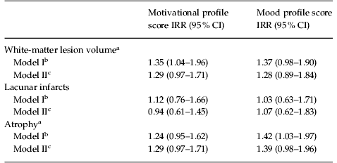
Patients in the highest quartile of progression of WML volume (>0.08% of ICV increase in WML volume) had significantly greater motivational profiles scores than patients in the lower three quartiles of WML progression in model I (Table 3), independent of progression of lacunar infarcts or atrophy. The association attenuated and became NS in model II. When individual depressive symptoms were distinguished, greater progression of WML volume increased the risk of anhedonia (RR 1.44, 95% CI 1.05–1.98), appetite disturbance (RR 1.55, 95% CI 1.00–1.40), concentration problems (RR 1.68, 95% CI 1.24–2.29) and psychomotor retardation (RR 2.43, 95% CI 1.35–4.35) in model II.
Results of regression analyses with progression of white-matter lesion volume, lacunar infarcts and atrophy as independent, and depressive symptom profiles as dependent variables (n=553)

IRR, Incidence rate ratio; CI, confidence interval; ICV, Intracranial volume; MMSE, Mini-Mental State Examination.
a Highest quartile versus other quartiles (white-matter lesions, >0.08% increase in total white-matter lesion volume (% of ICV); atrophy, >1.70% decrease in brain parenchymal fraction). White-matter lesions, lacunar infarcts, and atrophy measures were entered in the same model.
b Adjusted for age, gender, education and MMSE.
c Additionally adjusted for physical functioning, antidepressant use, smoking, alcohol intake, body mass index, hyperlipidaemia, hypertension and diabetes mellitus.
Progression of lacunar infarcts between baseline and follow-up (8% of patients) was not significantly associated with greater motivational and mood profile scores (Table 3), or with individual symptoms (data not shown).
Patients in the highest quartile of progression of atrophy (>1.70% decrease in brain parenchymal fraction) had significantly greater mood profile scores than patients in the lower three quartiles of atrophy progression in model I (Table 3). The association attenuated and became NS in model II. Associations of progression of atrophy with individual symptoms were not statistically significant (data not shown).
Diagnosis of atherosclerotic disease at inclusion
Additional adjustment for diagnosis of atherosclerotic disease at inclusion resulted in similar effect estimates and significance levels, except associations of lacunar infarcts in deep white matter (RR 1.86, 95% CI 0.98–3.53) and the thalamus (RR 1.89, 95% CI 0.86–4.16) with psychomotor retardation attenuated and were no longer statistically significant.
Executive functioning
A 1 s.d. decrease in z score of executive functioning was not significantly associated with greater motivational or mood profile scores in model II (data not shown). When individual symptoms were distinguished, a 1 s.d. decrease in z score of executive functioning was associated with a 1.28 (95% CI 1.01–1.62) times increased risk of psychomotor retardation only in model II.
Entering executive functioning to the model with cerebral changes and depressive symptoms did not change the estimates or significance levels, except for a modest attenuation of the association of lacunar infarcts in deep white matter and the thalamus with psychomotor retardation (RR 2.53, 95% CI 1.41–4.52; RR 2.51, 95% CI 1.17–5.37).
Exclusion of patients with major depressive disorder
Excluding patients with major depressive diagnosis (n=35) resulted in similar effect estimates and significance levels (data not shown), except that associations of cortical atrophy with appetite disturbance (RR 1.11, 95% CI 0.87–1.41), and progression of WML volume with appetite disturbance (RR 1.28, 95% CI 0.78–2.12) were attenuated and no longer significant.
Exclusion of patients with cortical or large subcortical infarcts
Excluding of patients with cortical or large subcortical infarcts on MRI (n=87) resulted in similar estimates and significance levels, except that associations of lacunar infarcts in deep white matter (RR 1.68, 95% CI 0.74–3.82) and the thalamus (RR 1.85, 95% CI 0.65–5.25) with psychomotor retardation were attenuated and no longer statistically significant.
Discussion
In this cohort of patients with symptomatic atherosclerotic disease, we observed that lacunar infarcts in deep white-matter tracts were associated with greater motivational as well as mood sumscores. When we differentiated between individual depressive symptoms, most symptoms associated with WMLs and lacunar infarcts in the deep white matter were characteristic of motivational problems, while those associated with cortical brain atrophy were more diverse. Furthermore, patients with greatest progression of WML volume were also at increased risk of motivational symptoms.
To our knowledge, this is the first study investigating associations of WML volume, lacunar infarcts and atrophy at different locations with the risk of characteristic depressive symptom profiles. Further, studies examining lesion progression and the risk of depressive symptom profiles have not yet been published. Other strengths include the large number of patients and the extensive information on small-vessel and degenerative MRI changes. Also, the use of volumetric assessment of WMLs and atrophy provided more precise and objective estimates than visual rating scales (Yoshita et al. Reference Yoshita, Fletcher and DeCarli2005; van den Heuvel et al. Reference van den Heuvel, ten Dam, de Craen, Admiraal-Behloul, van Es, Palm, Spilt, Bollen, Blauw, Launer, Westendorp and van Buchem2006), and enabled us to detect small volume changes over time more accurately.
A limitation of this study is that we did not distinguish other locations of atrophy in more detail, including the prefrontal cortex. In addition, depressive symptoms were assessed with the PHQ-9 and not with structured interviews. The low overall PHQ-9 score may have narrowed the contrast between patients with depressive symptoms and those without. The large sample size should, however, be sufficient to detect significant relationships between structural changes and depressive symptoms, even if the contrast between subjects was relatively small. Also, we investigated relationships of structural brain changes with individual depressive symptoms, irrespective of symptom frequency. Finally, we cannot draw firm conclusions regarding the direction of causality between the progression of structural changes and depressive symptoms, because depressive symptoms were only assessed at follow-up.
The ‘vascular depression’ hypothesis postulates that location of cerebrovascular lesions in prefrontal structures or their modulating pathways could predispose to vulnerability for late-life depression (Alexopoulos et al. Reference Alexopoulos, Meyers, Young, Campbell, Silbersweig and Charlson1997a; Krishnan et al. Reference Krishnan, Hays and Blazer1997), with clinical presentation of a lack of interest, psychomotor retardation and greater disability. Possible underlying neuropathological mechanisms include the disruption of frontal-subcortical pathways by subcortical ischaemic lesions (Taylor et al. Reference Taylor, Steffens and Krishnan2006), or by a combination of vascular and age-related degenerative disease (Alexopoulos, Reference Alexopoulos2001; Alexopoulos et al. Reference Alexopoulos, Kiosses, Klimstra, Kalayam and Bruce2002).
Direct evidence supporting the hypothesis that disruption of frontal-subcortical pathways by small-vessel or degenerative changes is associated with a motivational symptom profile is scarce, because most studies investigating whether vascular disease is associated with a characteristic depressive symptom profile did not use MRI. One study reported that depressed elderly with clinically defined risk factors for vascular depression showed more psychomotor retardation, and less agitation and guilt feelings than depressed elderly without such risk factors (Alexopoulos et al. Reference Alexopoulos, Meyers, Young, Kakuma, Silbersweig and Charlson1997b). However, two population-based studies could not demonstrate any significant differences in symptom patterns between depressed subjects with and without vascular risk factors or vascular disease, except for greater overall disability (Licht-Strunk et al. Reference Licht-Strunk, Bremmer, van Marwijk, Deeg, Hoogendijk, de Haan, van Tilburg and Beekman2004; Naarding et al. Reference Naarding, Tiemeier, Breteler, Schoevers, Jonker, Koudstaal and Beekman2007). A possible explanation for these discrepancies could be that cerebrovascular lesions directly predispose to a vulnerability for depressive symptoms, and that these lesions are not associated with extracerebral atherosclerosis (Lee et al. Reference Lee, Kim, Chung, Kim, Ahn and Lee2010).
Only one previous study in community-living elderly distinguished the location of small-vessel changes and individual depressive symptoms (Nebes et al. Reference Nebes, Vora, Meltzer, Fukui, Williams, Kamboh, Saxton, Houck, DeKosky and Reynolds2001). This study suggested that WML volume, located in deep white matter, increased the risk of impaired motivation, concentration and decision making, but lacunar infarcts and atrophy were not assessed. Other MRI studies either investigated associations of small-vessel changes or atrophy with depression sumscores and not with symptom characteristics, or only assessed small-vessel or atrophic severity and not location. For instance, one study reported higher levels of apathy among depressed patients with higher severity of WMLs or grey-matter lesions (Krishnan et al. Reference Krishnan, Taylor, McQuoid, MacFall, Payne, Provenzale and Steffens2004). In this study, other characteristic symptoms associated with small-vessel lesions could not be identified. Since depressive symptoms were examined within patients with clinical depression, it is possible that the contrast between individual depressive symptoms was much smaller than in subjects not selected on their depression status.
Our findings provide support for a relationship between disruption of frontal-subcortical pathways by small-vessel lesions and motivational depressive symptoms, insofar that WMLs and lacunar infarcts located in projecting deep white-matter tracts were associated with several individual symptoms characteristic of motivational problems, which are in agreement with symptoms that have been previously associated with disruption of cognitive/behavioural circuits (i.e. the anterior cingulate cortex, dorsolateral prefrontal cortex and orbitofrontal cortex) (Zgaljardic et al. Reference Zgaljardic, Borod, Foldi and Mattis2003). Additional analyses showed that associations of lacunar infarcts in deep white matter and the thalamus with psychomotor retardation were partially explained by concurrent large-vessel disease. In addition to lesion location, greater progression of WML volume increased the risk of mainly motivational symptoms, including anhedonia, concentration problems and psychomotor retardation. These findings indeed suggest that higher WML volume is not only associated with, but could be a preceding mechanism contributing to an increased risk of motivational depressive symptoms. Interestingly, associations of subcortical small-vessel lesions with motivational symptoms did not change when patients with major depressive disorder were excluded. Although we did not measure apathy, these findings suggest that symptoms associated with disruption of frontal-subcortical pathways could be part of a motivational syndrome that resembles apathy, rather than a ‘vascular depression’ subtype.
Few subjects (1.9%) in our sample scored below the normal range of global cognitive function (MMSE <24) (Folstein et al. Reference Folstein, Folstein and McHugh1975). In this respect, our population had, on average, normal age-adjusted cognitive performance. Recently, we associated the presence of lacunar infarcts in deep white-matter tracts with poorer executive functioning in patients with symptomatic atherosclerotic disease (Grool et al. Reference Grool, Van der Graaf, Mali and Geerlings2011). Our current findings suggest that although poorer executive functioning is associated with psychomotor retardation, it is not an intermediate factor in the pathway between small-vessel lesions in the deep white matter and motivational symptoms.
The controversies in findings from previous studies investigating the ‘vascular depression’ hypothesis incited the recent debate on the role of cerebral small-vessel disease in the development of depressive symptoms in later life. A recent population-based study found that atherosclerosis was not associated with incident depression, and suggested that depression may contribute to, rather than result from, vascular burden, or that both result from a mutual underlying mechanism (Newson et al. Reference Newson, Hek, Luijendijk, Hofman, Witteman and Tiemeier2010). However, this study did not use MRI markers of cerebral small-vessel disease. Our findings indicate that it is important to examine location and progression of cerebral small-vessel disease, and to distinguish motivational and mood symptoms rather than investigating depressive sumscores, and suggest that disruption of frontal-subcortical pathways by small-vessel lesions leads to a symptom profile characteristic of motivational problems, also in the absence of major depressive disorder. Additional analyses showed that large-vessel disease could also play a role in these associations. Atrophy was associated with a more mixed pattern of motivational and mood symptoms. In view of the large number of associations investigated in this study, some associations may be due to chance and additional studies are needed to confirm our findings and to further clarify the direction of causation between disruption of frontal-subcortical pathways by small-vessel lesions and motivational symptoms.
Acknowledgements
We gratefully acknowledge the members of the SMART Study Group of UMC Utrecht: A. Algra, M.D., Ph.D., Julius Center for Health Sciences and Primary Care and Rudolf Magnus Institute for Neurosciences, Department of Neurology; P. A. Doevendans, M.D., Ph.D., Department of Cardiology; D. E. Grobbee, M.D., Ph.D. and G. E. H. M. Rutten, M.D., Ph.D., Julius Center for Health Sciences and Primary Care; L. J. Kappelle, M.D., Ph.D., Department of Neurology; F. L. Moll, M.D., Ph.D., Department of Vascular Surgery; F. L. J. Visseren, M.D., Ph.D., Department of Vascular Medicine.
This study was supported by a programme grant from the Netherlands Heart Foundation (NHF: project no. 2007B027). The NHF had no involvement in the study design; in the collection, analysis and interpretation of the data; in writing of the report; and in the decision to submit the paper for publication.
Declaration of Interest
None.