INTRODUCTION
Neuroimaging studies are increasingly being used to examine changes in brain activation patterns after left-hemisphere stroke and aphasia. Despite several studies, understanding of the complex process of cortical reorganization of language-related brain regions during recovery from aphasia and the effects of therapeutic interventions on brain systems involved in language processing is limited.
A basic issue relates to whether language improvement during recovery and rehabilitation is sustained by left-hemisphere zones spared by the lesion, by recruitment of homologous right-hemisphere regions, or both. Several studies in recovering aphasic patients have reported shifts in activation to homologous right-sided territories and have interpreted these as compensatory (Buckner et al., 1996; Cardebat et al., 1994; Ohyama et al., 1996; Thulborn et al., 1999; Weiller et al., 1995). Recently, however, the role of right-hemisphere activation during functional imaging has been questioned, suggesting that it is a maladaptive response reflecting loss of active transcallosal inhibition (Belin et al., 1996; Blank et al., 2003; Rosen et al., 2000).
In fact, levels of activation in homologous right-hemisphere regions do not correlate with measures of verbal production (Fernandez et al., 2004; Naeser et al., 2004; Perani et al., 2003; Rosen et al., 2000) or comprehension (Breier et al., 2004; Fernandez et al., 2004). Rather, better recovery has been associated with greater left-hemisphere activation (Karbe et al., 1998a,b; Miura et al., 1999; Warburton et al., 1999). Furthermore, inhibitory (slow) rTMS to the anterior portion of the right frontal operculum, aimed to suppress this maladaptive right-hemisphere response, has a beneficial effect on the more widespread bihemispheric neural network for naming (Naeser et al., 2005).
The apparent contribution of right- and left-hemisphere activity to language recovery may depend on when it is measured following stroke onset. Right-hemisphere activation seems to occur early during recovery (Fernandez et al., 2004; Heiss et al., 1999; Xu et al., 2004) and may depend on the site of the left-hemisphere lesion (Abo et al., 2004; Xu et al., 2004). Right-hemisphere participation may also be more relevant when there is greater damage to the left-hemisphere language areas (Cao et al., 1999; Heiss et al., 1999; Karbe et al., 1998a,b).
Studies that directly assess functional anatomical changes occurring with language therapy are emerging; yet, there remains no consensus even on the most basic question of laterality. Left-sided activations have been reported following a positive response to the intervention with Melodic Intonation Therapy (MIT; Belin et al., 1996), phonological training via reading aloud (Small et al., 1998), repetitive naming of semantically related pictures (Cornelissen et al., 2003), and memorization of articulatory gestures followed by repetition, reading aloud, and picture naming of a core of words (Leger et al., 2002). Both Leger et al. (2002) and Small et al. (1998) found that posttherapy patterns were consistent with task-dependent activity in healthy adults, while Cornelissen et al. (2003) found increased activity in the perilesional cortex. In contrast, others have found increased activity in right homologues of the anterior (Thompson, 2000) and posterior (Musso et al., 1999; Thompson, 2000) language areas with therapy.
Several issues about these imaging studies and the reported alterations in functional activation following therapy deserve comment. First, no study so far has reported more than a few patients at any one time (as we do here). Second, the neurobiological measures differ across studies. Combining individual differences with task differences leads to a wide variety of possible outcomes.
Clearly, many questions remain regarding recovery and the effect of rehabilitation on patterns of language organization. The purpose of this study is twofold. First, we describe the language status of two patients with chronic aphasia and compare their functional neuroanatomical response to that of a group of control subjects on two tasks that typically recruit different patterns of activation. Second, we describe the aphasic subjects' behavioral and functional neuroanatomical response to treatment targeted at both language production and auditory comprehension. We studied these patients before and after therapy on both reading aloud and audiovisual story comprehension.
The current literature on aphasia recovery and response to therapy suggests that best recovery is associated with brain activation patterns similar (e.g., degree of lateralization) to that of healthy controls. Certainly, when a region has little neural tissue or poor connectivity with other regions, alternative circuits (e.g., involving adjacent or homologous regions) subserving similar or compensatory functions will play a greater role. Given these generalities, we hypothesized first that the patient with better language ability would have an activation pattern both before and after treatment that would be more like that of a control group than the patient with less skill. Second, we hypothesized that more positive responses to language treatment would be associated with increased activity in undamaged left perisylvian areas, while weaker positive responses would be associated with increased activity in homologous regions of the right perisylvian areas.
METHODS
Participants
Two right-handed patients with chronic aphasia following left middle cerebral artery (MCA) distribution ischemic stroke participated in functional magnetic resonance imaging (fMRI) scanning sessions before and after 24 sessions of rehabilitation therapy. Subjects were similar in etiology of stroke (carotid dissection), acute language profile, length of time after onset, and demographic variables. Both were native English speakers, college educated, with Broca's aphasia, diagnosed clinically based on behavioral criteria shortly after stroke onset. Both participants were strongly right-handed (Oldfield, 1971), had normal hearing, normal corrected vision, and were more than 1 year after onset.
Subject 1 was a 54-year-old man with occlusion of the left internal carotid artery secondary to dissection 14 months before entry. Subject 2 was a 49-year-old man with similar dissection 21 months prior. The participants had differential injury to the cerebral cortex and underlying white matter in the left MCA distribution: Subject 1 had more frontal lobe and anterior temporal lobe involvement than Subject 2, who had more involvement of the parietal lobe and posterior superior temporal region.
Four control subjects (ages 19–34; three women) were right-handed native speakers of English with no neurological or psychiatric history. All subjects gave oral and written consent, and the Institutional Review Boards of the Biological Science Division of The University of Chicago and Northwestern University approved the study.
Language Intervention
Both subjects with aphasia received 24 treatment sessions, 2–3 times weekly. Subject 1's treatment lasted 9 weeks; Subject 2's lasted 12 weeks. The therapeutic technique used multisensory auditory and visual–verbal information to facilitate primarily production of connected language, but also auditory and reading comprehension. It emphasized reading aloud (in unison with the therapist) of written stimuli, 15–30 words long (typically two to three sentences, at a sixth grade reading level). Treatment was as follows: Subjects listened to a stimulus twice, while simultaneously looking at the sentences on an index card or computer screen; subjects read the stimulus aloud together with the therapist—this was repeated twice; for each sentence, subjects were asked to identify two or three randomly selected single words and read each word aloud; finally, the subject read the entire stimulus aloud again in unison with the therapist. Stimuli were sentences of varied vocabulary items and grammatical structures, presented with natural speech prosody. During an hour of treatment, subjects typically practiced 30 different stimuli. Over 24 sessions, 90 stimuli were available for practice. The current therapy approach derives from previous stroke therapy work by our group in oral reading (Cherney et al., 1986; Cherney, 1995, 2004) and in hand motor imitation (Buccino et al., 2002, 2006).
Functional MRI
Control subjects and aphasic participants underwent identical fMRI procedures, with the aphasic patients doing so both before and after therapy. All imaging sessions were performed identically. Spiral BOLD functional scans were acquired at 3T (Noll et al., 1995) with a standard quadrature head coil. Volumetric T1-weighted scans (120 axial slices, 0.9375 × 0.9375 × 1.5 mm resolution) were acquired to provide high-resolution anatomical images.
Subjects performed two tasks developed to tap the modalities being trained in therapy: reading aloud (oral-reading) and audiovisual story comprehension (story-comprehension). Additionally, we expected the tasks to primarily recruit different cortical regions—the frontal cortex for reading aloud and the temporal cortex for story-comprehension.
For oral-reading, subjects saw sentences in which a word was highlighted in red every 1.5 seconds. Participants were instructed to read the word aloud. There were two reading blocks lasting 72 seconds, separated by a 16.5-second rest period. Performance during scanning was monitored to ensure that participants provided verbal responses to the stimuli.
For story-comprehension, participants watched and listened to high-resolution video clips of a storyteller telling simplified versions of Aesop's fables in a single run consisting of four stories, each approximately 45 seconds long and separated by a 15-second period of rest, for a total of 1 minute per story. The storyteller was seated with face, trunk, and hands visible. No overt motor response was required during scanning. However, after completion of the scanning session, participants were interviewed about the stimuli. They reported being engaged by the stories. They answered specific yes/no questions about the stories' events, although they were not instructed that they would be doing so. All participants accurately responded regarding details of the stories, indicating that they did attend to them.
For both tasks, participants viewed the stimuli through a mirror that allowed them to see a projection screen at the end of the scanning bed. For story-comprehension, audio stimuli were delivered at 85 dB SPL through headphones containing MRI-compatible electromechanical transducers (Resonance Technologies, Inc., Northridge, CA). Test–retest reliability for story-comprehension was good for both healthy subjects (r = .9) and stroke subjects (r = .86; Chen & Small, 2006). Test–retest reliability data for oral-reading was not available, but we anticipate it to be at least as robust as the more passive story-comprehension task.
A total of 118 whole brain images were collected for oral-reading and 190 images for story-comprehension. Each functional imaging acquisition consisted of 27 5-mm spiral gradient echo T2* images, with a whole volume collected every 1.5 seconds in the axial plane.
Image analysis consisted of several steps following reconstruction of raw spiral k-space data into image space. Images were spatially registered in three-dimensional space by transformation of each of the time points and corrected for head movement, using AFNI (Cox, 1996). Effective in-plane resolution was 3.75 × 3.75 × 5 mm.
The two sessions for each subject were coregistered using the same approach. Time series data for oral-reading were spatially blurred using a 6 mm full-width half-max Gaussian template. Next, the imaging data for each block were analyzed, using a standard multiple linear regression analysis. For oral-reading, there was one regressor of interest, three other behavioral regressors, and nine additional regressors including the mean, linear, and quadratic trends, and the six motion parameters from the motion correction procedure. The regressor of interest was a waveform with similarity to the hemodynamic response, generated by convolving a gamma-variant function with the onset time and duration of each block. Data for story-comprehension were analyzed similarly.
To establish an objective level of “activation,” a Monte Carlo Simulation was run to establish an individual voxel significance level and cluster size threshold (Forman et al., 1995). This procedure established that using a cluster size of three voxels and a cluster connection radius of 5.2, an individual voxel p value of 3e-05 would establish the desired whole brain alpha (p ≤ .05). To create functional images, these parameters were used to form data sets containing significant data satisfying the cluster criteria for each of the two scans (before and after) for each of the two subjects. Signal-to-noise ratio, calculated using the mean of the time series divided by the corresponding standard deviation (Parrish et al., 2000), was the same across sessions for both subjects.
To facilitate anatomical parcellation and comparison across time and between individuals, anatomical images were transformed (using Freesurfer) to a two-dimensional representation for cortical surface-based analysis (Dale et al., 1999; Fischl et al., 1999). Anatomical images were first segmented, inflated, and registered to a sphere, which allows for more accurate registration into a common space. A surface representation of the cortical white matter was then generated (Desikan et al., 2006), onto which the functional images were mapped (using SUMA). Because of the distortions associated with inflating and segmenting large lesions in Freesurfer, stroke brains were first processed using the Virtual Brain Transplant method (Wier et al., 2006). Anatomical scans from separate sessions were coregistered to provide an accurate composite anatomical scan. Lesions were delineated manually, and analogous regions from the opposite hemisphere were transplanted into the lesioned area. The transplanted volume was then morphed (using Xmorph) to reduce the abnormalities in perilesional intensity caused by the transplant. This new transplanted volume was then processed and parcellated using Freesurfer.
To transform the functional data into the coordinate space of the surface domain, we calculated percent signal change values for each whole-brain functional data set. Next, these percent signal change images were mapped directly onto the surface domain. The functional images were then smoothed on the surface with a full-width half-max Gaussian kernel of 3.
Finally, we used a standard parcellation scheme (Fischl et al., 2004) to compute the mean percent signal change, the standard deviation of this value, and the maximum percent signal change for each region, as well as the total number of active voxels in the region.
RESULTS
Pretreatment Language Testing
Although both subjects demonstrated relatively good recovery following the acute stage, Subject 2 had recovered better language than Subject 1. Subject 1 presented with a moderate Broca's aphasia characterized by some apraxia of speech, slow and effortful productions, difficulty formulating complete grammatical sentences, and anomia. Subject 2's output more closely resembled an anomic aphasia, with frequent pauses, use of nonspecific words (“thing,” “here,” “there”) and attempts at circumlocution; however, he used only very simple grammatical constructions and continued to have difficulty with comprehension of complex grammatical structures.
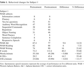
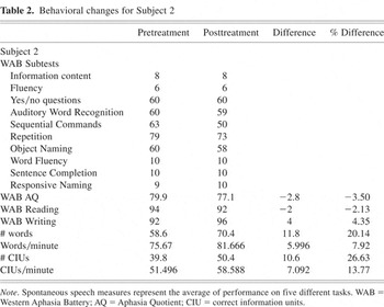
These differences were reflected in test scores. Language testing included the Western Aphasia Battery (WAB; Kertesz, 1982) and measures of spontaneous speech elicited by picture description and picture story sequences (Nicholas and Brookshire, 1993). The measures of spontaneous speech were analyzed for productivity [number of words and correct information units (CIU)] and efficiency (words per minute and CIUs per minute). Language test results are shown in Tables 1 and 2.
Behavioral changes for Subject 1

Behavioral changes for Subject 2

Subject 1 had an Aphasia Quotient (AQ) of 67.4, while Subject 2 had an AQ of 79.9. Subject 1's reading and writing scores were 82 and 74, respectively, while Subject 2 achieved WAB Reading and Writing scores of 94 and 92, respectively. Spontaneous speech measures were averaged across five tasks (i.e., the WAB picture description and four discourse tasks comprising single composite picture descriptions and picture story sequences from Nicholas & Brookshire, 1993). Results indicated that Subject 2 spoke more quickly than Subject 1 (75.67 words/min compared with 55.184 words/min). He also produced more CIUs overall and at a faster rate (39.8 CIUs and 51.496 CIUs per minute) than Subject 1 (29.2 CIUs and 23.026 CIUs per minute).
Posttreatment Language Testing
Tables 1 and 2 also summarize language performance after therapy. For Subject 1, language performance improved in all language modalities as evidenced by a change of almost 10 points (14%) on the WAB AQ (Kertesz, 1982), and changes of 6 points (7%) and 16 points (22%), respectively, on the WAB reading and writing. The gains in the WAB AQ resulted mainly from improved performance on repetition and object naming. Language changes were reflected in Subject 1's spontaneous discourse. He produced an increased number of both words and correct information units (CIUs) following treatment (approximately 50% in both cases), however, rate of speech (words per minute) and rate of CIU production both decreased (11% and 13%, respectively).
Subject 2 did not show improvement on any WAB subtests. However, he demonstrated improved discourse production after treatment with increases in number of words (20%) and correct information units (27%). He also increased efficiency of performance, with slightly higher speech rate (7%) and more CIUs per minute (14%).
Brain Activation
For this investigation, we selected eight cortical regions of interest (ROIs) that approximated the motor and language areas. These ROIs are (1) inferior frontal gyrus and middle frontal gyrus, including pars orbitalis and pars triangularis; (2) ventral premotor area and pars opercularis; (3) dorsal premotor area; (4) sensorimotor cortex, including precentral gyrus, postcentral gyrus, and paramedian gyrus; (5) anterior superior temporal gyrus; (6) posterior superior temporal gyrus; (7) planum temporale and transverse temporal gyrus; and (8) inferior parietal lobule.
Imaging results are presented separately for each subject. For each imaging task, the pretreatment volume of activation for both left and right hemispheres are compared with the averaged volume and pattern of activation for the normal controls. Then changes in activation from pretreatment to posttreatment are presented.
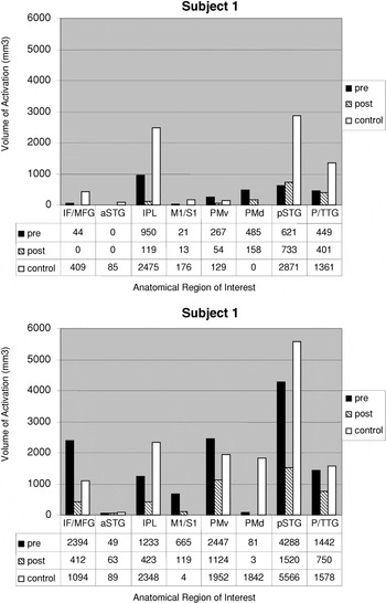
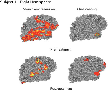
Subject 1, who demonstrated less recovery from the acute stage, and who had more frontal and anterior temporal injury, showed almost no left- or right-hemisphere activity on oral-reading. Only right superior temporal gyrus and left anterior frontal and middle frontal gyri showed any activation. For story-comprehension, the pattern of left-hemisphere recruitment was more like the control group, although overall activation was less. Activated areas included left inferior parietal, left posterior superior temporal, and left planum temporale and transverse temporal regions. In the right hemisphere, all ROIs activated in the control group were also active in Subject 1. In fact, activation in right anterior frontal and middle frontal gyri, and in right ventral premotor region was greater in Subject 1 than in the averaged control group.
Following treatment resulting in favorable language changes, activation on oral-reading continued to be minimal in the left hemisphere. However, large changes occurred in all right-hemisphere ROIs except dorsal premotor. Activation was greatest in right anterior inferior frontal and middle frontal gyri and anterior superior and posterior superior temporal gyri—these areas are homologous to the left-hemisphere areas activated in the controls. For story-comprehension, Subject 1 demonstrated overall less activity in both left and right hemispheres from pretreatment to posttreatment, except for left posterior superior temporal gyrus, where activity increased somewhat.
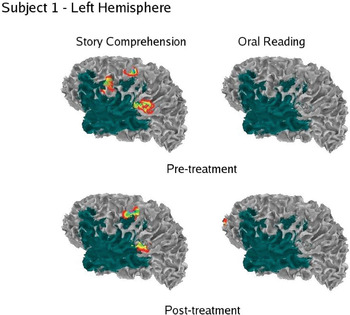
Figure 1 displays oral-reading volumes of activation for each ROI in left and right hemispheres, respectively, for Subject 1 (pre- and posttreatment) and the control group. Figure 2 displays story-comprehension volumes of activation for each ROI in left and right hemispheres, respectively, for Subject 1 (pre-and posttreatment) and the control group. Figures 3 and 4 are brain maps illustrating, for each task, changes in cortical activation from pretreatment to posttreatment—on Figures 3 and 4, areas of activation can be seen in relation to lesion site.

Subject 1: Pretreatment and posttreatment volume of activation in left (above) and right (below) hemispheres for oral-reading (reading aloud words). Volume of activation for eight regions of interest are compared with that of a control group of four subjects. Regions of interest include (1) inferior frontal gyrus and middle frontal gyrus, including pars orbitalis and pars triangularis (IF/MFG); (2) premotor ventral premotor area and pars opercularis (PMv); (3) dorsal premotor area (PMd); (4) sensorimotor cortex, including the precentral gyrus, postcentral gyrus, and the paramedian gyrus (M1/S1); (5) anterior superior temporal gyrus (aSTG); (6) posterior superior temporal gyrus (pSTG); (7) planum temporale and transverse temporal gyrus (P/TTG); and (8) inferior parietal lobule (IPL).

Subject 1: Pretreatment and posttreatment volume of activation in left (above) and right (below) hemispheres for story-comprehension. Volume of activation for eight regions of interest are compared with that of a control group of four subjects. Regions of interest include (1) inferior frontal gyrus and middle frontal gyrus, including pars orbitalis and pars triangularis (IF/MFG); (2) premotor ventral premotor area and pars opercularis (PMv); (3) dorsal premotor area (PMd); (4) sensorimotor cortex, including the precentral gyrus, postcentral gyrus, and the paramedian gyrus (M1/S1); (5) anterior superior temporal gyrus (aSTG); (6) posterior superior temporal gyrus (pSTG); (7) planum temporale and transverse temporal gyrus (P/TTG); and (8) inferior parietal lobule (IPL).

Subject 1: Left-hemisphere brain maps showing pretreatment (above) and posttreatment (below) activation for oral-reading (reading words aloud) and story comprehension.

Subject 1: Right-hemisphere brain maps showing pretreatment (above) and posttreatment (below) activation for oral-reading (reading words aloud) and story comprehension.
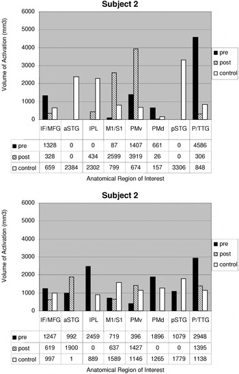
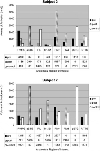
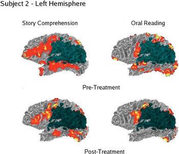
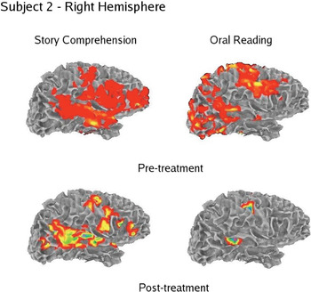
Subject 2, who showed more recovery after the acute stage and who had more injury to the posterior temporal and parietal regions, showed much less left-hemisphere activation on oral-reading than controls. However, more activation was noted in specific ROIs, including left anterior inferior and middle frontal gyri, dorsal premotor region, and planum temporale and transverse temporal regions. In the right hemisphere, overall activation was greater than overall activation in the controls. For story-comprehension, Subject 2's overall left-hemisphere activation resembled that of the controls. However, differences included more activation in anterior inferior frontal and middle frontal gyri, and in dorsal and ventral premotor regions. Less activation occurred in inferior parietal, posterior superior temporal, and planum temporale and transverse temporal gyrus—these results are not unexpected because these are Subject 2's lesion sites. For the right hemisphere, the absence of activation in posterior superior temporal gyrus and premotor dorsal areas differed from that of controls.
Following treatment that resulted in little change on WAB measures, but some positive changes in discourse production and efficiency, overall activation decreased bilaterally on oral-reading. However, increased activation was noted in left primary motor and sensory and left ventral premotor areas. Right-hemisphere activation increased in anterior superior temporal gyrus and ventral premotor, with less activation in right anterior inferior and middle frontal gyri, dorsal premotor, posterior superior temporal gyrus, and inferior parietal lobe. On story-comprehension, increased left-hemisphere activation was evident, particularly in anterior superior temporal gyrus, planum temporale and transverse temporal gyrus, and ventral and dorsal premotor regions. Increased right-hemisphere activation was noted in anterior superior temporal gyrus, primary motor and sensory areas, and dorsal premotor region. Decreased right-hemisphere activation occurred in inferior and middle frontal gyri, inferior parietal lobule, and planum temporale and transverse temporal gyrus. Of interest, both left and right posterior superior temporal gyri showed no activity posttreatment, which contrasts with the pattern of activity noted in the controls.
Figure 5 displays oral-reading volumes of activation for each ROI in left and right hemispheres, respectively, for Subject 2 (pre- and posttreatment) and the control group. Figure 6 displays story-comprehension volumes of activation for each ROI in left and right hemispheres, respectively, for Subject 2 (pre- and posttreatment) and the control group. Figures 7 and 8 illustrate, for each task, changes in cortical activation from pretreatment to posttreatment—on these brain maps, areas of activation can be seen in relation to lesion site.

Subject 2: Pretreatment and posttreatment volume of activation in left (above) and right (below) hemispheres for oral-reading (reading aloud words). Volume of activation for eight regions of interest are compared with that of a control group of four subjects. Regions of interest include (1) inferior frontal gyrus and middle frontal gyrus, including pars orbitalis and pars triangularis (IF/MFG); (2) premotor ventral premotor area and pars opercularis (PMv); (3) dorsal premotor area (PMd); (4) sensorimotor cortex, including the precentral gyrus, postcentral gyrus, and the paramedian gyrus (M1/S1); (5) anterior superior temporal gyrus (aSTG); (6) posterior superior temporal gyrus (pSTG); (7) planum temporale and transverse temporal gyrus (P/TTG); and (8) inferior parietal lobule (IPL).

Subject 2: Pretreatment and posttreatment volume of activation in left (above) and right (below) hemispheres for story-comprehension. Volume of activation for eight regions of interest are compared with that of a control group of four subjects. Regions of interest include (1) inferior frontal gyrus and middle frontal gyrus, including pars orbitalis and pars triangularis (IF/MFG); (2) premotor ventral premotor area and pars opercularis (PMv); (3) dorsal premotor area (PMd); (4) sensorimotor cortex, including the precentral gyrus, postcentral gyrus, and the paramedian gyrus (M1/S1); (5) anterior superior temporal gyrus (aSTG); (6) posterior superior temporal gyrus (pSTG); (7) planum temporale and transverse temporal gyrus (P/TTG); and (8) inferior parietal lobule (IPL).

Subject 2: Left-hemisphere brain maps showing pretreatment (above) and posttreatment (below) activation for oral-reading (reading words aloud) and story comprehension.

Subject 2: Right-hemisphere brain maps showing pre-treatment (above) and posttreatment (below) activation for oral-reading (reading words aloud) and story comprehension.
DISCUSSION
This study first compared the functional neuroanatomical response in two patients with chronic aphasia to control subjects and then investigated changes in brain activation following language treatment of the same type and amount. Aphasic subjects were similar regarding stroke etiology, reported acute language profile, time after onset, and demographic variables. However, Subject 1 had more anterior injury, while Subject 2 had more posterior injury.
Recovery in Aphasia
A caveat regarding data interpretation relates to differences in the age of the control subjects compared with those with aphasia. Because there may be an age-related shift in the amount, magnitude, and pattern of activation, our data need to be interpreted with caution. Nevertheless, results suggest that right-hemisphere activation patterns, approaching the pattern of right-hemisphere activation in control subjects, might be important for better recovery. At first glance, these results appear inconsistent with previous notions that better recovery is associated with greater left-hemisphere activation (Karbe et al., 1998a, 1998b; Miura et al., 1999; Warburton et al., 1999) and that increased right-hemisphere homologous activity reflects a maladaptive response (Belin et al., 1996; Blank et al., 2003; Naeser et al., 2005; Rosen et al., 2000).
Consideration of lesion site (Abo et al., 2004; Cao et al., 1999; Xu et al., 2004) and the type of imaging task may explain these findings. Although their lesions were large and similar in size, Subject 1's lesion was more anterior while Subject 2's lesion was more posterior. In control subjects, oral-reading recruited more anterior left-hemisphere regions, while story-comprehension recruited more posterior left-hemisphere regions. Therefore, Subject 1's lesion interfered with typical left-hemisphere activation associated with oral-reading, while Subject 2's lesion interfered with typical left-hemisphere activation associated with story-comprehension.
Additionally, absence of a right to left functional shift commonly associated with late recovery in chronic aphasia may relate to the level of language skills achieved. It has been suggested that a shift in hemispheric functional activity is possible only when language recovery is more complete (Belin et al., 1996). Although both subjects showed language improvement, complete language recovery did not occur.
Impact of Rehabilitation in Chronic Aphasia
Although we present behavioral and neurophysiological findings on only two subjects, our results contribute to the continued debate regarding hemispheric changes during rehabilitation. Because both subjects had chronic aphasia, we had anticipated that a positive response to language treatment might be associated with increased perilesional left-hemisphere activity. Clearly, this did not occur; rather, the change in the pattern of accompanying brain activation from pretreatment to posttreatment was task-dependent and lesion-dependent. Therefore, results highlight the importance of considering the nature of the fMRI task and the degree of injury in areas that are important for the task function. Additionally, baseline level of performance and the nature of the training should also be considered.
Oral Reading
The oral-reading task executed during functional brain imaging closely resembled the training intervention performed repeatedly during treatment. In comparing functional brain images of the subjects on this task, the pattern of activity from pretreatment to posttreatment was actually opposite for each subject, with increased overall activity for Subject 1, particularly in the right hemisphere, and decreased overall cortical activity for Subject 2.
For Subject 1, who showed oral reading problems at the start of the study and improved during therapy, increased right-hemisphere activity involved primarily the homologue of the significantly damaged Broca's area. Importantly, this increase was task-dependent and did not occur with story-comprehension (see below). Furthermore, this subject had considerable damage to areas known to be important for oral speech, including left inferior and middle frontal gyri and premotor cortex, but had sparing posteriorly. Only a small amount of perilesional tissue was available for functional recruitment. Considering the substantial increases in activation on this task and not on story-comprehension, and the concomitant moderate language improvement, it is possible that these positive language changes relate to the right-hemisphere contribution.
Subject 2 presents a quite different picture, because he began the study with relatively good language skill and did not change substantially. His lesion was more posterior, with sparing of left inferior and middle frontal gyri. Thus, in this case, aphasia therapy could theoretically stimulate functional recruitment of the inferior and middle frontal regions, assuming no physiological obstacles, such as severe white matter damage and/or diaschisis.
The contrasting behavioral and activation changes for Subjects 1 and 2 from pretreatment to posttreatment may have been mediated by differences in baseline language performance and subsequent effects of the intervention. For Subject 1, oral reading difficulty was primarily due to speech production deficits. The effect of treatment was to improve motor planning with subsequent increased activation in those areas (either left-hemisphere or homologous right-hemisphere regions) responsible for motor planning. For Subject 2, with his more posterior lesion involving the parietal area, the effect of the intervention may have been to improve phonological processing, including verbal working memory. The parietal lobe, especially the inferior parietal lobule, forms part of a network of brain areas that mediate short-term storage and retrieval of phonologically coded verbal material (Jonides et al., 1998). The oral reading treatment may have provided support and practice in phonological processing, including phonological short-term memory processes. For Subject 2, enhanced phonological processing may have decreased the overall processing load associated with the task, which may account for the decreased activation in some ROIs.
These neuroimaging results are compatible with the behavioral changes seen in both subjects. Subject 1 demonstrated changes on the WAB AQ, primarily in repetition and naming, both of which could have resulted from improved motor planning and production. Such improvements would also be reflected in discourse production. Subject 2 did not show changes on the WAB AQ, possibly because WAB subtests are not particularly sensitive to phonological processing deficits. However, with improved verbal working memory, we would anticipate changes in the ability to hold in working memory conceptual/semantic information and formulate appropriate sentences with resultant improved discourse.
Treatment intensity differences may also have contributed to differences in the subjects' response. Although amount of treatment was the same (24 sessions), intensity of treatment differed, with Subject 1 receiving treatment over 9 weeks and Subject 2 receiving treatment over 13 weeks. Recent studies have suggested that intensity of treatment is an important variable impacting outcomes (Bhogal et al., 2003; Pulvermuller et al., 2001; Meinzer et al., 2005).
Story Comprehension
Changes in brain activation differed across the two participants, with decreased activation in most regions for Subject 1 and increased activation bilaterally for Subject 2. Size and location of their respective brain lesions provide some explanatory insight into this difference. Story comprehension is known to involve significant processing in temporal regions. Historically, the most important part of this network was considered to be the posterior portion of the left superior temporal gyrus, although current thinking is that the superior and middle temporal gyri bilaterally, and especially the superior temporal sulci, play an important role (Dronkers et al., 1995, 2004). The lesion in Subject 2 involved more of the left temporal lobe and was more posterior than that of Subject 1.
This study has addressed the relationship between rehabilitation and changes in brain activation. Like other similar investigations, there are limitations, including the small number of subjects studied. Certainly, we cannot make grand conclusions about brain organization or reorganization from two isolated cases. Yet, when these imaging data are taken into consideration in conjunction with differences in behavior and the nature of the lesions, the data are provocative. In particular, they argue that different patterns of brain activation result partly from lesion location, from baseline performance and change, and from the particular task performed.
Without associated electrophysiological data, it is speculative to make conclusions about the nature of the physiology underlying the observed changes. Increased activation can occur from neural activity of both an inhibitory or excitatory type. Thus, in reporting increased activation in one region or another (e.g., right-hemisphere homologues), it is not clear whether this finding represents greater activity of inhibitory neurons or excitatory neurons or both. Furthermore, we do not have measures of perfusion and, thus, quantification of lesion location is limited to well-defined structural lesions, without regard to the much larger area of diminished functioning that could be seen on metabolic imaging (Hillis et al., 2001; Metter, 1991).
Nevertheless, the present study reinforces the notion of individual variability in anatomy and functionality of individuals with aphasia, and that these differences, as well as task differences, should be considered when interpreting the effects of an intervention on language and language-related brain activity. Given this variability, to enhance our understanding of recovery and rehabilitation, and the contributions of each brain region (and each hemisphere) to this process, it is important in future research to analyze, compare, and contrast findings from large numbers of cases with different lesion sites and different levels of language skills, using a variety of fMRI tasks at multiple time points after stroke.
ACKNOWLEDGMENTS
This study was supported in part by grants H133G010098 and H133G 040269 from the National Institute on Disability and Rehabilitation Research, U.S. Department of Education (to L.R.C.) and by grant R01 DC007488 from the National Institute on Deafness and Other Communication Disorders (to S.L.S.). The authors extend thanks to Edna Babbitt, Dawn Papachronis, and Jodi Oldani for their assistance with language assessment and treatment; to Patrick Wong for assistance with development of the fMRI tasks; and to Helen Wier and Nameeta Lobo for their help with data analysis. There is no potential conflict of interest affecting this manuscript. Information in this study is new and original and has not been published previously.