Introduction
Congenital cholesteatoma is rare, accounting for a small percentage of all cholesteatomas. This ‘bad skin in the middle-ear cleft’ classically presents as an asymptomatic white mass behind an intact tympanic membrane. However, not all congenital cholesteatomas arise in the middle ear. Cases have been reported in the petrous apex, cerebellopontine angle, mastoid cavity and external auditory canal; therefore, presentation is variable.Reference Lee, Hong, Park and Jung1
We present a case of cholesteatoma, presumably congenital, presenting low down in the left occipital bone, with no effect on middle-ear function. The particulars of this case would appear to make it unique in the medical literature. However, it is acknowledged that neurosurgeons operate on epidermoid cysts, a rare sub-group of which occupy an intradiploic site within the various cranial bones. The question is raised as to whether these lesions actually represent different disease processes or are simply two names for the one condition.
Case report
A 52-year-old woman presented with a headache located deeply and centrally. This failed to improve with cranial osteopathy or with any conventional analgesia. The patient also noticed a lump behind her left ear, but was otherwise asymptomatic.
A computed tomography (CT) scan was arranged by the general practitioner, which demonstrated a lesion in the left mastoid region. This finding prompted a magnetic resonance imaging (MRI) scan to help determine the nature of the lesion, and also a referral to an ENT specialist, one year after the onset of the headache.
The subsequent otological examination was unremarkable, with no hearing loss, nor any history of trauma or surgery to the area.
Imaging
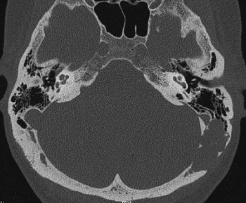
Magnetic resonance imaging as well as high resolution CT scanning of the skull base was performed. Computed tomography (Figure 1) demonstrated a solitary, well circumscribed, lytic lesion sited posteriorly in the mastoid portion of the left temporal bone and extending into the occipital bone. Both temporal bones were well pneumatised and the mastoid air cells and middle ears were clear. The lesion breached the outer and inner cortex of the temporal bone and occipital bone without eliciting a periosteal reaction.
High resolution, axial computed tomography scan through the skull base at the level of the cochlea, demonstrating a sharply demarcated, lytic lesion on the left mastoid and occipital bone. Note the close relationship with the mastoid air cells anteriorly.

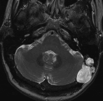
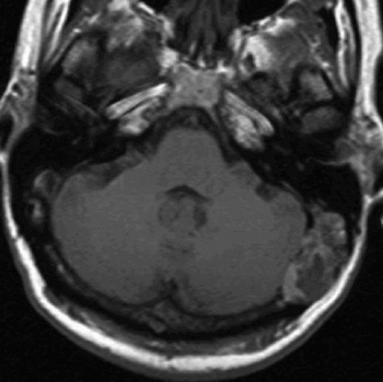
The lesion returned a relatively uniform high signal on T2-weighted MRI (Figure 2), and an irregular, peripheral high signal around central areas of lower signal on T1-weighted MRI (Figure 3). There was no enhancement following intravenous administration of gadolinium-based contrast material (not shown). The combination of the ‘punched-out’ appearance of the lesion, lack of contrast enhancement and absence of a periosteal reaction made a malignant lesion of bone (either primary or metastatic) very unlikely. Benign lesions that could have these features included Langerhans' cell histiocytosis of bone (eosinophilic granuloma) and cholesteatoma. However, the location was highly unusual for an acquired or congenital cholesteatoma.
Axial, T2-weighted magnetic resonance imaging scan at the same level as Figure 1, demonstrating a fairly uniform high signal within the lesion. There is no invasion of the cerebellum or cerebellar oedema to suggest extension through the dura.

Unenhanced, axial, T1-weighted magnetic resonance image demonstrating heterogeneous signal within the lesion.

Surgery
A decision was made to perform a surgical excision, in order to enable definitive histological diagnosis. The surgical appearance was of a large cholesteatoma (later confirmed histologically) lying on the posterior fossa dura, the transverse sinus and the postero-superior sigmoid sinus of the left mastoid and lower part of the occipital bone. The matrix was adherent to the dura, so complete removal could not be safely achieved. The post-auricular mass was also removed (subsequently confirmed to be a reactive lymph node). The middle ear was entirely normal, and recovery was uneventful.
Discussion
The point of interest in this case is what determined the diagnosis. Our specimen was sent for histopathological analysis with a remark questioning whether the lesion was a cholesteatoma. This was because, as ENT surgeons accustomed to operating on middle-ear cholesteatomas, this was how the lesion appeared macroscopically, to our eyes. The histopathology report stated that the specimen comprised keratinising squamous epithelium with keratin, in keeping with a diagnosis of cholesteatoma, and so this diagnosis was confirmed. However, what would have been the outcome had the same specimen been removed by a neurosurgeon and sent for analysis, questioning whether it was an epidermoid cyst? There would appear to be no histopathological differentiation between the two diagnoses. Reports on cases of giant intradiploic epidermoid cysts include nothing that would not be suitable for a report on a cholesteatoma specimen.Reference Constans, Mede, De Divitiis, Donzelli and Maiuri2 Similarly, there is no radiological differentiation to be made, with the radiologist confirming that the differential diagnosis, from the scans obtained, included an epidermoid cyst.
There are no reports of a congenital cholesteatoma occurring at the site described in this case report. In the ENT literature, Canalis et al. reported a few cases of intradiploic cholesteatomas, two of which involved the occipital bone, but these had their epicentre more superiorly, at the occipitoparietotemporal junction (the asterion).Reference Canalis, Shapiro, Lufkin and Becker3 These lesions grew large, destroying the surrounding cranial table and demonstrating predominantly intracranial growth. Their matrix was intimately attached to dura, preventing their complete excision.
In the neurosurgical literature, reports of intradiploic epidermoid cysts are similarly rare, with two cases reported in the occipital bone.Reference Ciappetti, Artico, Raco and Gagliardi4 In 1829, Cruveilhier coined the term ‘tumeurs perlées’ for intracranial lesions with a pearl-like appearance.Reference King5 In 1838, Mueller coined the term ‘cholesteatoma’ for lesions containing cholesterol crystals, and described a sub-group arising within the skull bones (thought to develop within the cranial tables), referred to as intradiploic cholesteatomas.Reference Canalis, Shapiro, Lufkin and Becker3 In 1854, von Remak conceived the idea of these lesions arising from embryonic epithelial cell rests, hence the term ‘epidermoid’. Subsequent differentiation was made, depending on the location of the lesion, by Horrax in 1922. Intradural lesions were termed meningeal cholesteatomas, differentiating them from the middle-ear variety that lacked a primary meningeal attachment. In the same year, Cushing referred to such an extradural lesion as an ‘epidermal cholesteatoma’. What is common to all these lesions would appear to be their histopathology. They are described as containing masses of epithelial debris resulting from slow accumulation of desquamated cells from the epithelial layer of the lining of the lesion. Or, more eloquently, ‘beautiful, lustrous, silky, pearly-white’ lesions, with an inner ‘amorphous, grumous, nonhomogeneous mass of crumbling, soft, caseous material, some of which is murky-white … dingy yellowish-brown … or brownish-green’.Reference King5
Regarding presenting symptoms, cholesteatoma demonstrates independent growth and so its expanding mass usually accounts for symptom development. A recent series of rare cases of cholesteatomas arising solely in the mastoid showed that most of these presented with pain or were found incidentally.Reference Warren, Bennet, Wiggins, Saltzman, Blevins and Shelton6 It is thought that pain may arise once the bony cortex is breached and the disease involves the periosteum. This is in contrast to middle-ear congenital cholesteatomas, which are more likely to present with a conductive hearing loss and, as a result, will present earlier. Mastoid congenital cholesteatoma has presented as a post-auricular mass, being initially misdiagnosed as a sebaceous cyst; removal was thus attempted under local anaesthesia. Only at the time of surgery was the underlying bony defect evident and the true diagnosis made.Reference Sand, Bechara, Altmeyer, Hoffmann, Scholz and Sand7 Headache and pain are common clinical features of intradiploic epidermoid cysts.Reference Ciappetti, Artico, Raco and Gagliardi4
• Congenital cholesteatoma can arise outside the middle ear, although it has never previously been reported in the lower occipital bone, as in this case
• Neurosurgeons operate on epidermoid cysts, which can rarely be intradiploic within the cranial bones; two such cases have been reported in the occipital bone
• The imaging (computed tomography and magnetic resonance imaging) and histopathology results of the presented case were compared with those described for intradiploic epidermoid cysts. The two lesions were indistinguishable, both radiologically and histopathologically. It would appear that these two conditions are one and the same disease, named differently between specialties
• The treatment of such lesions is surgical removal. When dura is involved, it can be left intact to minimise complications, as imaging can be used subsequently to monitor the patient
There are various theories as to how congenital cholesteatoma arises. It may arise from disorders of embryogenesis, with the persistence of epithelial rests that fail to involute.Reference Persaud, Hajioff, Trinidade, Khemani, Bhattacharyya and Papadimitrious8 These are always found in the anterosuperior mesotympanum, the most common site for the disease, but are also potentially found in other regions of the middle-ear cleft. Similar theories are proposed for the pathogenesis of epidermoid cysts, i.e. epithelial remnants detaching from the neural groove and becoming deposited in ectodermal structures (brain and skin).Reference Ciappetti, Artico, Raco and Gagliardi4 The migration theory suggests that ectodermal tissue moves from the external ear into the middle ear at a stage in embryogenesis when the connective tissue of the tympanic ring fails to prevent this. The amniotic fluid contamination theory has now been largely discredited as a cause. Cranial suture development is a prolonged process, from early embryogenesis to the third decade of life. Fontanelles arise where cranial bones meet and will eventually close. At this point, there is a risk of trapping ectodermally derived squamous cells, and Canalis et al. suggested that this is the true pathogenesis behind intradiploic cholesteatomas, which they suggested should more accurately be termed congenital implantation cholesteatomas.Reference Canalis, Shapiro, Lufkin and Becker3
In terms of treatment, Cushing stated that the aim was to completely remove the tumour together with its capsule, which needed dissection from both the bone and the dura.Reference Ciappetti, Artico, Raco and Gagliardi4 This might necessitate removal of some of the dura itself, with subsequent repair, so that further progression of the erosive lesion would not occur. Whilst we acknowledge the tendency for such a lesion to potentially recur if residual tissue is left behind, we now benefit from high quality imaging which allows us to monitor patients post-operatively. This enables a more conservative surgical procedure (i.e. the dura is left intact) and a lower complication profile, while still retaining the ability to detect recurrence should it occur and to act accordingly
Conclusion
We would conclude that intradiploic congenital cholesteatoma and intradiploic epidermoid cysts are one and the same condition. Histopathologically and radiologically, cholesteatoma and epidermoid cysts at any site are indistinguishable. The relevance of the site lies in the development of particular symptoms and the varying approaches required for removal. Clarifying the nomenclature would help to avoid confusion, and would allow all relevant sources to be accessed when the subject is researched.