Introduction
Anisakis simplex is a common nematode of marine invertebrates, fish and mammals. Accidental injestion by human beings provokes the disease know as anisakiasis. Based on the location of the lesion, this disease is divided into gastric, intestinal and heterologous anisakiasis (Ishikura et al., Reference Ishikura, Kikuchi, Nagasawa, Ooiwa, Takamiya, Sato, Sugane and Sun1993). Diagnosis of anisakiasis has generally been based on antibody detection using the whole Anisakis larval antigen but several cross-reactions have appeared (Lorenzo et al., Reference Lorenzo, Iglesias, Audícana, García, Pardo, Sanmartin and Ubeira1999). Consequently, this disease has often been misdiagnosed as appendicitis, as acute abdomen, gastric tumour or cancer, ileitis, cholecystitis, diverticulitis, tuberculous peritonitis, cancer of the pancreas or Crohn's disease (Sakanari & McKerrow, Reference Sakanari and McKerrow1989). Previously, in order to improve the specificity and sensitivity of the immunodiagnosis of human anisakiasis, Rodero et al. (Reference Rodero, Jiménez, Chivato, Laguna and Cuéllar2001) developed a method of affinity chromatography for the purification of species-specific antigens from A. simplex third-stage larvae (L3) and subsequently, evaluated by ELISA (enzyme-linked immunosorbent assay), using sera from immunized animals and patients suffering other parasitic diseases (Rodero et al., Reference Rodero, Jiménez and Cuéllar2002, Reference Rodero, Chivato, Muro and Cuéllar2005).
The aim of the present study was to evaluate the A. simplex antigen purified by affinity chromatography, using the Western blot technique. Although this technique is less sensitive than ELISA, it allows the identification of immune-reactive proteins present in antigens used in the assay.
Materials and methods
Anisakis simplex L3 antigen [A. simplex crude extract (CE) antigen], Ascaris suum and Toxocara canis adult antigens (A. suum and T. canis CE antigens) were used (Welch et al., Reference Welch, Symons and Dobson1983; Águila et al., Reference Águila, Cuéllar, Fenoy and Guillén1987; Cuéllar et al., Reference Cuéllar, Fenoy, del Águila and Guillén1990; Perteguer & Cuéllar, Reference Perteguer and Cuéllar1998). Twenty-four human anti-Anisakis sera were obtained from the ‘Servicio de Alergia del Hospital del Aire de Madrid’ and were assayed usup the Pharmacia CAP system RAST FEIA (Fluoro-enzyme-immunoassay) (Pharmacia AB, Uppsala, Sweden). The results of this fluoroimmunoassay were reported in ku/ℓ and converted to CAP ‘scores’ of 0( < 0.35), 1(0.35–0.7), 2(0.7–3.5), 3(3.5–17.5), 4(17.5–50), 5(50–100) and 6(>100). All sera showed positive CAP values to Anisakis by FEIA, which varied from CAP = 2 (41.6%), CAP = 3 (25%), CAP = 4 (12.5%), CAP = 5 (12.5%) to CAP = 6 (8.3%) (Rodero et al., Reference Rodero, Chivato, Muro and Cuéllar2005). A column was prepared with Protein A affinity isolated IgG anti-A. simplex, coupled to cyanogen bromide (CNBr)-activated Sepharose 4B (Pharmacia Biotech). The A. simplex CE antigen was loaded on to the column and the bound antigens were eluted. This antigen (called A. simplex PAK antigen) was then loaded on to a column prepared with anti-A. suum rabbit IgG. Unbound (A. simplex free of A. suum antigens, called PAS antigen) proteins were washed, and the bound proteins were eluted (cross-reacting A. suum antigens, called EAS antigen) (Rodero et al., Reference Rodero, Jiménez and Cuéllar2002, Reference Rodero, Chivato, Muro and Cuéllar2005). Following sodium dodecyl sulphate–polyacrylamide gel electrophoresis (SDS-PAGE) of antigens (Hames, Reference Hames, Hames and Rickwood1986; Laemmli, Reference Laemmli1970), the protein bands were transferred on to a nitrocellulose membrane (Pharmacia). The membrane was blocked prior to immune recognition by incubation with human sera diluted 1/10, and incubated with goat anti-human Igs (total immunoglobulin), IgM, IgG or IgA horseradish peroxidase conjugate (Biosource International, Camarillo, California, USA). For IgE determination, human sera were diluted 1/2 and incubated with a murine monoclonal antibody against an epsilon human IgE chain (IgG1κ, E21A11; INGENASA, Madrid, Spain), followed by goat anti-mouse IgG1 (gamma) horseradish peroxidase conjugate (CALTAG Laboratories, Burlingame, California, USA) (Rodero et al., Reference Rodero, Chivato, Muro and Cuéllar2005).
Results and discussion
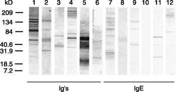
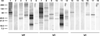
High molecular weight proteins (209–134 kDa) were immune-recognized by 70.8% of sera in the case of the Ig and IgM antibodies (figs 1 and 2). Similarly, Lorenzo et al. (Reference Lorenzo, Romaris, Iglesias, Audícana, Alonso, Leiro and Ubeira2000) assayed sera from patients diagnosed with Anisakis sensitization by an ELISA capture, and observed reactions with the 134 and 154 kDa proteins in 92% and 100%, respectively, of sera. One hundred per cent of our sera recognized a 60 kDa protein, such a response being the highest for Igs and IgM antibodies. When IgG was tested, 37.5% of sera reacted with a protein of around 40 kDa, and 12% with a protein around 25 kDa. These latest sera had the highest CAP values (two sera with CAP = 5 and the other with CAP = 6) (figs 1 and 2). With IgE determination, 10 of 24 sera reacted with proteins with high molecular weights (84 kDa). In nine sera, a reactive band appeared around 40 kDa and only one serum reacted with a protein of 25 kDa and another with a 14 kDa protein (fig. 1).
Immune-recognition patterns of patients diagnosed with Anisakis sensitization; total immunoglobulin (Igs) and immunoglobulin E (IgE). Lanes: 1 and 7, Anisakis simplex crude extract; 2 and 8, Toxocara canis crude extract; 3 and 9, Ascaris suum crude extract; 4 and 10, PAK antigen (A. simplex antigen purified by means of a column of IgG anti-A. simplex); 5 and 11, PAS antigen (PAK antigen purified by means of a column of IgG anti-A. suum); 6 and 12, EAS antigen (eluted from the anti-A. suum column after PAK purification).

When the CE T. canis antigen was tested, one-third of the sera reacted with a triplet of 40–84 kDa and 29.2% with an 84 kDa protein; and, finally, when the Igs were tested 41.7% and 33.3% of sera reacted with the 32 and 25 kDa bands, respectively. For the IgE determination, reactive bands at 184, 60 and 40 kDa appeared in only three patients (fig. 1).
In the present study, cross-reactions were lower against the CE A. suum antigen. Only 50% of sera reacted with a doublet of around 40 kDa present in the CE A. suum antigen when the Igs, IgM and IgG antibodies were tested, and one-third in the case of IgA antibodies. A 60 kDa protein was recognized by 16.7% of sera when Igs and IgG immunoglobulins were tested. All sera reacted with proteins of high molecular weights between 209 and 134 kDa. The Igs and the IgG antibodies reacted with a protein of 25 kDa in 29.2% of sera, and only 4.2% immune-recognized the 35 kDa protein (figs 1 and 2). Moneo et al. (Reference Moneo, Caballero, Gómez, Ortega and Alonso2000) and Arrieta et al. (Reference Arrieta, del Barrio, Vidarte, del Pozo, Pastor, González, Cardaba, Rojo, Minguez, Cortesano, Gallardo, Aceituno, Palomino, Vivanco and Lahoz2000) purified and cloned, respectively, proteins from A. simplex with similar masses. With the IgE determination, two sera recognized a protein of around 40 kDa, three sera reacted with proteins of around 184 kDa and only one serum reacted with a 60 kDa protein (fig. 1).
The following results were obtained when the PAK and the PAS antigens were evaluated. Intense bands were detected with the Igs and IgM antibodies, and responses were higher in sera with the lowest anti-A. simplex CAP values. These results indicate that low anti-A. simplex Igs levels may not exclude Anisakis sensitization. When PAK and especially PAS antigens were used, the main bands reacting with Igs, IgG and IgM antibodies showed up at 60, 40 and 25 kDa. The 60 kDa band was recognized by 83.3% and 79.2% of patient sera, respectively (figs 1 and 2).
Finally, when the EAS antigen was tested, 83.3% of assayed sera immuno-recognized the 14 kDa protein when the Igs, IgG and IgM immunoglobulins were measured (figs 1 and 2). This suggests that the 14 kDa protein shares common and strong epitopes in A. simplex and A. suum.
Immune-recognition patterns of patients diagnosed with Anisakis sensitization; immunoglobulin M (IgM), immunoglobulin G (IgG) and immunoglobulin A (IgA). Lanes: 1, 7 and 13, Anisakis simplex crude extract; 2, 8 and 14, Toxocara canis crude extract; 3, 9 and 15, Ascaris suum crude extract; 4, 10 and 16, PAK antigen (A. simplex antigen purified by means of a column of IgG anti-A. simplex); 5, 11 and 17, PAS antigen (PAK antigen purified by means of a column of IgG anti-A. suum); 6, 12 and 18, EAS antigen (eluted from the anti-A. suum column after PAK purification).

Surprisingly, when IgE levels were determined against the CE of A. simplex, only 58.3% of sera reacted with some bands, even when the A. simplex sensitization was confirmed in all patients under study. A possible cause of the small proportion of positive sera detected by immunoblot, compared to the CAP system, could be attributed to a higher amount of antigens used in this latter technique compared with immunoblot. This could also be the reason for an increase in cross-reactions and thus in the detection of false positives. This assumption was reinforced when purified antigens were tested. Against the PAK antigen we did not detect any IgE-positive sera, while against the PAS antigen 41.7% of sera reacted with the three different proteins. Nevertheless, it should be noted that the PAK and PAS antigens were purified using IgG antibodies, and thus IgE-specific epitopes could have been partially lost during the purification process (fig. 1).
The most intense bands detected in both PAK and PAS antigens were the 60, 40, the 30–35 and 25 kDa proteins. In the purification process, a doublet around 60 kDa appeared in the A. simplex PAK antigen, which was maintained with a high intensity in the PAS antigen (Rodero et al., Reference Rodero, Jiménez and Cuéllar2002). A 60 kDa band was clearly detected by IgA antibodies in the A. simplex CE, the PAK and PAS antigens (figs 1 and 2), while this band was not present in A. suum CE and EAS or T. canis CE antigens. Previously Rodero et al. (Reference Rodero, Chivato, Muro and Cuéllar2005) observed that serum Igs from Loa loa, Ascaris lumbricoides and Schistosoma mansoni patients reacted with the 60 kDa protein of the A. simplex antigen. With the PAK antigen, immune recognition against the 60 kDa protein was enhanced in intensity with L. loa and A. lumbricoides sera. This reaction disappeared with S. mansoni serum and appeared using T. canis, Echinococcus granulosus and Fasciola hepatica sera. Against the PAS antigen, immune recognition of the 60 kDa protein was only observed in the case of L. loa, A. lumbricoides and T. canis sera. This 60 kDa protein could be related to a 67 kDa Anisakis antigen recognized by the UA2 monoclonal antibody described by Iglesias et al. (Reference Iglesias, Leiro, Santamarina, Sanmartin and Ubeira1997). This IgMκ recognized both components of an excretion–secretion antigen preparation and antigens in the excretory cell and oesophageal glands of A. simplex L3. Likewise, Nakata et al. (Reference Nakata, Yamamoto and Yamamoto1990) demonstrated humoral IgE and IgG responses to a 60 kDa molecular weight protein in sera from patients with acute gastrointestinal anisakiasis. These antibodies were Anisakis-specific because they did not react with the similar molecular weight protein of A. suum homogenates and they also reacted with a 60 kDa Anisakis excretory–secretory protein. The description of a band of about 60 kDa has been associated with the consumption of raw fish, and this may have a diagnostic value since it was detected in patients with anisakiasis (del Rey et al., Reference del Rey, Valero, Mayorga, Gómez, Torres, Hernández, Ortiz and Lozano2006). When we obtained the PAS antigen, the concentrations of the 60, 40, 25 and 30–35 kDa proteins increased (Rodero et al., Reference Rodero, Jiménez and Cuéllar2002). The immune recognition of these proteins increased to 79.2%, 45.8%, 25% and 25%, respectively.
In summary, we have obtained and characterized an A. simplex purified antigen (PAS), enriched in A. simplex-specific molecules triggering an anti-IgG response in patients sensitized with this parasite. In addition, we have been able to detect a putative 60 kDa A. simplex-specific antigen in the CE, PAK and PAS A. simplex extracts. The detection of this band with IgG and IgA antibodies seems to be highly specific and sensitive and thus could potentially constitute a new tool to improve the diagnosis of anisakiasis. In conclusion, in the purification process of the A. simplex larval crude extract, the proteins implicated in cross-reactions with Ascaris and Toxocara were eliminated, with an important concentration of the proteins responsible for the induction of a specific IgE.
Acknowledgements
This research was supported by SAF98–0072 (Comisión Interministerial de Ciencia y Tecnología). Marta Rodero is supported by a fellowship from International Pharmaceutical Immunology, ASAC Pharmaceutical Immunology, S.A.