INTRODUCTION
Signal transduction molecules play central roles in regulating developmental processes, such as morphogenesis, organogenesis and cell differentiation in all organisms (Gerhart, 1999; Freeman, 2000). These molecules are organized into ‘pathways’ which represent a coordinated network of cell-surface receptors and intracellular molecules, playing specialized roles in sensing environmental stimuli and transducing signals produced to regulate cellular processes, including gene expression and cytoskeletal dynamics (Schenk and Snaar-Jagalska, 1999; Freeman, 2000). Signalling pathways have been studied extensively in a range of vertebrates and invertebrates (Gerhart, 1999; Schenk and Snaar-Jagalska, 1999), including the free-living nematode Caenorhabditis elegans (see Sternberg, 1993; Hanna-Rose and Han, 2000; Patterson and Padgett, 2000). The latter invertebrate is particularly well suited for studies of such pathways because of the ease of propagation and culture, the ability to disrupt developmental pathways through genetic manipulation and the detection of resultant phenotypic effects on the nematode (Bürglin et al. 1998; Plasterk, 1999; Aboobaker and Blaxter, 2000; Wixon et al. 2000).
Various studies of C. elegans have focused on cell-fate patterning and early embryonic development (e.g. Mickey et al. 1996; Bowerman et al. 1997; Leung et al. 1999; Berkowitz and Strome, 2000). Cell fate patterning was found to be highly dependent on the establishment of correct cell polarity in the early blastomere (Gonczy and Hyman, 1996; Rose and Kemphues, 1998; Bowerman, 2000; Golden, 2000), a process critical for survival, and considered to be conserved for a broad range of organisms (Doe and Bowerman, 2001). A particularly important group of molecules implicated in the establishment and maintenance of cell polarity is the PAR proteins (the name being derived from “partition defective” in asymmetric cell division; Kemphues et al. 1988) (reviewed by Macara, 2004; Nance, 2005; Munro, 2006; Suzuki and Ohno, 2006). In C. elegans, it has been demonstrated that the inhibition of their activity in mediating cell polarity can lead to defects in development (hence, “partition defective”) (Kemphues et al. 1988; Guo and Kemphues, 1995). In spite of the knowledge of PAR proteins for C. elegans and other model organisms (such as Drosophila melanogaster and Xenopus laevis), until recently, there had been no information available for parasitic nematodes of socio-economic importance. Insights into the function of these proteins and their pathways could improve the understanding of the molecular aspects of parasite development and survival, and, importantly, may provide a basis for developing novel intervention strategies against parasites by identifying targets for the disruption of such pathways.
The purpose of the present article is to (1) review key aspects of PAR proteins in the development of C. elegans and other model organisms, with an emphasis on PAR-1, (2) describe and discuss recent advances in the understanding of a PAR-1 homologue from the economically important parasitic nematode, Haemonchus contortus, of small ruminants (Veglia, 1915; Georgi and Georgi, 1990; Nikolaou and Gasser, 2006), and (3) propose avenues for future research in this exciting area of developmental biology in parasitic nematodes.
EARLY EMBRYONIC DEVELOPMENT IN THE FREE-LIVING NEMATODE
The life-cycle of C. elegans is well described (Wood, 1988; Riddle et al. 1997; Hope, 1999) and is summarized in Fig. 1. This nematode undergoes development in a soil environment as a free-living organism, but it can be readily cultured in vitro and maintained in the laboratory. Early studies elucidated its development through the determination of the embryonic (Sulston et al. 1983) and post-embryonic (Sulston and Horvitz, 1977) cell lineages.

Fig. 1. Comparison of the life-cycle of the (A) parasitic nematode Haemonchus contortus with that of the (B) free-living nematode Caenorhabditis elegans. Developmental stages: L1, first larval stage; L2, second larval stage; L3, third larval stage; xL3, exsheathed third larval stage; L4, fourth larval stage. Features of development: E, embryogenesis; G, growth phase; F, feeding phase (F1, bacteria; F2, blood); S, sexual differentiation; R, reproduction. * Denotes the potential for arrested development.
Once an egg is fertilized, a series of cytoplasmic rearrangements occurs which organizes the 1-cell embryo (P0) into distinct anterior and posterior poles (the latter being defined by the site of sperm penetration; Goldstein and Hird, 1996). Upon completion of the first cell cycle, division occurs asymmetrically in a plane perpendicular to the anterior-posterior axis to create 2 unequal daughter cells AB (larger) and P1 (smaller). Each of these cells and their subsequent daughter cells have a characteristic length of cell cycle and axis of division (see Figs 2 and 3). At the 2-cell stage, the AB cell divides first, symmetrically along the anterior-posterior axis to generate the daughter cells ABa and ABp, followed by a division of P1 asymmetrically and in a division plane perpendicular to the division of AB and the anterior-posterior axis, to create the daughters EMS (larger) and P2 (smaller). From the 4-cell stage, the descendents of AB continue to divide symmetrically, whereas both EMS and P2 divide asymmetrically. EMS generates the daughter cells MS (larger) and E (smaller), and P2 generates the daughter cells C (larger) and P3 (smaller). Subsequent cell divisions of MS, E and C utilize a symmetrical division pattern. The P3 cell undergoes the final round of asymmetric cell division early in development to produce the daughter cells D and P4, which then divide symmetrically. Gastrulation begins after the generation of P4.

Fig. 2. The embryonic cell lineage of Caenorhabditis elegans leading to the generation of the 6 founder cells (underlined). Adapted from Sulston et al. (1983).

Fig. 3. The early embryonic cell divisions of Caenorhabditis elegans which lead to the 4-cell stage. Specific cell–cell inductive signalling occurs between P2 and EMS for the specification of the MS and E cell lineages (see Fig. 2). In wild-type embryos, asymmetric cell division of the 1-cell embryo leads to the correct distribution of maternal factors in subsequent daughter cells, permitting cell–cell inductive signalling at the 4-cell stage. However, in par-1 mutants, correct distribution of maternal factors is not achieved due to a symmetrical first cell division; therefore, inductive signalling does not occur, which affects the specification of subsequent cell lineages (e.g. 2 MS-like cells are formed rather than an MS and E cell, leading to the lack of an intestine; see Fig. 2).
AB, MS, E, C, D and P4 are embryonic blast cells, known as the ‘6 founder cells’, which lead to the production of various organ and tissue cell lineages in C. elegans (Fig. 2). The AB lineage produces 389 cells, predominantly of neuronal and hypodermal tissues (ectoderm), the first half of the pharynx and some of the body muscle tissues (mesoderm). The MS lineage produces 80 cells, predominantly of body muscle, the second half of the pharynx, coelomocytes (putative primative blood cells; Maduro et al. 2001), the somatic gonad precursors (mesoderm), and also some neurons (ectoderm). The E lineage produces 20 cells, exclusively of the intestine (endoderm). The C lineage produces 47 cells, predominantly of body wall muscle (mesoderm), and some hypodermal and neuronal tissues (ectoderm), and the D lineage produces 20 body wall muscle cells (mesoderm). The P4 lineage produces 2 cells that represent exclusively the germ-line. The total number of cells (n=558) makes up a newly-hatched L1 hermaphrodite.
The specification of the ‘6 founder cells’ is controlled by maternally-derived proteins (see Bowerman, 1998). Much is known about these initial patterning steps, but they are not fully understood and, in some cases, are represented by hypothetical models based soley on experimental observations of both autonomous and cell–cell inductive signalling. The P lineage (P0 through to P4) and their daughters (excluding EMS) are specified by cell autonomous processes, primarily through a polar rearrangement of the cytoplasm which leads to the selective distribution and/or degradation of maternal transcription factors and asymmetric cell division. The maternal transcription factors are specific for the activation of zygotic gene transcription relating to organ and/or tissue patterning and differentiation (see Newman-Smith and Rothman, 1998). Cell–cell interactions are required between EMS and P2 for the establishment of asymmetry in EMS and the correct specification of the E lineage (Goldstein, 1992, 1993; see Fig. 3), and these are mediated by the Wnt and mitogen-activated protein kinase (MAPK) signalling pathways (Rocheleau et al. 1997; Thorpe et al. 1997; Meneghini et al. 1999; Schlesinger et al. 1999; Lo et al. 2004). A number of cell–cell interactions have also been identified which specify the fate of descendants of the AB lineage, which also lead to the establishment of the dorsal-ventral and left-right embryonic axes (Hutter and Schnabel, 1995a,b) and are mediated by the abnormal germline proliferation-1 (GLP-1)/Notch signalling pathway (Hutter and Schnabel, 1994; Mango et al. 1994; Mello et al. 1994; Mickey et al. 1996; Shelton and Bowerman, 1996).
Hence, based on this information, the disruption of any process relating to the specification of early blastomere fate has a negative impact on the development of specific organs and/or tissues. One of the earliest steps sensitive to disruption is the establishment of correct anterior-posterior axis in the P0 cell (1-cell stage). Mutations affecting genes involved in early cytoplasmic rearrangements lead to a symmetrical first cell division, alternations of the cell cycle period and division plane of daughter cells, and mislocalization of maternal factors (Kemphues et al. 1988; see Fig. 3). The end result is a disruption of signalling events between the EMS and P2 blastomeres leading to defects in the patterning of the intestinal (E) and germ-line (P) lineages (Kemphues et al. 1988; see Fig. 3). The extent of the resultant defects in development depend on the severity of gene function impairment, where ‘null’ function leads to an excess number of pharyngeal/body wall muscle cells and a total absence of intestinal cells (embryonic lethal), whereas ‘partial’ function leads to an absence of a germ-line (sterility) (Kemphues et al. 1988). Further studies into the molecular mechanism controlling this first step of early fate specification indicate that this process involves molecules which function in the regulation of the cytoskeleton (see Nance, 2005). These molecules have been subsequently linked, not only to downstream developmental processes, but also to cell maintenance and survival in the terminal differentiation phase in a diverse range of species.
PAR PROTEINS IN AND OTHER ORGANISMS AND THEIR INVOLVEMENT IN CELL POLARITY AND THE CYTOSKELETON, WITH AN EMPHASIS ON PAR-1
Involvement of PAR proteins in the embryonic development of C. elegans
In total, 6 par genes have been identified (Fig. 4). The par-1 and par-4 genes code for putative serine/threonine protein kinases (STKs) (Guo and Kemphues, 1995; Watts et al. 2000). The par-3 and par-6 genes code for novel PDZ domain-containing proteins (Etemad-Moghadam et al. 1995; Hung and Kemphues, 1999). The par-2 gene codes for a novel zinc finger (RING domain) and ATP-binding protein of unknown function (Levitan et al. 1994), and the par-5 gene codes for a 14-3-3 protein (Morton et al. 2002). Therefore, the PAR proteins are considered to form part of a complex network of signal transduction pathways which control the cytoskeleton (Kemphues, 2000; Wodarz, 2002; Macara, 2004; Wiggin et al. 2005). Early studies of genetic pathways suggested that the PAR proteins function in a common process (Kemphues et al. 1988; Kirby et al. 1990; Levitan et al. 1994; Cheng et al. 1995). However, more recent studies indicate that the process of cytoplasmic organization in the early embryo is not linear (Bowerman et al. 1997; Crittenden et al. 1997; Cuenca et al. 2003; Cheeks et al. 2004; Hao et al. 2006).

Fig. 4. The PAR proteins of Caenorhabditis elegans. (A) Localization of the PAR proteins in the wild-type 1-cell embryo. (B) Schematic representation of the domain profile of each PAR protein, drawn to scale (see Levitan et al. 1994; Etemad-Moghadam et al. 1995; Guo and Kemphues, 1995; Hung and Kemphues, 1999; Watts et al. 2000; Morton et al. 2002). (C) The functional domains present in the PAR proteins. Amino acid (aa).
The polarization of the 1-cell embryo by the PAR proteins into distinct anterior and posterior domains (see Fig. 4) is proposed to occur in 2 phases (Cuenca et al. 2003). The first polarization phase is called the ‘establishment phase’ and is mediated by the asymmetric destabilization and flow of the actomyosin cytoskeleton (e.g. non-muscle myosin-2 [NMY-2] and F-actin), initiated from the point of sperm entry (posterior) towards the anterior pole of the 1-cell embryo (Munro et al. 2004). The cytoplasmic and cortical flow generated by this process facilitates the anterior localization of proteins, including PAR-3 and PAR-6 which, together, act in a protein complex with the C. elegans homologue of atypical protein kinase C (aPKC), protein kinase C-3 (PKC-3), to promote this flow (Cheeks et al. 2004; Munro et al. 2004). PAR-4, muscle excess proteins-5 and -6 (MEX-5 and MEX-6, respectively) also appear to facilitate cytoplasmic and cortical flows (Cheeks et al. 2004). Prior to the initiation of flow, both PAR-1 and PAR-2 are excluded from the cortex by the PAR-3/PAR-6/PKC-3 complex via a mechanism which involves the phosphorylation of PAR-2 by PKC-3 (Hao et al. 2006). The progression of cytoplasmic and cortical flow leads to a decrease in the levels of the PAR-3/PAR-6/PKC-3 complex in the posterior, allowing cortical localization of PAR-2 in this region (Cheeks et al. 2004). PAR-2 then recruits PAR-1 to the posterior cortex through an unknown mechanism (Hao et al. 2006). The localization of the PAR-1 protein is also dependent on PAR-3 and PAR-5 (Etemad-Moghadam et al. 1995; Guo and Kemphues, 1995; Boyd et al. 1996; Morton et al. 2002). PAR-5 is also required for the establishment phase (Cuenca et al. 2003; Hao et al. 2006).
The second polarization phase is called the ‘maintenance phase’, and involves the continual exclusion of the PAR-3/PAR-6/PKC-3 complex from the posterior by PAR-2 (Cheeks et al. 2004; Munro et al. 2004), an activity which is mediated by its RING domain (Hao et al. 2006). PAR-1 then appears to play a role in the localization and/or stabilization of posterior maternal factors, such as skinhead-1 (SKN-1; Bowerman et al. 1992, 1993; Guo and Kemphues, 1995) and those present in P-granules which specify the germ-line (e.g. pharynx and intestine in excess-1 [PIE-1]; Reese et al. 2000). In addition, this localization/stabilization activity by PAR-1 has been suggested to also be the result of excluding the anterior proteins MEX-5 and MEX-6, which otherwise function in the transcriptional repression of the germ-line (Schubert et al. 2000; Cuenca et al. 2003; Cheeks et al. 2004). In spite of this model of polarization, many aspects are not fully understood at the molecular level. Nonetheless, it has been suggested that, of all PAR proteins, PAR-1 plays the most direct role in mediating the asymmetric localization of maternal proteins (Cuenca et al. 2003; Cheeks et al. 2004).
The PAR-1 protein of C. elegans
Following the 1-cell stage, the localization of PAR-1 can be detected along all cell–cell contacts at the 4-cell stage, consistent with a proposed involvement in mediating cell–cell interactions (Guo and Kemphues, 1995). Localization then shifts baso-laterally by the end of the 4-cell stage to promote roles in gastrulation and cell adhesion (Nance et al. 2003). Antibody staining for PAR-1 becomes less pronounced after the generation of the P4 daughter cells, and is no longer detectable by the onset of morphogenesis (Guo and Kemphues, 1995). However, PAR-1 is required for at least one aspect of morphogenesis – the morphogenesis of the vulva (Hurd and Kemphues, 2003). The exact molecular activity of PAR-1 in mediating these processes has not been determined, but mutations within the putative kinase domain known to be essential for activity (considered to be conserved for all kinases; see Hanks et al. 1988) has provided some insight (see Guo and Kemphues, 1995). It was found that, although the cortical localization of PAR-1 is not dependent on its kinase activity, its function in development, however, requires it, as mutations which destroy the activity of the kinase domain lead to the typical par-1 phenotype (Guo and Kemphues, 1995; see Fig. 3).
The function of PAR-1 in other organisms
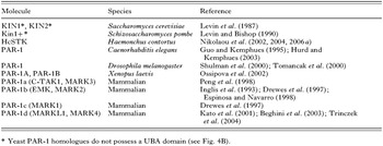
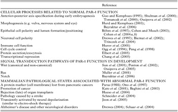
The putative kinase domain within PAR-1 classifies it as member of a large subfamily of STKs (designated PAR-1/microtubule-affinity regulating kinase [MARK]; see Table 1) which all appear to be involved in the establishment and maintenance of cell polarity (Drewes et al. 1998). Many of these PAR-1 homologues have been partially characterized at the molecular and biochemical levels. Roles in anterior-posterior axis specification and/or embryonic patterning have been established in both D. melanogaster (see Shulman et al. 2000; Tomancak et al. 2000; Bayraktar et al. 2006) and X. laevis (see Ossipova et al. 2002). Similar roles in the embryonic development in mammals have only recently been investigated (Moore and Zernicka-Goetz, 2005; Vinot et al. 2005), but it has been noted that PAR-1 can play roles in growth and fertility (Bessone et al. 1999; Hurov et al. 2001). PAR-1 also appears to participate in many cytoskeletal-mediated processes in various cell types (see Table 2): epithelial cell polarity and lumen formation/positioning (e.g. Böhm et al. 1997; Cohen and Musch, 2003; Cohen et al. 2004a,b), neuronal cell polarity (e.g. Drewes et al. 1997; Biernat et al. 2002; Trinczek et al. 2004), immune cell function (Hurov et al. 2001), cell cycle control (Ogg et al. 1994; Peng et al. 1998), protein secretion/exocytosis (Elbert et al. 2005) and transcriptional repression (Chang et al. 2005). To date, PAR-1 kinases have been implicated in the Wnt (canonical and non-canonical), Ras and Notch signal transduction pathways (see Muller et al. 2001; Sun et al. 2001; Penton et al. 2002; Ossipova et al. 2005; Bayraktar et al. 2006; Table 2) – pathways which have importance in signalling during development (Gerhart, 1999). Since PAR-1 homologues function in a number of essential cellular processes, it is not surprising that a number of pathological states have been associated with their disfunction in mammals (see Table 2). PAR-1/MARK has been identified as a protein marker at the cell membrane which is lost from cancers of the pancreas (Parsa, 1988; Ono et al. 1997), and is suggested to be involved in the promotion of cancer (Kato et al. 2001; Beghini et al. 2003). PAR-1/MARKs have also been implicated in the (late) rejection of organ transplants (Hueso et al. 2004) and the pathology caused by a stroke (Schneider et al. 2004). It is also transiently activated by neural depolarization, similar to that produced in electro-shock therapy (Jeon et al. 2005).


The structure and biochemistry of PAR-1
All PAR-1 STKs have similar protein domain profiles (see Fig. 4B), which include an N-terminal kinase domain, a putative tyrosine-dependent membrane-targeting motif, a ubiquitin-associated (UBA) domain, a spacer domain (known to mediate membrane localization; Vaccari et al. 2005) and a C-terminal tail domain (also known as the kinase-associated 1 [KA1] domain; Hofmann and Bucher, 1996). The UBA and tail domains are implicated in mediating protein–protein interactions (Guo and Kemphues, 1996; Hofmann and Bucher, 1996; Bertolaet et al. 2001a,b; Wilkinson et al. 2001). The tail domain is also implicated in the negative regulation (auto-inhibition) of kinase activity (Elbert et al. 2005). The PAR-1/MARK STKs are relatively closely related to another subfamily of STKs (e.g. synapses of amphids defective-1, SAD-1; Crump et al. 2001) based on the kinase domain. However, homology is restricted to this domain alone and these STKs do not possess the other functional domains, such as the UBA and KA1-type C-terminal tail domains, which are characteristic of PAR-1 kinases, and consequently, may have different functional and regulatory modes.
The UBA domain is considered to bind ubiquitin, which has been described as being a form of reversible post-translational protein modification (analogous to phosphorylation mediated by protein kinases), capable of regulating the localization, activity, substrate binding and degradation of proteins (Schnell and Hicke, 2003). The type of ubiquitination often determines the ‘fate’ of a protein, whereby ‘poly-ubiquitination’ usually targets a protein for degradation by the 26S proteasome, whereas ‘mono-ubiquitination’ signals other cellular activities, such as the endocytic trafficking of cell surface receptors and DNA repair (see Haglund et al. 2003). The UBA domain is capable of binding both poly- and mono-ubiquitin, as well as ubiquitinated proteins (Buchberger, 2002; Di Fiore et al. 2003).
Despite PAR-1 being a protein with a UBA domain (Hofmann and Bucher, 1996), the role of the domain in this and related kinases has not yet been adequately explored. Ubiquitin-binding has been demonstrated for the PAR-1 of D. melanogaster through a protein–protein interaction study (see Giot et al. 2003). However, it has not yet been shown that these interactions are mediated via the UBA domain; and it should be noted that no interaction has yet been identified in a similar study of C. elegans (see Li et al. 2004). In yeast, the UBA domain of Rad23, a protein involved in DNA repair, has been reported to play a role in the inhibition of protein degradation by binding ubiquitin to prevent chain extension (i.e. substrate-linked multi-ubiquitination; Chen et al. 2001). This mechanism may represent the way in which PAR-1 protects proteins specific to the germ-line in early embryogenesis (e.g. PIE-1, posterior segregation-1 [POS-1], MEX-1, MEX-5 and MEX-6; see Reese et al. 2000; DeRenzo et al. 2003), which are otherwise selectively degraded in all cells external to the P lineage (Pellettieri et al. 2003).
Two recent studies (Jaleel et al. 2006; Panneerselvam et al. 2006) examined the structure of a partial mammalian homologue of PAR-1 (kinase and UBA domains). Both of them suggested that the UBA domain does not take on the conventional conformation capable of binding ubiquitin. Rather, the UBA domain interacts directly with one of the two lobes of the catalytic domain to positively regulate kinase activity (the N- or C-terminal lobe; Jaleel et al. 2006; Panneerselvam et al. 2006). Also, Panneerselvam et al. (2006) revealed a structure similar to the ‘common docking’ (CD) motif of MAPKs in the region between the catalytic and UBA domains, previously suggested to be a putative membrane-targeting motif (Drewes et al. 1998). These finding have functional implications in relation to these domains in PAR-1/MARK STKs. However, given that a considerable portion of the protein was absent from each of these structure models, there may be considerable variation in the true nature of the structure, indicated by variation in the interaction of the UBA domain with the 2 lobes of the catalytic domain (see Jaleel et al. 2006; Panneerselvam et al. 2006).
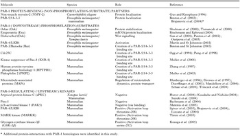
The KA1 tail domain of C. elegans has been demonstrated to function in mediating protein–protein interactions via an interaction-cloning screen of a library (Guo and Kemphues, 1996). The interacting protein was identified as NMY-2, a component of the actin cytoskeleton which acts in the polarization in the early embryo (see Munro et al. 2004; Table 3). The interaction with the KA1/tail domain of PAR-1 involves the NMY-2 domain region spanning the last 607 amino acids (aa) from the C-terminus, and was subsequently confirmed in solution and whole C. elegans extracts (Guo and Kemphues, 1996). Also, NMY-2 co-localizes with PAR-1 within the embryo and has been reported to play a role in the establishment of anterior-posterior polarity, producing similar par-like phenotypes through RNA interference (RNAi) (Guo and Kemphues, 1996). NMY-2 is also required for cytokinesis and the localization of the PAR-1, PAR-2 and PAR-3 proteins, but is itself unaffected by par mutations (Guo and Kemphues, 1996; Cuenca et al. 2003; Cheeks et al. 2004; Munro et al. 2004).

Phosphorylation-substrates of PAR-1
Despite the requirement of kinase activity to mediate PAR-1 function in the early development of C. elegans (see Guo and Kemphues, 1995), no protein substrate has been directly identified as a target for phosphorylation. This is not the case in D. melanogaster. C. elegans and D. melanogaster differ with respect to how the anterior-posterior axis is specified during embryogenesis. For instance, in C. elegans, specification occurs following fertilization and is mediated by the actin cytoskeleton, whereas in D. melanogaster, it occurs during oogenesis and is mediated by the microtubule cytoskeleton (Pellettieri and Seydoux, 2002). However, there are some recent indications that there are more similarities between the two species. For example, there appears to be some dependence on the actin cytoskeleton for the posterior localization of PAR-1 in D. melanogaster at the seventh stage of oogenesis (Doerflinger et al. 2006). Another common mechanism is the selective control of maternal factor stability (see Pellettieri and Seydoux, 2002). In D. melanogaster, one way in which PAR-1 directly regulates the stability of a protein is by phosphorylation (see Table 3). An example of this process is the stabilization of the ‘unstable protein’ Oskar (Osk), which is translated at the posterior of the oocyte once PAR-1 mediates the localization of oskar mRNA (Shulman et al. 2000; Tomancak et al. 2000; see Table 3). The phosphorylation of Osk by PAR-1 prevents its degradation and allows it to accumulate at the posterior pole for subsequent patterning of the germ-line (Benton and St Johnston, 2002; Riechmann et al. 2002). However, this model of the regulation of protein stability by phosphorylation appears to contrast that proposed for PAR-1 in C. elegans, in that protein stability may be mediated by the UBA domain via the inhibition of substrate-linked multi-ubiquitination (Pellettieri et al. 2003), although this model has yet to be tested experimentally. However, it has been suggested that the degradation of Osk in D. melanogaster is likely to be mediated by the ubiquitin-proteosome pathway (DeRenzo and Seydoux, 2004). Therefore, there may be similarities, which await discovery.
Many other substrates of PAR-1 phosphorylation have been determined in D. melanogaster (Table 3). The protein Exuperantia (Exu), which is a mediator of the localization of bicoid mRNA (an anterior oocyte determinant), is phosphorylated by PAR-1 at 2 sites – region A (serine-438 and serine-440) and region B (serine-457) (Riechmann and Ephrussi, 2004). Phosphorylation of the protein Dishevelled (Dsh) by PAR-1 (while in a complex with Dsh) was found to activate the canonical Wnt/beta-catenin signalling pathway and inhibit an alternative Dsh pathway for planar cell polarity (PCP) – the Jun kinase (JNK) signalling (non-canonical Wnt) pathway (Sun et al. 2001). Therefore, PAR-1 can act as a molecular ‘switch’ between 2 alternative Wnt signalling pathways which control the development of many species (canonical Wnt/beta-catenin or non-canonical PCP/JNK pathways), and this activity by PAR-1 appears to be conserved between X. laevis and humans (Sun et al. 2001). However, it has been reported that Dsh phosphorylation by PAR-1 can signal the PCP (non-canonical) pathway (Penton et al. 2002); this is further supported by a study of X. laevis, where the pathway decision is mediated by different isoforms of PAR-1 (Ossipova et al. 2005). Therefore, the phosphorylation of Dsh is not considered to be specific for canonical Wnt/beta-catenin signalling. In C. elegans, PAR-1 has yet to be directly implicated in Wnt signalling, although similar defects in the P2-EMS cell–cell induction are observed for various mutations in both systems (e.g. the muscle excess phenotype which results from both EMS daughters adopting an MS-like fate in par-1, mom-1, mom-2 and mom-5 mutant embryos; see Guo and Kemphues, 1995; Rocheleau et al. 1997; Thorpe et al. 1997; Fig. 3).
In D. melanogaster and other organisms, PAR-1-phosphorylation plays a widespread role in the regulation of protein–protein interactions with PAR-5/14-3-3 by creating 14-3-3-binding sites (Benton et al. 2002; see Table 3). PAR-1 also appears to be responsible for delivering these phosphorylated protein substrates directly to PAR-5/14-3-3, via protein binding. One such protein is PAR-3/Bazooka (Baz). It has been proposed that the creation of a Baz/14-3-3 complex inhibits the formation of the Baz/PAR-6/aPKC complex linked to the establishment of polarity (Benton and St Johnston, 2003). Direct interactions between PAR-1 and PAR-5 have yet to be verified in C. elegans, although they seem probable based on the requirement of PAR-5 for the localization of PAR-1 (see Morton et al. 2002).
The regulation of 14-3-3 protein–protein interactions is the mechanism which forms the basis of cell-cycle control (see Tables 2 and 3). The mammalian homologue Cdc25C-associated kinase 1 (C-TAK1) phosphorylates, and therefore negatively regulates the protein Cdc25C, a phosphatase which promotes the entry into mitosis (M phase) (Ogg et al. 1994; Peng et al. 1998). Phosphorylated Cdc25C binds to PAR-5/14-3-3, keeping its localization sequestered to the cytoplasm to maintain the cell cycle in the interphase (a non-dividing state). The negative regulation of C-TAK1, for example, by the kinase Pim-1 (see Table 3), leads to the translocation of unbound Cdc25C into the nucleus and transition of the cell-cycle into the G2/M phase (Bachmann et al. 2004). Similar mechanisms of C-TAK1 phosphorylation-mediated control of PAR-5/14-3-3 protein binding and localization have also been described for the kinase suppressor of Ras-1 (KSR-1) (Muller et al. 2001), human protein-tyrosine phosphatase homology 1 (HPTPH1; Zhang et al. 1997) and plakophilin 2 (PKP2), where C-TAK1 modulates its localization between desmosomes (positive) and the nucleus (negative) (Muller et al. 2003).
The model of KSR-1 regulation by C-TAK1 is currently considered to be consistent with PAR-1 activity in vulval morphogenesis in C. elegans. Vulval induction is mediated by the Ras signalling pathway, of which PAR-1 appears to be a negative regulator (Kao et al. 2004; Yoder et al. 2004). The exact molecular nature of this action of PAR-1 has not yet been determined, but the genetic pathway involves the protein suppressor of Ras mutations-6 (SUR-6, the PR55/B regulatory subunit of protein phosphatase 2A), SUR-7 (a zinc ion transporter) and KSR-1 (Kao et al. 2004; Yoder et al. 2004). In mammals, KSR-1 is directly phosphorylated by C-TAK1 in the absence of Ras signalling, to promote its association with PAR-5/14-3-3 and localization to the cytoplasm (Muller et al. 2001; Yoder et al. 2004). Upon stimulation of the Ras pathway, C-TAK1 phosphorylation is reduced and KSR-1 (unable to bind PAR-5/14-3-3) localizes to the plasma membrane to associate with Ras signalling components (Muller et al. 2001). In this scenario, it is suggested that SUR-6 acts in opposition to PAR-1 activity through the dephosphorylation of KSR-1 (Kao et al. 2004), and SUR-7 acts to reduce the level of zinc ions, because they appear to promote the phosphorylation of KSR-1 (Yoder et al. 2004). It remains to be determined whether KSR-1 is a PAR-1 substrate in C. elegans and how zinc ions mediate the phosphorylation of KSR-1, including whether they directly or indirectly positively effect PAR-1 activity.
The mammalian PAR-1 homologue MARK has been reported to phosphorylate the microtubule-associated proteins (MAPs) Tau, MAP2, MAP4 and Doublecortin (Dcx) at KXGS motifs within their microtubule-binding domains (see Table 3). The effect of this phosphorylation is the detachment of MAPs from mictotubules, thereby increasing microtubule dynamics (Illenberger et al. 1996; Drewes et al. 1997; Schaar et al. 2004; Trinczek et al. 2004) and facilitating the transport of molecules (Mandelkow et al. 2004). The hyper-phosphorylation of MAPs, such as Tau, is implicated in neurological disease (i.e. Alzheimer's disease; see Drewes, 2004; Table 2). It has recently been suggested that abnormal phosphorylation by MARKs (resulting from an upregulation of kinase activity) can prime MAPs for hyper-phosphorylation by other kinases, allowing these MAPs to accumulate into aggregates and tangles to cause neuronal toxicity (Drewes, 2004; Schaar et al. 2004). A cascade of ‘Tau toxicity’ is also described for D. melanogaster (see Nishimura et al. 2004), although there is some contradiction regarding the mechanism of microtubule regulation in the ‘normal’ physiological state. In D. melanogaster, PAR-1 phosphorylation of MAPs results in mictotubule stabilization rather than destabilization, as occurs in mammals (cf. Drewes et al. 1997; Doerflinger et al. 2003). Recently, the p21-activated kinase 5 (PAK5) was described as being a negative regulator of PAR-1/MARK activity in mammals, by directly binding to the PAR-1/MARK catalytic domain, in order to prevent its activation by upstream kinases via steric hindrance (Matenia et al. 2005; Table 3). Since the effect of this binding (in cell culture) is an increase in microtubule stability at the expense of actin stability (which increases actin dynamics), this interaction presents an interesting ‘point of cross-talk’ between the regulation of the microtubule and actin cytoskeletons (Matenia et al. 2005). At this stage, it is still somewhat unclear whether PAR-1 plays a direct role in the regulation of the microtubule cytoskeleton in C. elegans.
PAR-1-regulating (‘upstream’) kinases
There are conflicting reports on the kinase cascade associated with PAR-1 (Table 3). The PAR-4/LKB1 protein of D. melanogaster has been reported to be a substrate for phosphorylation by PAR-1, which mediates its activation (Martin and St Johnston, 2003). However, this ‘linear pathway’ appears to directly contrast that reported for mammals, in which PAR-4/LKB1 has been found to be the ‘upstream kinase’ activating PAR-1 (Spicer et al. 2003). This latter model is supported by other studies which have implied that PAR-4/LKB1 activated PAR-1/MARK by phosphorylation (Brajenovic et al. 2004; Lizcano et al. 2004). However, reported mutants of par-1 and par-4/lkb1 in C. elegans and D. melanogaster are consistent with PAR-1 being the upstream kinase (see Martin and St Johnston, 2003). Therefore, the linear pathway model which places PAR-1 upstream of PAR-4/LKB1 is applicable, at least for invertebrates, although direct evidence has yet to be presented for C. elegans. Atypical PKC, MARK kinase (MARKK, a member of the Ste20 kinase family, also known as the ‘one thousand and one’ protein kinase [TAO-1]; Hutchison et al. 1998) and glycogen synthase kinase-3β (GSK-3β) have also been implicated as upstream kinases of PAR-1 in mammals and X. laevis, where aPKC regulates the localization of PAR-1 during development via binding to PAR-5 (Timm et al. 2003; Kusakabe and Nishida, 2004; Suzuki et al. 2004; Kosuga et al. 2005). Atypical PKC has also been reported to negatively regulate the kinase activity of PAR-1 in humans (Hurov et al. 2004), and a similar pathway has also been reported for both C. elegans and D. melanogaster (see Gotta, 2005). ‘Upstream kinases’ PAR-4/LKB1, MARKK and GSK-3β phosphorylate PAR-1/MARK within the activation loop of its catalytic domain to promote the activation of kinase activity: threonine-208 by PAR-4/LKB1 and MARKK, and serine-212 by GSK-3β (Timm et al. 2003; Lizcano et al. 2004; Kosuga et al. 2005). The latter contrasts the suggestion that phosphorylation of the serine-212 site has a negative effect on kinase activity (Timm et al. 2003).
In summary, PAR proteins are important regulators of early development, acting in a number of processes that regulate the cytoskeleton, including the cytoplasmic localization of maternally derived cell fate determinants. Of the 6 PAR proteins which have been partially characterized, PAR-1 appears to play or mediate the most direct role in these processes (Cuenca et al. 2003; Cheeks et al. 2004). PAR-1 also features in similar, prominent roles in the early development of D. melanogaster and X. laevis (see Shulman et al. 2000; Tomancak et al. 2000; Ossipova et al. 2002; Kusakabe and Nishida, 2004). Interestingly, investigations into mammalian homologues have indicated that PAR-1 also has broader cellular roles, such as the regulation of microtubule dynamics, which appear to impact on the growth of organisms and the integrity of a number of cell types (e.g. Drewes et al. 1997; Bessone et al. 1999; Hurov et al. 2001). While PAR-1 has been characterized in C. elegans, the wealth of molecular and biochemical information has been drawn from homologues from other species, such as D. melanogaster, X. laevis and mammals (e.g. Drewes et al. 1997; Sun et al. 2001; Riechmann et al. 2002; Benton and St Johnston, 2003; Timm et al. 2003; Hurov et al. 2004; Kusakabe and Nishida, 2004). Some current evidence suggests that PAR-1 acts to mediate cell polarity through different signalling pathways in different species (e.g. Sun et al. 2001; Penton et al. 2002; Ossipova et al. 2005; Bayraktar et al. 2006). Alternatively, this evidence emphasizes the versatility of PAR-1, in being able to act as a point of cross-talk between a number of different pathways, not only in regulating cell polarity but also in other functions which are regulated by the cytoskeleton (Brajenovic et al. 2004). Hence, PAR-1 is a key molecule involved in important developmental and survival ‘mechanisms’ of an organism, but there are major knowledge gaps in fundamental aspects of PAR-1 at the molecular level, which make this an exciting research focus.
NEED FOR COMPARATIVE STUDIES BETWEEN AND PARASITIC NEMATODES
Despite all of the research on signal transduction pathways in C. elegans, there has been very limited study in this area for key parasitic nematodes of socio-economic importance, such as H. contortus. Given that the complete cell lineage of C. elegans has been mapped and its genome fully sequenced (The C. elegans Sequencing Consortium, 1998), this free-living nematode has been considered as a model for the study of parasitic nematodes (see Blaxter et al. 1998; Bürglin et al. 1998; Aboobaker and Blaxter, 2000; Gasser and Newton, 2000; Geary and Thompson, 2001; Hashmi et al. 2001; Gilleard, 2004). However, biological differences in their life histories (i.e. C. elegans is free-living and exists as a population of hermaphrodites and males, in contrast to many dioecious parasitic nematodes) make it conceivable that some signalling pathways and their regulation will be divergent (e.g. Viney et al. 2005).
The development of C. elegans arises from an ‘invariant pattern’ of cell division which produces daughter cells with a specific fate, either to terminally differentiate into a cell-type within a tissue or to undergo cell death (see Sulston and Horvitz, 1977; Sulston et al. 1983; Sulston, 1988). This pattern of cell lineage is controlled by a number of processes, including autonomous and inductive signalling (see Bowerman, 1998). Hermaphrodites and males have the same cell lineage and only differ in their terminal states where a different pattern of cell death occurs (e.g. formation of the male 1-armed gonad versus the 2-armed gonad of the hermaphrodite, and male-specific neuroanatomy). Interestingly, differences in the embryonic cell lineage, particularly in the generation, migration and death of blast cells, have been reported to be associated with divergent morphologies (e.g. in buccal capsule and reproductive organs) among different species of nematodes (see Felix and Sternberg, 1996; Sternberg and Felix, 1997; Sommer, 2000; Schierenberg, 2001).
The genes that regulate early embryogenesis (such as the par genes) are of particular interest in the context of nematode evolution. This is due to their role in mediating the orientiation of the mitotic spindle and, consequently, cell–cell contacts at the 4-cell stage (see Goldstein, 2001). A mutation in these genes which causes a change in the division plane and orientation of cells at the 4-cell stage, often ‘mimic’ the normal cell division and orientation pattern observed for other species of nematode (see Goldstein, 2001). Given that the PAR proteins have been considered to be conserved across a diverse range of species and function in similar processes (Ahringer, 2003; Baas et al. 2004; Macara, 2004), this may also apply to parasitic nematodes.
The validity of using C. elegans as a basis for the study of similar developmental processes in parasitic nematodes will depend on their level of relatedness. With respect to H. contortus and other parasitic nematodes of the order Strongylida, there is some support for a relatively close relationship with C. elegans based on both molecular data and preliminary observations of early embryonic development (see Vanfleteren et al. 1994; Fitch and Thomas, 1997; Blaxter et al. 1998; Goldstein et al. 1998; Couthier et al. 2004). Indeed, specification of the intestine (the E lineage) in H. contortus appears to be similar to that of C. elegans (Couthier et al. 2004). Similarities also exist in the structure of the sensory neuroanatomy of the L1 and L3 of H. contortus and equivalent stages of C. elegans (Li et al. 2000, 2001). Therefore, the application of the knowledge of developmental biology in C. elegans could assist in the study of economically important parasitic nematodes, such as H. contortus. However, these molecules still need to be studied in the context of a parasitic system, in order to address function(s) specifically absent from the free-living system. Several recent studies give first insights into a PAR-1 homologue from H. contortus.
PROGRESS USING THE BLOOD-FEEDING PARASITIC NEMATODE
Characterization of HcSTK, a Caenorhabditis elegans PAR-1 homologue from Haemonchus contortus
Recently, a putative serine/threonine protein kinase (called HcSTK) from the parasitic nematode Haemonchus contortus was isolated and characterized (Nikolaou et al. 2002). Based on amino acid sequence similarity and conservation in protein subdomains, HcSTK clearly belonged to the PAR-1/MARK STK subfamily (Guo and Kemphues, 1995; Drewes et al. 1997, 1998).
The size (4–5 kb) of hcstk cDNAs and their transcription in different developmental stages and organs of H. contortus seemed to be comparable with the par-1 gene of C. elegans (cf. Jiang et al. 2001). The size variation (~1 kb) between some transcripts suggested that hcstk was expressed differentially among some life-cycle stages and/or tissues, consistent with studies of Emk/MARK in vertebrates (Böhm et al. 1997; Drewes et al. 1997; Espinosa and Navarro, 1998; Bessone et al. 1999). In addition, a ~710 bp hcstk transcript predicted to encode a truncated version of the α or β HcSTK isoform contained a region of the catalytic domain (subdomains I–V) which was temporally upregulated in adult H. contortus. Considering that this region is associated with ATP-binding for a range of organisms (Hanks and Hunter, 1995), the ~710 bp hcstk transcript was interpreted to play a regulatory role in H. contortus. However, further study is needed to test this proposal.
Various forms of hcstk cDNA, with alternate translation start sites in the header (LH and SH) domains, differential trans-splicing (LH1 and LH2) of the LH header domain and selective splicing in the spacer (SA and SB) domains were identified (Fig. 5). Southern blot analysis of H. contortus genomic DNA suggested that a single gene is transcribed and processed by alternative splicing under the control of multiple promoters, which is consistent with evidence for the par-1 genes of C. elegans and D. melanogaster (see Shulman et al. 2000; Tomancak et al. 2000). Although the significance of different HcSTK isoforms is presently unknown, differences in intracellular localization had been reported for MARK isoforms in the rat (Drewes et al. 1997), and variation in substrate recognition and biological function has been described for the different isoforms of mammalian MARK4 (Kato et al. 2001; Schneider et al. 2004). Additionally, specific differences in localization of PAR-1 isoforms in D. melanogaster have recently been correlated with a specific signal present in the sequence of an alternatively utilized exon (Doerflinger et al. 2006).

Fig. 5. (A) Subdomain profiles for the HcSTK isoforms α, β, γ and δ transcribed in Haemonchus contortus L3 (diagram adapted from Drewes et al. 1998). LH1 and LH2 (‘long headers’) represent 2 different N-termini in both the α and β isoforms. SH (‘short header’) represents the N-terminus of the δ isoform. SA represents the first splice region in the spacer domain; SB is the second. An asterix marks the location of a ‘minor’ alternative splice site of exon 17 (6 bp change in exon size). The catalytic domain includes the putative tyrosine-dependent, membrane-targeting motif. UBA indicates the ubiquitin-associated domain. White rectangles or squares with grey borders represent indeterminate subdomains. (B) Genomic organization of hcstk isoforms of H. contortus compared with par-1 of Caenorhabditis elegans. For hcstk, the isoform α possesses a long header and the SA spacer exon, and the conceptual isoform γ with a δ 5′-end possesses a short header, an extended exon 17 and the SB spacer exon. Isoform a (Accession no. U22183) and isoform b (Accession no. U40858) represent par-1. A new isoform (designated c) has been recently identified in C. elegans (only partially confirmed by cDNAs; Accession no. CAJ85745), which initiates as a short, unique exon within intron 5 (indicated by the asterisk), and continues as isoform a (WormBase, http://www.wormbase.org, release WS158, 26 May 2006). Diagrams are drawn approximately to scale (in bp). For both hcstk and par-1, solid boxes indicate constitutive exons, whereas shaded boxes relate to alternatively spliced exons. The cDNA encoding lengths are 3474 bp and 3081 bp for the hcstk isoforms α and γ/δ, respectively, and 3576 bp and 3288 bp for par-1 isoforms a and b, respectively.
Relative conservation in domains known to be functional (the catalytic and C-terminal tail domains) existed between HcSTK and other members of the PAR-1/MARK STK subfamily (Guo and Kemphues, 1995; Böhm et al. 1997; Drewes et al. 1997, 1998; Shulman et al. 2000). Such conservation is associated predominantly with residues that are considered to be linked to kinase activity (Hanks et al. 1988), supported by crystal structure studies indicating that all protein kinases fold into similar core structures and carry out catalysis using the same or similar mechanism(s) (Hanks and Hunter, 1995; Taylor et al. 1995). The catalytic domain of HcSTK was predicted to possess a tyrosine-dependent, membrane-targeting motif (Drewes et al. 1998) with a cluster of 3 negatively charged residues, characteristic of other STK subfamily homologues. The C-terminal tail domain (also designated KA1) (Hofmann and Bucher, 1996) was predicted to contain 2 amphipathic helices, which are implicated in protein–protein interactions in various eukaryotes (Drewes et al. 1997). In particular, the tail domain of C. elegans PAR-1 is known to interact with NMY-2 (Guo and Kemphues, 1996), suggesting that HcSTK may bind to a similar myosin in H. contortus. Variation in the amino acid sequence of the tail domain (and the N-terminal header domain) among STK homologues may be linked to differences in substrate specificity, where homologues bind and/or phosphorylate a related or different set of cytoskeletal elements.
Genomic organization and transcriptional analysis of hcstk
Nikolaou et al. (2004) studied the organization and the transcription (by real-time PCR) of the hcstk gene in H. contortus, allowing comparisons to be made with par-1 of C. elegans (see Guo and Kemphues, 1995). The hcstk gene was ~26·7 kb in size, from the LH1 initiating methionine to the TAG stop codon and contained 26 exons (Fig. 5). Variation in splicing was inferred for 2 coding regions (the N-terminal header and the spacer domains), the least conserved regions of the PAR-1/MARK STK subfamily (Drewes et al. 1998; Shulman et al. 2000; Nikolaou et al. 2002; Ossipova et al. 2002). The N-terminal header of HcSTK was represented by the LH1, LH2 and SH domains (Fig. 5). The former 2 were related in sequence and represented the initiation of transcription from different promoters, as defined by the presence of SL1 sequence at 2 different 5′-end positions in the cDNA corresponding to genomic sequences upstream from the initiation site (LH1) of exon 1 and within exon 2 (LH2) (Nikolaou et al. 2002). While the sequence of the trans-3′ splice site (i.e. the site of splice leader addition or ‘outron’; Blumenthal and Steward, 1997) for LH1 was not studied, the outron sequence of LH2 conformed to the C. elegans consensus (Blumenthal and Steward, 1997) and to the H. contortus 3′-acceptor splice site consensus defined for hcstk (Nikolaou et al. 2004).
The hcstk and par-1 genes were both ~30 kb in size (Fig. 5) and predicted to produce transcripts of similar coding sizes (~3–3·5 kb, minus the UTRs). The inferred proteins had similar domain profiles, and the catalytic and C-terminal tail (functional) regions were relatively conserved (Nikolaou et al. 2002). Despite these similarities, the hcstk gene contained 26 exons compared with 17 for par-1 (Fig. 5). The conservation in the structure of the N-terminal header and C-terminal regions (last 6 exons) and the nucleotide similarities flanking some exon-exon boundaries suggested that the 9 additional exons in hcstk relate to the division of 2 large exon regions in par-1 (Fig. 5). This proposal is supported in that exons 6–12 of hcstk possess (individually) 70–81% nucleotide identity to exon 7 of par-1, and 63% between a portion of exon 17 of hcstk and exon 11 of par-1. Also, identities of 56–83% were recorded between exon 5 of hcstk and exon 6 of par-1, and between exons 22 and 24–26 of hcstk and exons 13 and 15–17 of par-1, respectively. Nevertheless, there was no significant sequence identity among the introns of the two genes.
The hcstk gene was more complex in structure than par-1, due to the size of intron 1, a greater number of exons and additional types of alternative splicing. These features were interpreted to relate to differences in the level of regulation and processing required for the transcription of hcstk in H. contortus compared with C. elegans. Both nematodes have considerably different life-cycles and biology (see Fig. 1), H. contortus being dioecious (males and females) and C. elegans existing as hermaphrodites and males. While C. elegans spends its entire life-cycle in the soil as a bacteriovore, H. contortus, although bacteriophagous and free-living in the first larval stages, infects its host at the 3 stage, and then completes its development to become a blood feeder in the stomach (abomasum) of its host (small ruminant).
The transcriptional profile of hcstk indicated that HcSTK related mainly to key cellular events in the L1 and late L4 stages of H. contortus (see Nikolaou et al. 2004). Based on the C. elegans cell lineage model (Sulston, 1988), considered to be similar to that of H. contortus (see Couthier et al. 2004), many cellular activities/changes occur in these 2 stages. For C. elegans, it is known that cells of the hypodermis, neurons, muscle and intestine undergo post-embryonic cell division at the L1 stage and these cell lineages are completed at the L4 stage, including those linked to sexual morphogenesis (which begins at the L3) and maturation (Sulston, 1988). Specifically, PAR-1 of C. elegans plays roles in mediating embryonic asymmetric cell division (Guo and Kemphues, 1995) and morphogenesis of the vulva (Hurd and Kemphues, 2003). The increased transcription of hcstk in the L1 and late L4 stages of H. contortus seems to be consistent with such processes in C. elegans, although microarray analysis suggests that the transcription of the par-1 gene (cosmid H39E23.1) of C. elegans is also up-regulated in the L2 and L3 stages (cf. Jiang et al. 2001).
Tissue localization and prokaryotic expression of an active form of HcSTK
Following the genomic and transcription analyses, Nikolaou et al. (2006a) prokaryotically expressed a recombinant protein of 47 kDa, comprising the region common to all currently characterized isoforms of HcSTK that included the predicted kinase and UBA domains. HcSTK was localized to the cytoplasm, compartmentalized around numerous nuclei of the cells in the ovarian and intestinal tissues of H. contortus adults. This was different from the predominantly peripheral (cell cortex/membrane) localization pattern for PAR-1 of C. elegans in early embryonic and developing vulval cells (see Guo and Kemphues, 1995; Hurd and Kemphues, 2003). Although, it is possible that the localization pattern may be distinct in different cell types, the localization of HcSTK observed was similar to that reported for human PAR-1b/MARK2 when co-expressed with constitutively-activated aPKC in HeLa (cervical epithelial) cells (Hurov et al. 2004). The aPKC acts upstream of PAR-1b via phosphorylation to negatively regulate kinase activity as well as membrane localization, leading to a redistribution of PAR-1b from the cell membrane to a distinct cellular compartment around the nucleus (Hurov et al. 2004). The site phosphorylated on PAR-1b (threonine-595) in the spacer domain has been proposed to be conserved (Hurov et al. 2004) and was indeed the same in sequence between C. elegans PAR-1 and HcSTK.
The kinase activity of a recombinant HcSTK protein component (47 kDa) was shown, utilizing an assay based on the synthetic peptide (TR1) containing the phosphorylation recognition motif (KXGS) of the mammalian homologous kinase MARK (Nikolaou et al. 2006a). The recombinant molecule was able to phosphorylate TR1; its activity was reduced in the absence of the peptide and partially inhibited in the presence of neutralizing, affinity-purified, polyclonal rabbit anti-HcSTK antibody. Since the kinase activity of HcSTK is linked to the KXGS motif, it is possible that it may be able to auto-phosphorylate a site in the spacer domain (KSGS motif, amino acids 764–767 of isoform α [LH1 initiation]), which is similar to that of human C-TAK1 (Bachmann et al. 2004). Presently, it is not known what effect phosphorylation in the spacer domain has on the activity and/or localization of the molecule. However, an effect on localization has been reported for the PAR-1 of D. melanogaster, which is phosphorylated by aPKC within the spacer domain at the conserved site analogous to that described for human PAR-1b/MARK2 (Vaccari et al. 2005).
The localization of HcSTK around nuclei in the cytoplasm of ovarian and intestinal cells may reflect its involvement in a pathway of cytoskeletal regulation for the establishment and/or maintenance of cell polarity, or another process, such as the regulation of the cell-cycle (e.g. Ogg et al. 1994; Peng et al. 1998; Bettencourt-Dias et al. 2004). In C. elegans, the localization of PAR-1 appears to be regulated in a cell-cycle-dependent manner in the 1-cell embryo (cf. Kemphues et al. 1988; Guo and Kemphues, 1995). Although the localization of HcSTK is consistent with a role requiring close proximity to the nucleus, the identification of phosphorylation substrates and binding partners is required to infer function(s). Further studies should establish the functional roles of individual HcSTK isoforms expressed at different stages of development in H. contortus.
CONCLUDING REMARKS
The main reason for limited progress in the understanding of the molecular aspects of development in parasitic nematodes of animals appears to be the inability to culture and maintain them throughout their complete life-cycle in vitro. This makes it difficult to apply many functional genomic and reverse genetic approaches in parasitic nematodes, such as H. contortus. Nonetheless, recent studies describe some progress toward understanding PAR-1 homologues in parasitic nematodes.
An initial investigation (Nikolaou et al. 2002) suggested that the PAR-1 homologue in H. contortus, HcSTK, is involved in one or more conserved signal transduction pathways. However, subsequent studies (Nikolaou et al. 2004, 2006a) have revealed substantial differences between HcSTK and the homologous molecule from C. elegans. Compared with the par-1 gene of C. elegans, the hcstk gene has 9 additional exons and 3 additional alternatively spliced regions, indicating that the splicing processes/mechanisms for the 2 genes are quite distinct. Despite relative amino acid sequence conservation in the functional domains between HcSTK and PAR-1 (Nikolaou et al. 2002), the differences in intron-exon structure and RNA processing between the 2 nematode genes suggest that these molecules differ in their biological function. Also, the localization of HcSTK to a cytoplasmic compartment around the nuclei of ovarian and intestinal cells differs from the peripheral (cell membrane/cortex) localization reported for PAR-1 in C. elegans. A recombinant portion of HcSTK has been shown to display protein kinase activity, which directs phosphorylation to KXGS motifs, consistent with other members of the PAR-1/MARK STK subfamily. This activity has yet to be shown for PAR-1 in C. elegans, which has hindered efforts to identify downstream targets of phosphorylation and provide a molecular basis for the activity of PAR-1 in both early embryonic development and in the morphogenesis of the vulva (Guo and Kemphues, 1995; Hurd and Kemphues, 2003). The molecular differences between HcSTK and PAR-1 may relate to the reproductive and other biological differences between the nematodes, and further studies could explore these differences via the use of functional genomic tools (see Kuwabara and Coulson, 2000; Brooks and Isaac, 2002).
Recent studies (Nikoalou et al. 2002, 2004, 2006a–c) provide a starting point for future investigations into the downstream components of the proposed HcSTK signalling pathway in H. contortus and form the basis for molecular and biochemical studies in other nematodes. An additional key point relating to the C. elegans pathway is the link between the cytoskeleton and the function of PAR-1, as demonstrated by a direct molecular interaction with NMY-2 (Guo and Kemphues, 1996). Thus, exploring an interaction between NMY-2 and HcSTK in H. contortus may be relevant in elucidating functional aspects of the signalling pathway (Nikoalou et al. 2006b). This requires the isolation and characterization of a range of myosins representing this group of cytoskeletal molecules (see Nikoalou et al. 2006c). Elucidating the precise functional roles of both HcSTK and myosins in specific signalling pathways in H. contortus and other parasitic nematodes is interesting fundamentally, and also has the potential to lead to new intervention strategies for these pathogens via the specific interference with key developmental processes.
While the main focus of recent studies of HcSTK in H. contortus has been on determining fundamental molecular aspects, significant applied implications could also flow from such work, which may lead to biotechnological outcomes. Importantly, there is a major need for the development of new nematocidal compounds and approaches for controlling parasitic nematodes, because of the serious problems with anthelmintic resistance (e.g. Wolstenholme et al. 2004). Developing novel intervention strategies, based on a detailed understanding of molecular developmental processes in parasites (such as signalling pathways) has significant potential. More broadly, the recent advances in genomic technologies provide opportunities in this area (Newton et al. 2002; Nisbet et al. 2004). For instance, significant recent progress has been made in silencing genes in parasitic nematodes employing RNAi (Aboobaker and Blaxter, 2004; Issa et al. 2005; Geldhof et al. 2006; Kotze and Bagnall, 2006). The application of this approach could provide insights into signalling pathways and could also assist in identifying new targets for therapeutic intervention. Also, the technological advances occurring in the field of proteomics will allow the analysis of proteins expressed within short time-frames, or within organs or micro-environments of a parasite, such as those involved in the parasite-host interplay. Hence, the application of genomic and proteomic approaches should provide new insights into molecular developmental processes in parasitic nematodes and may have implications for developing novel ways of treating and controlling parasitic nematodes by blocking or disrupting signal transduction pathways.
The authors thank Dadna Hartman and Paul Presidente for helpful discussions and comments on the draft manuscript. Support from Novartis Animal Health Australasia Pty Ltd, the Department of Primary Industries (DPI) Victoria, Australia, and the Australian Research Council (LP0346983 and LP0667795 to R.B.G.) is gratefully acknowledged. S.N. was the recipient of a postgraduate scholarship from The University of Melbourne and a ‘top-up award’ from Novartis Animal Health Australasia Pty Ltd.