INTRODUCTION
The supramarginal gyrus (SMG; Brodmann Area 40), is a portion of the inferior parietal lobe (IPL) that is known to be involved in several cognitive functions, including speech repetition (Baldo, Katseff, & Dronkers, Reference Baldo, Katseff and Dronkers2012), auditory short-term memory (Buchsbaum & D’Esposito, Reference Buchsbaum and D’Esposito2009), and phoneme segments sequencing (Gelfand & Bookheimer, Reference Gelfand and Bookheimer2003). SMG is also involved in gestural production, as imitation (Caspers, Zilles, Laird, & Eickhoff, Reference Caspers, Zilles, Laird and Eickhoff2010; Mengotti et al., Reference Mengotti, Corradi-Dell’Acqua, Negri, Ukmar, Pesavento and Rumiati2013), tool use (Ishibashi, Pobric, Saito, & Lambon Ralph, Reference Ishibashi, Pobric, Saito and Lambon Ralph2016), and knowledge supporting tool use, namely, manipulation knowledge (Andres, Pelgrims, & Olivier, Reference Andres, Pelgrims and Olivier2013) and mechanical knowledge (Reynaud, Lesourd, Navarro, & Osiurak, Reference Reynaud, Lesourd, Navarro and Osiurak2016).
According to the gesture engram theory (Buxbaum, Reference Buxbaum2001; Rothi, Ochipa, & Heilman, Reference Rothi, Ochipa and Heilman1991), activation of manipulation knowledge is a prerequisite for using familiar tools and is assumed to be located in the left IPL (Haaland, Harrington, & Knight, Reference Haaland, Harrington and Knight2000; Van Elk, Reference Van Elk2014). However, recent results obtained from transcranial magnetic stimulations (TMS) challenge the predictions made by this theory. Indeed, Pelgrims, Oliver, and Andres (Reference Ishibashi, Lambon Ralph, Saito and Pobric2011) used two manipulation judgement tasks supposed to assess manipulation knowledge, that is, a hand-object and a hand configuration tasksFootnote 1 and found that a virtual lesion in the left SMG interfere only with the hand configuration task. This result suggests that distinct cortical pathways may sustain manipulation knowledge.
According to the gesture engram theory, if manipulation knowledge was stored in the left IPL, a virtual lesion in the left SMG should impact any manipulation judgement tasks. Moreover, this finding questions the predictions made by another tool use theory, namely, the technical reasoning theory (Osiurak & Badets, Reference Osiurak and Badets2016; Osiurak, Jarry, & Le Gall, Reference Osiurak, Jarry and Le Gall2010). For the technical reasoning theory, the left IPL sustains mechanical knowledge (i.e., tool-object relationship) rather than manipulation knowledge (i.e., hand-tool relationship), thus a virtual lesion made in the left IPL should not interfere with any manipulation judgement tasks.
Here, we examine the results from other virtual lesion studies and recent functional magnetic resonance imaging (fMRI) meta-analysis on tool use to shed a new light on the assumptions made by the gesture engram and technical reasoning theories.
LEFT SMG AND THE GESTURE ENGRAM THEORY
In the long history of apraxia, the ability to use tools has been linked to the activation of gesture engrams or manipulation knowledgeFootnote 2 (Buxbaum, Reference Buxbaum2001; Rothi et al., Reference Rothi, Ochipa and Heilman1991). Manipulation knowledge is conceived as stored knowledge of motor skills and contains invariant and characteristic features of a given gesture. For instance, for a hammer, the stored aspect describes the canonical hand posture for holding and acting with a hammer (e.g., Chaminade, Meltzoff, & Decety, Reference Chaminade, Meltzoff and Decety2005). Thus, a prerequisite for tool use may be the activation of manipulation knowledge (Niessen, Fink, & Weiss, Reference Niessen, Fink and Weiss2014).
In broad terms, manipulation knowledge stores knowledge about correct posture and manipulation of a specific tool. This knowledge is assumed to be located in the left IPL (Buxbaum, Reference Buxbaum2001; Buxbaum, Kyle, Grossman, & Coslett, Reference Buxbaum, Kyle, Grossman and Coslett2007; Haaland, Harrington, & Knight, Reference Haaland, Harrington and Knight2000; Niessen, Fink, & Weiss, Reference Niessen, Fink and Weiss2014; Rothi et al., Reference Rothi, Ochipa and Heilman1991; Van Elk, Reference Van Elk2014). So, a lesion in this area would lead to representational apraxia, characterized by impairment in the use of familiar objects (Buxbaum, Sirigu, Schwartz, & Klatzky, Reference Buxbaum, Sirigu, Schwartz and Klatzky2003). From the results obtained using TMS (Andres et al., Reference Andres, Pelgrims and Olivier2013; Pelgrims et al., Reference Pelgrims, Olivier and Andres2011), one may conclude that left SMG would be the locus of, or at least highly involved in the processing of manipulation knowledge. However, in a recent lesion study (Buxbaum, Shapiro, & Coslett, Reference Buxbaum, Shapiro and Coslett2014), it was found that postural and kinematics components of gestural action are processed in distinct cortical areas.
Indeed, the authors observed that kinematic aspects of gestures rely on inferior parietal and frontal regions, whereas the postural aspects of gestures rely on left posterior temporal lobe. In the study of Andres et al. (Reference Andres, Pelgrims and Olivier2013), the participants had to judge whether the two presented tools needed the same hand posture, but not the same kinematic movement. Thus, given the results from Buxbaum et al. (Reference Buxbaum, Shapiro and Coslett2014), a virtual lesion of the left posterior MTG should lead to a deficit in retrieving the correct hand posture. However, it is not the case because a virtual lesion made in the left posterior MTG did not interfere with hand configuration task, whether the correct posture had to be retrieved (Andres et al., Reference Andres, Pelgrims and Olivier2013).
Moreover, it was shown that a virtual lesion made in the left SMG interfered with a hand configuration task but not with an object-hand task (Pelgrims et al., Reference Pelgrims, Olivier and Andres2011), suggesting that manipulation knowledge may be supported by distinct cortical pathways. At first glance, this result may be explained by the recent development of engram theory, suggesting that posture aspect of gesture is processed in left posterior MTG and kinematic aspect of gesture in left IPL (Buxbaum et al., Reference Buxbaum, Shapiro and Coslett2014). However, in a recent study with left brain damage patients (Kalénine, Buxbaum, & Coslett, Reference Kalénine, Buxbaum and Coslett2010), the authors used a spatial recognition task very close to the hand-object task used in Pelgrims et al. (Reference Pelgrims, Olivier and Andres2011), and found that a damage to the IPL significantly predicts spatial gesture recognition performance. Thus, a virtual lesion made in the left SMG would have impacted the object-hand task performance (Pelgrims et al., Reference Pelgrims, Olivier and Andres2011), but once again it is not the case.
To sum up, the gesture engram theory predictions, even in its recent development, do not fit with results obtained using TMS, questioning the role of the left SMG in manipulation knowledge.
LEFT SMG AND THE TECHNICAL REASONING THEORY
The technical reasoning theory assumes that people reason about the physical object properties to solve everyday life activities. This reasoning is based on mechanical knowledge (e.g., cutting, lever, or percussion), which is thought to be non-declarative (Osiurak et al., Reference Osiurak, Jarry and Le Gall2010; Osiurak, Jarry, & Le Gall, Reference Osiurak, Jarry and Le Gall2011; Osiurak & Lesourd, Reference Osiurak and Lesourd2014). Mechanical knowledge is based on the understanding of opposition existing between properties of tools and objects. For example, understanding the cutting action relies on the understanding of the relative opposition between one thing possessing the properties “abrasiveness” and “hardness” versus another one possessing the opposite properties (e.g., Lesourd, Baumard, Jarry, Le Gall, & Osiurak, Reference Lesourd, Baumard, Jarry, Le Gall and Osiurak2016). This kind of knowledge is required for allocentric relationships (i.e., tool-object relationship), that is, when we have to focus on the relation between a tool and an object. This knowledge is assumed to be supported by the ventral–dorsal system and particularly the left IPL (Goldenberg & Hagmann, Reference Goldenberg and Hagmann1998; Goldenberg & Spatt, Reference Goldenberg and Spatt2009; Jarry et al., Reference Jarry, Osiurak, Delafuys, Chauviré, Etcharry-Bouyx and Le Gall2013; Osiurak et al., Reference Osiurak, Jarry, Allain, Aubin, Etcharry-Bouyx, Richard and Le Gall2009; Osiurak, Jarry, Lesourd, Baumard, & Le Gall, Reference Osiurak, Jarry, Lesourd, Baumard and Le Gall2013).
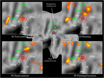
The IPL is made of several areas including the SMG. It has been shown that the left SMG does not sustain a unique functional area but several functional areas, that is, the anterior SMG (aSMF/PFt) and the posterior part of SMG (PF) (Orban & Caruana, Reference Orban and Caruana2014). In a recent neuroimaging meta-analysis (Reynaud et al., Reference Reynaud, Lesourd, Navarro and Osiurak2016), the area PF was found to be preferentially activated when participants have to focus on how a tool has to be used appropriately with an object (i.e., mechanical knowledge; Figure 1a).
Flat-map representation of a left hemisphere (PALS-B12: Population-Average, Landmark- and Surface-based human cortical atlas; Van Essen, Reference Van Essen2005), using Caret, version 5.65 (http://brainmap.wustl.edu/caret.html; Van Essen et al., Reference Van Essen, Drury, Dickson, Harwell, Hanlon and Anderson2001). On the center, are represented, after conversion from Montreal Neurological Institute to Talairach coordinates (Lacadie, Fulbright, Constable, & Papademetris, Reference Lacadie, Fulbright, Constable and Papademetris2008), virtual lesions made during two manipulation judgment tasks, that is, a hand configuration task (Andres et al., Reference Andres, Pelgrims and Olivier2013; x=−59, y=−32, z=43; Pelgrims et al., Reference Pelgrims, Olivier and Andres2011; x=−58, y=−30, z=43; red circles) and a manipulation-matching task (Ishibashi et al., Reference Ishibashi, Lambon Ralph, Saito and Pobric2011; x=−38, y=−41, z=45, yellow circle); and during a motor imagery task (Pelgrims et al., Reference Pelgrims, Andres and Olivier2009; x=−56, y=−48, z=43; blue circle). On panels a, b, c, and d are represented Automatic Likelihood Estimation (ALE) maps, obtained by Reynaud et al. (Reference Reynaud, Lesourd, Navarro and Osiurak2016) and viewed on a PALS-B12 left hemisphere atlas surface configuration (Van Essen, Reference Van Essen2005) in four conditions of tool use: tool-centered (a); hand-centered (b); planning (c); and planning/execution (d). The virtual lesions obtained in hand configuration tasks are also depicted on the four panels with a little red circle and a larger one which takes into account the spatial resolution intrinsic to the TMS (~0.5–1 cm; Thielscher & Kammer, Reference Thielscher and Kammer2002; Toschi et al., Reference Toschi, Welt, Guerrisi and Keck2008). Depicted regions represent (1) IPL: aSMG, anterior portion of SMG, which largely overlaps with the cytoarchitectonic area PFt of SMG; PF, PFm, PFop, PFt, and PFc, five cytoarchitectonic areas located in the left IPL, approximately at the position of BA 40 on the SMG; and (2) IPS: phAIP, putative human homologue of anterior intraparietal area; DIPSA, anterior dorsal intraparietal sulcus (Orban & Caruana, Reference Orban and Caruana2014; see also Peeters, Rizzolatti, & Orban, Reference Peeters, Rizzolatti and Orban2013).

In broad terms, area PF would be the locus of stored mechanical knowledge. As it can be seen in Figure 1, the virtual lesions made in the left SMG falls in PF area (Andres et al., Reference Andres, Pelgrims and Olivier2013; Pelgrims et al., Reference Pelgrims, Olivier and Andres2011). In line with the technical reasoning theory, PF area is involved in the processing of tool-object relationships, that is, the link between tools and objects (i.e., mechanical knowledge). Nevertheless, a virtual lesion in PF leads to the inability of judging hand configurations, that is, an inability to focus on the link between the hand and the tool (i.e., tool-hand relationship). Thus, the predictions made by the technical reasoning theory fail to fit with this result given that hand-centered relationships should be processed in the rostral part of the intraparietal sulcus (i.e., DIPSA; Reynaud et al., Reference Reynaud, Lesourd, Navarro and Osiurak2016; Figure 1b) but not in PF.
However, Pelgrims et al. (Reference Pelgrims, Olivier and Andres2011) did not find any interference effect following a virtual lesion made in PF in a hand-object task, where participants were asked to judge whether a handgrip was appropriate for a given tool. Moreover, focusing on the handgrip to be performed is associated with IPS but not with PF activations (Figure 1b; Reynaud et al., Reference Reynaud, Lesourd, Navarro and Osiurak2016). Thus, this result seems to be in line with the predictions made by the technical reasoning hypothesis, which assumes that tool-hand relationships are encoded in the dorso–dorsal stream (Osiurak & Badets, Reference Osiurak and Badets2016; Reynaud et al., Reference Reynaud, Lesourd, Navarro and Osiurak2016).
In this part, we found that the results reported in recent virtual lesions studies might be at odds with technical reasoning predictions. Nevertheless, we will see in the next part that some arguments are rather in agreement with the technical reasoning theory than with the gesture engram theory.
LEFT IPL: AN INTERTWINED REGION SUSTAINING SEVERAL COGNITIVE PROCESSES
“Functionally Distinct” Sub-regions Within the Left SMG
In recent tool use studies, we saw that the left SMG was organized according to functionally distinct sub-regions (i.e., aSMG/PFt and PF; Orban & Caruana, Reference Orban and Caruana2014; Reynaud et al., Reference Reynaud, Lesourd, Navarro and Osiurak2016). Moreover, it seems that the same pattern can be observed for other cognitive functions. As it has been evoked at the very beginning of the introduction, the left SMG is not involved only in tool use tasks but it also supports other cognitive functions including phonological processing. In a recent work, four “functionally distinct” regions were reported, all within the left SMG (Oberhuber et al., Reference Oberhuber, Hope, Seghier, Parker Jones, Prejawa, Green and Price2016). The authors found that in ventral SMG, an anterior sub-region was associated with articulatory sequencing and a posterior sub-region was associated with auditory short-term memory. In dorsal SMG, a posterior sub-region was associated with integration of lexical and sub-lexical information whereas in anterior dorsal SMG, activations were higher for both pseudo-word reading and object naming. This result confirms with other cognitive processes that the left SMG is a brain area made of several “functionally distinct” sub-regions.
PF Area and Motor Imagery
A potential interpretation of the link between left SMG and hand configuration task observed in Andres et al. (Reference Andres, Pelgrims and Olivier2013) comes from another TMS study where the left SMG was stimulated while participants were asked to make a hand laterality judgment task (Pelgrims, Andres, & Olivier, Reference Pelgrims, Andres and Olivier2009). Hand laterality judgement task implicitly triggers motor imagery and is associated with bilateral activations (more consistent in the right hemisphere; Hétu et al., Reference Hétu, Grégoire, Saimpont, Coll, Eugène, Michon and Jackson2013). As can be seen in Figure 1, we cannot exclude an overlap between virtual lesions made during hand configuration tasks (Andres et al., Reference Andres, Pelgrims and Olivier2013; Pelgrims et al., Reference Pelgrims, Olivier and Andres2011; Figure 1, red circles) and during hand laterality judgement tasks (Pelgrims et al., Reference Pelgrims, Andres and Olivier2009; Figure 1, blue circle). In other words, a virtual lesion made in the same area may lead to a deficit in both hand configuration and hand laterality judgment tasks, which requires, for the latter, motor imagery processing (de Lange, Helmich, & Toni, Reference de Lange, Helmich and Toni2006; Parsons, Reference Parsons1994). Thus, a motor imagery impairment may prevent the mental simulation of a given action (Decety, Reference Decety1996) and may explain the deficit in the hand configuration task, without evoking the need for manipulation knowledge activation.
Left SMG and Left IPS: Close Adjacent Areas, Distinct Functions
Virtual lesions made in PF are located near the junction of three areas of interest, namely the anterior part of the SMG (i.e., aSMG/PFt), the intraparietal sulcus (IPS) and PF. Given that IPL regions are highly inter-twined and given the inter-subject variability and the spatial resolution intrinsic to the TMS (approximately 0.5–1 cm; Thielscher & Kammer, Reference Thielscher and Kammer2002; Toschi, Welt, Guerrisi, & Keck, Reference Toschi, Welt, Guerrisi and Keck2008), one could not exclude that virtual lesions made in left SMG encompassed more functional areas than only PF (as it is represented by the larger red circles in Figures 1a–d), and consequently disrupted cognitive processes supported by IPS and aSMG/PFt.
For instance, the virtual lesion may have included the left IPS (i.e., phAIP), corroborating findings that activations are found in the left IPS (phAIP, DIPSA) in situations where planning is required (see also Przybylski & Króliczak, Reference Przybylski and Króliczak2017; for aSMG/PFt activation following planning of functional grasps) (Figure 1c) and when participants were asked to focus on the handgrip to be performed (i.e., hand-centered task; Figure 1b). The IPS is widely involved in the extraction of object affordances (Buccino et al., Reference Buccino, Vogt, Ritzl, Fink, Zilles, Freund and Rizzolatti2004) and might also play a role in motor simulation, by allowing people to anticipate tool-hand relationships (Jeannerod, Reference Jeannerod1994).
Finally, the virtual lesion may encompass the left aSMG/PFt which is activated when participants have to plan both the handgrip and the mechanical interaction (Figure 1d), but is not activated in situations where participants have to focus on either the mechanical interactions between the tool and the object (i.e., tool-object relationship; Figure 1a) or the handgrip to be performed (i.e., tool-hand relationship; Figure 1b). Thus, aSMG/PFt area might be an integrative area between information coming from IPS (i.e., production system) and information coming from PF (i.e., mechanical knowledge), which, if disrupted, would lead to a deficit in a hand configuration task.
Manipulation Knowledge Outside the Left IPL
In line with the gesture engram theory, manipulation knowledge depends on the left IPL (Boronat et al., Reference Boronat, Buxbaum, Coslett, Tang, Saffran, Kimberg and Detre2005; Buxbaum, Reference Buxbaum2001) and more specifically on the left SMG (Humphreys & Lambon Ralph, Reference Humphreys and Lambon Ralph2015). However, as it can be seen in Figure 1, a virtual lesion made in the left IPS (Ishibashi, Lambon Ralph, Saito, & Pobric, Reference Ishibashi, Lambon Ralph, Saito and Pobric2011; x=−38, y=−41, z=45, yellow circle) leads to an impairment of a manipulation judgment task. In this task, participants were asked to choose the word-target (e.g., staple) that had the same way of manipulation as the probe (e.g., scissors) among two foils. Thus, a virtual lesion made elsewhere than in left IPL can disrupt a manipulation judgment task, which is not predicted by the engram theory. On the contrary, the technical reasoning theory predicts that the left IPS is involved in situations where either planning (Figure 1c) or focusing on tool-hand relationships (Figure 1b) is required. Moreover, neuroimaging studies found activations in left IPS for manipulation judgment tasks (Canessa et al., Reference Canessa, Borgo, Cappa, Perani, Falini, Buccino and Shallice2008). In addition, the results obtained by Ishibashi and coworkers (Reference Ishibashi, Lambon Ralph, Saito and Pobric2011) can also suggest that virtual lesions made in the left SMG could have affect other areas of parietal cortex.
CONCLUSION AND PERSPECTIVES
To summarize, the results obtained in recent TMS studies suggest that a virtual lesion in the left SMG, and more precisely in PF, disrupts the cognitive processes that underlie the ability to judge the correct hand posture relative to a specific tool. Moreover, it points out the limitation for the gesture engram theory and the technical reasoning theory to fully explain this result. The gesture engram theory fails to make clear predictions about the cortical structures that may store the hand posture associated with a tool. The technical reasoning theory fails to explain why PF, which should be involved in hand-object relationships, would also be involved in egocentric relationships (i.e., hand-tool relationship). Thus, the result reported here is seriously challenging both theories of tool use considered here. However, the predictions made by the technical reasoning theory seem to be more consistent than gesture engram theory predictions given the results of TMS studies reviewed here.
To go beyond this limitation, we suggest that the virtual lesion made in PF is at the junction of major areas necessary for the processing of the ability to use tools (i.e., aSMG/PFt, PF and phAIP). The observation that the left SMG is organized following several “functionally distinct areas” also in other cognitive functions (e.g., phonological processing; Oberhuber et al., Reference Oberhuber, Hope, Seghier, Parker Jones, Prejawa, Green and Price2016), may corroborate our proposal. Thus, one could not exclude that this virtual lesion has disrupted a cortical network involved in the ability to use tools rather than only one functional area supporting hand posture knowledge. Indeed, there is a wide range of variation of TMS within left SMG and these stimulations are likely to affect nearby peri-Sylvian regions as well, such as superior temporal gyrus (Kraemer, Hamilton, Messing, Desantis, & Thompson-Schill, Reference Kraemer, Hamilton, Messing, Desantis and Thompson-Schill2014). Moreover, we also found that manipulation knowledge which relies on left IPL, according to the gesture engram theory, can be disrupted by a virtual lesion made in the left IPS (Ishibashi et al., Reference Ishibashi, Lambon Ralph, Saito and Pobric2011).
This result raises two conclusions: first, the engram theory fails to explain this pattern, whereas it is fully explained by the technical reasoning theory (Osiurak & Badets, Reference Osiurak and Badets2016; Reynaud et al., Reference Reynaud, Lesourd, Navarro and Osiurak2016). Second, the left SMG is not the locus of stored manipulation knowledge, as lesions in other regions should produce manipulation judgment tasks impairment. This demonstration is easily transposable to the hand configuration task reported here.
A potential explanation for the contradictory findings reported here may be such that TMS and fMRI studies analyzed in this work used different paradigms. However, the fMRI results discussed here were obtained with a meta-analysis (35 studies, 60 experiments; Reynaud et al., Reference Reynaud, Lesourd, Navarro and Osiurak2016) that aims to characterize the cortical networks involved in different context of tool use (e.g., tool-hand or tool-object relations) whatever the specificity of the paradigms used in each study. Moreover, this meta-analysis contains manipulation judgement tasks (e.g., Canessa et al., Reference Canessa, Borgo, Cappa, Perani, Falini, Buccino and Shallice2008) close to the TMS studies considered here (e.g., Pelgrims et al., Reference Pelgrims, Olivier and Andres2011). It is, therefore, unlikely that the difference between fMRI and TMS paradigms may explain on its own the contradictory results reported here.
In the field of tool use, TMS is a powerful technique to observe cognitive impairment following virtual lesions made in specific cortical areas. For instance, using TMS, Andres et al. (Reference Andres, Pelgrims and Olivier2013) demonstrated that the ability to infer a context of use or a hand posture from tool perception relies on distinct processes, performed in the temporal and parietal regions, respectively. TMS becomes more powerful when applied on coordinates of activation sites reported by functional imaging (e.g., Pelgrims et al., Reference Pelgrims, Andres and Olivier2009). Indeed, a brain area may be considered as essential for a task/cognitive process when (1) this area is found to be activated in fMRI, and (2) a brain lesion (e.g., TMS) in this area leads to a deficit in the same task. If TMS is a good method to investigate the link between cognitive processes and brain structures, it also has its limitations regarding the spatial resolution and the spread-out effects of the induced magnetic field. For instance, when targeting the IPL, it can be expected that both angular and supramarginal regions are stimulated. Thus, it may be particularly problematic when targeting a specific area in the left IPL which contains several “functionally distinct” areas (e.g., phonological processing: Oberhuber et al., Reference Oberhuber, Hope, Seghier, Parker Jones, Prejawa, Green and Price2016; or tool use: Reynaud et al., Reference Reynaud, Lesourd, Navarro and Osiurak2016).
To sum up, TMS is a good method to explore cerebral correlates of tool use but other techniques have to be used when testing neurocognitive models with specific hypotheses on distinct areas of the left IPL (e.g., PF and aSMG/PFt; Reynaud et al., Reference Reynaud, Lesourd, Navarro and Osiurak2016). For instance, stereo-EEG, an invasive technique, may be a good candidate because it allows intra-cerebral recordings of active cortical nodes with an excellent spatial resolution and permits to build four dimensional maps of human cortical processing (e.g., somato-sensory processing; Avanzini et al., Reference Avanzini, Abdollahi, Sartori, Caruana, Pelliccia, Casaceli and Orban2016).
ACKNOWLEDGMENTS
The authors declare that the research was conducted in the absence of any commercial or financial relationships that could be construed as a potential conflict of interest. This work was supported by grants from ANR (Agence Nationale pour la Recherche; Project “Démences et Utilisation d’Outils/Dementia and Tool Use”, ANR-2011-MALZ-006-03; Project “Cognition et économie liée à l’outil/Cognition and tool-use economy” ECOTOOL; ANR-14-CE30-0015-01), and was performed within the framework of the LABEX CORTEX (ANR-11-LABX-0042) of Université de Lyon, within the program “Investissements d’Avenir” (ANR-11-IDEX-0007) operated by the French National Research Agency (ANR).