




No CrossRef data available.
Article contents
Assessment of benzimidazole binding to individual recombinant tubulin isotypes from Haemonchus contortus
Published online by Cambridge University Press: 12 July 2001
Abstract
One α- and 2 β-tubulin isotypes (isotypes 1 and 2) from the parasitic nematode Haemonchus contortus were artificially expressed in E. coli and purified to obtain tubulin that was capable of polymerizing into microtubules. Binding of [14C] mebendazole (MBZ), a benzimidazole compound, to each individual unpolymerized isotype and to microtubules polymerized from recombinant α- and β-tubulin was assessed and Kd and Bmax values determined. Mebendazole bound to the individual tubulin isotypes with a stoichiometry of 1:1. Binding occurred with highest affinity to α-tubulin followed by β-tubulin isotype 2 and β-tubulin isotype 1 indicating that α-tubulin may play a role in benzimidazole binding to microtubules. Upon polymerization of α- and β-tubulin isotype 2 into microtubules the stoichiometry of binding increased to 2:1 (mebendazole:tubulin) while binding affinity remained the same. Mebendazole binding to α/β-isotype 1 microtubules remained unchanged following polymerization. The increase in the number of benzimidazole receptors on α/β-isotype 2 microtubules suggests the formation of a new benzimidazole receptor upon polymerization.
Information
- Type
- Research Article
- Information
- Copyright
- © 2001 Cambridge University Press
INTRODUCTION
The benzimidazoles are safe, broad-spectrum anthelmintics that have long been used to combat many nematode, trematode and cestode parasites occurring in humans and animals (McCracken & Lipkowitz, 1990). The benzimidazoles are believed to exert their effect in the parasite by binding to the tubulin protein and preventing it polymerizing into microtubules (Lacey, 1988). Tubulin molecules possess 3 major binding sites, the colchicine binding site, the taxol binding site and the vinblastine binding site. It is believed that the benzimidazole drugs bind to the colchicine binding site on the tubulin molecule (Lacey, 1988) forming a tubulin–benzimidazole complex which changes depending on the isotype of tubulin involved (Sackett, Bhattachararyya & Wolff, 1994). However, some evidence exists to the contrary, for example, microtubules from Caenorhabditis elegans containing the β-tubulin gene mec-7 are insensitive to benzimidazoles but sensitive to colchicine (Geary et al. 1992). When incubated with tubulin, colchicine, podophyllotoxin and several benzimidazoles cause tubulin to lose its ability to polymerize into microtubules at 37 °C. The tubulin–colchicine complex binds to the growing end of a microtubule preventing further assembly (Barrowman, Marriner & Bogan, 1984; Gill & Lacey, 1992; Sackett & Varma, 1993).
There has been much speculation regarding the position of the benzimidazole binding site on the tubulin molecule. The majority of studies have proposed that benzimidazoles bind to β-tubulin as mutations in the β-tubulin gene appear to confer benzimidazole resistance in several organisms (Kwa et al. 1993; Jung & Oakley, 1990; Katiyar et al. 1994). In addition, tubulin isotypes appear to differ from each other with respect to the C-terminal sequence on the β-tubulin subunit and different isotypes of β-tubulin have been found to differ with respect to their interactions with colchicine (Wolff et al. 1991; Banerjee & Luduena, 1992; Uppuluri et al. 1993). However, others have suggested that the binding site is situated; on α-tubulin (Serrano et al. 1984; Lubega & Prichard, 1991a); on α-tubulin and regulated by the β-subunit (Banerjee & Luduena, 1992); on both α- and β-tubulin (Floyd, Barnes & Williams, 1989); or at the α/β-tubulin interface (Lubega & Prichard, 1991a; Sackett, Bhattachararyya & Wolff, 1994). It is also possible that β-tubulin residues are necessary for benzimidazole binding but do not actually form part of the binding site (Geary et al. 1992).
Recently we have produced individual isotypes of pure recombinant Haemonchus contortus tubulin, one α- and two β-tubulins (β8–9 and β12–16 or isotypes 1 and 2), using an E. coli expression system. This has allowed the measurement of benzimidazole binding to individual tubulin isotypes which was not previously possible using methods relying on the purification of tubulin from its original source. When combined under appropriate conditions, these recombinant α- and β-tubulin isotypes have previously been shown to be capable of polymerizing into microtubules. The extent of this polymerization and the type of structures that form is altered in the presence of albendazole (a benzimidazole compound), which produces a significant decrease in polymerization and results in the formation of abnormal microtubules (Oxberry et al. 2001). In the present study, benzimidazole binding to each unpolymerized recombinant H. contortus tubulin isotype, as well as to polymerized recombinant microtubules, was assessed in order to gain knowledge about the benzimidazole binding site on the tubulin molecule. This type of information can be used to design more effective anthelmintics, to increase the effectiveness of the current benzimidazole compounds, and to help understand mechanisms of benzimidazole resistance.
MATERIALS AND METHODS
Transformation and induction of gene expression
One α-tubulin (Oxberry et al. 2001) and two β-tubulin (isotypes 1 and 2, representing different β-tubulin isoforms (Geary et al. 1992; Lubega et al. 1993) gene constructs in pTrp2 were introduced into E. coli and the corresponding proteins expressed as described by Oxberry et al. (2001). Briefly, following heat shock, positive E. coli transformants (strains Jm101 and Jm109) were selected on ampicillin plates and a single colony selected to be grown to mid-log phase in Luria-Bertani (LB) medium containing 100 μg/ml ampicillin and 1 mg/ml tryptophan. The cells were harvested and grown for a further 3–4 h in minimal medium (Sambrook, Fritsch & Maniatis, 1989) in the absence of tryptophan, harvested again, lysed by sonication on ice (16 cycles of 9 sec bursts with 6 sec pauses in a Vibra Cell VCX400 sonicator (Sonics and Materials Inc.) set on an amplitude of 20%) and centrifuged at 8000 g for 15 min at 4 °C. Tubulin-containing inclusion bodies were collected in the pellet, incubated in 1 mg/ml DNAse (Lubega et al. 1993), solubilized in lysis buffer (50 mM Tris, 1 mM ethylenediamine tetraacetic acid (EDTA), 100 mM NaCl, 8 M urea, pH 8.0 (Sambrook et al. 1989), neutralized and the partially purified tubulin stored at −70 °C. A protease inhibitor cocktail (Complete Mini, Boehringer Mannheim) was used throughout the procedure.
Purification of tubulin using phosphocellulose
This procedure was based on that used by Williams & Lee (1982) and Correia, Baty & Williams (1987), as described by Oxberry et al. (2001). Briefly, partially purified tubulin isotypes were added to Whatman P11 phosphocellulose which had been equilibrated in column buffer (0.4 M piperazine-N,N-bis(2-ethanesulfonic acid) (PIPES), 50 mM MgSO4, 100 mM ethylene glycol bis(2-aminoethyl ether)-N,N,N′,N′ tetraacetic acid (EGTA), 2 mM dithioerythritol (DTE), pH 6.9), mixed for 20 min at 4 °C and centrifuged. Supernatants were collected and concentrated to approximately half their volume using 6–8000 molecular weight dialysis tubing placed in polyethylene glycol (8000 g/mole). The resulting tubulin was analysed for protein content and for purity by sodium dodecyl sulfate–polyacrylamide gel electrophoresis (SDS–PAGE) and Western blotting using monoclonal anti-α- and anti-β-tubulin antibodies (Amersham) then stored at −70 °C.
Polymerization of tubulin
Mebendazole binding was assessed for unpolymerized individual tubulin isotypes and for polymerized microtubules (α-tubulin with isotype 1 or with isotype 2 β-tubulin). Each tube containing polymerized tubulin was prepared by adding 10 μl of 1 mg/ml α-tubulin, 10 μl of 1 mg/ml β-tubulin, 19 μl of PEM buffer (0.1 M PIPES, 0.1 M NaOH, 1 mM EGTA, 1 mM MgSO4, pH 6.8 Lopes et al. 1997), 1 μl of 100 mM guanosine-5′-triphosphate (GTP) and incubated at 37 °C for 1 h.
Benzimidazole binding studies
Two sets of tubes were prepared containing 5 μl of [14C]mebendazole (Janssen), to give final concentrations of 0.1 to 1.5 μM, and 5 μl of dimethyl sulphoxide (set 1 – to determine total binding) or 5 μl of 16.6 mM unlabelled mebendazole (set 2 – to determine non-specific binding). Individual unpolymerized recombinant tubulin isotypes (40 μl of 1 mg/ml tubulin) or 40 μl of polymerized microtubules were added and the tubes mixed and incubated at 37 °C for 1 h. Mebendazole has been shown previously to have a high affinity for H. contortus tubulin at 37 °C (Lubega & Prichard, 1991b). A charcoal suspension containing 1.25% animal charcoal (Fisher Scientific) and 0.125% bovine serum albumin (ICN) in PEM buffer was incubated on ice on a rotating mixer for 1 h prior to use and 1 ml aliquots added to each tube. The tubes were centrifuged at 3000 g for 5 min at 4 °C. Then 500 μl of supernatant were removed and placed in 2.5 ml of Universol scintillation fluid (ICN) for counting on a Wallac WinSpectral 1414 liquid scintillation counter. DPM binding data were analysed using Graph Pad Prism, Version 2.0.
RESULTS
Phosphocellulose affinity chromatography yielded almost pure recombinant tubulin of each isotype which was capable of polymerizing into normal microtubules when α- and β-tubulin were mixed and incubated under appropriate conditions (Oxberry et al. 2001). The individual and polymerized tubulin isotypes were recognized by anti-α- and anti-β-tubulin monoclonal antibodies (Fig. 1).

Fig. 1. Western blot analysis of an SDS–PAGE gel using monoclonal α- and β-tubulin antibodies. Lane 1, molecular weight markers; lane 2, α-tubulin; lane 3, β-tubulin isotype 1; lane 4, β-tubulin isotype 2; lane 5, polymerized α/β-isotype 2 microtubules with tubulin dimer at 110 kDa.
Total binding and low affinity (non-specific) binding of [14C] mebendazole to purified recombinant tubulin was measured for each tubulin isotype at [14C] mebendazole concentrations of 0.1 to 1.5 μM. Low affinity binding, which ranged from 28 to 58% of total binding, was subtracted from total binding to give high affinity (specific) binding values which were graphed in terms of mmoles [14C] mebendazole bound per mole purified tubulin (Fig. 2). Kd and Bmax values were determined from this data using nonlinear regression analyses generated by GraphPad Prism 2.01 software (Table 1).

Fig. 2. Specific (high affinity) binding of various concentrations of [14C] mebendazole to ([bull ]) α-, ([dtrif ]) β-tubulin isotype 1 and ([utrif ]) β-tubulin isotype 2 soluble (unpolymerized) tubulin isotypes and ([squf ]) α/β-isotype 1 and ([lozf ]) α/β-isotype 2 polymerized tubulin.

Of the individual tubulin isotypes, mebendazole bound specifically with the highest affinity to α-tubulin (Kd = 1.35 μM) followed by β-tubulin isotype 2 and β-tubulin isotype 1 (Kd = 2.31 μM and 3.95 μM respectively). The differences between the Kds for the 3 isotypes were significant (P = 0.0278). β-tubulin isotype 1 showed a slightly higher Bmax (1267 mmoles MBZ bound/mole tubulin) compared with α-tubulin and β-tubulin isotype 2 (998 and 949.4 mmoles/mole respectively). Of the polymerized tubulin, mebendazole bound with similar affinity to microtubules containing α/β-isotype 1 and α/β-isotype 2 tubulin (Kd = 2.21 and 2.12 μM respectively). The number of binding sites on α/β-isotype 1 microtubules was similar to that of individual isotypes (1167 mmoles/mole). However, microtubules containing α/β-isotype 2 tubulin had approximately twice as many receptor sites as α/β-isotype 1 microtubules and individual isotypes (2334 mmoles/mole).
DISCUSSION
Of the 2 individual unpolymerized H. contortus β-tubulin isotypes examined, mebendazole bound β-tubulin isotype 2 with a higher affinity than β-tubulin isotype 1. However, α-tubulin bound mebendazole with a higher affinity than either of the 2 β-tubulin isotypes. Binding affinity to β-tubulin isotype 1 was increased upon polymerization into microtubules with α-tubulin while that of β-tubulin isotype 2 remained similar. The number of available receptor sites on the individual tubulin isotypes and on α/β-isotype 1 microtubules (as determined by Bmax) was fairly similar with mebendazole binding to tubulin occurring with a stoichiometry of approximately 1:1. However, receptor number was doubled following polymerization of α- and β-isotype 2 tubulin into microtubules.
It appears that the presence of α-tubulin or the configuration of the α- and β-tubulin isotypes within the α/β-isotype 2 microtubule structure results in an increased availability of high affinity mebendazole receptors. This idea is supported by the computer analysis of the binding data which chose a 2-binding site category over a 1-site category as the best fit to the data generated using polymerized tubulin.
β-tubulin isotypes 1 and 2 from H. contortus have both previously been implicated in benzimidazole binding (Lubega et al. 1994; Beech, Prichard & Scott, 1994; Kwa et al. 1993). The 2 β-tubulin isotypes are 79% identical at the nucleic acid level and 93.5% identical at the amino acid level. Most of the differences between the two exist at the carboxy terminal between residues 436 and 445 although other differences also occur, such as one at position 165, a site that has been implicated in benzimidazole binding and resistance (Geary et al. 1992; Jung & Oakley, 1990). The difference in the number of mebendazole binding sites on microtubules containing the different isotypes of β-tubulin suggests that the two isotypes may vary at a region(s) important in the formation of the benzimidazole binding site. The doubling of the number of binding sites on α/β-isotype 2 microtubules, combined with the similarity in binding affinity of mebendazole to both types of microtubules, indicates that the differences between the two β-tubulin isotypes result in a change in the number of receptors rather than in binding affinity. A possible theory to explain this would be that, upon polymerization of α- and β-isotype 2 tubulin into microtubules, a second binding site forms, possibly at the α/β-tubulin interface. The previously published theory of the formation of a new benzimidazole receptor site at the α/β-tubulin interface (Lubega & Prichard, 1991a; Sackett et al. 1994) is one that cannot be ruled out and warrants further investigation based on the results of the present study.
This research was supported by NSERC of Canada and Upjohn and Pharmacia Inc.
References
BANERJEE, A. & LUDUENA, R. F. (1992). Kinetics of colchicine binding to purified β-tubulin isotypes from bovine brain. Journal of Biological Chemistry 267, 13335–13339.Google Scholar
BARROWMAN, M. M., MARRINER, S. E. & BOGAN, J. A. (1984). The binding and subsequent inhibition of tubulin polymerisation in Ascaris suum (in vitro) by benzimidazole anthelmintics. Biochemical Pharmacology 33, 3037–3040.CrossRefGoogle Scholar
BEECH, R. N., PRICHARD, R. K. & SCOTT, M. E. (1994). Genetic variability of the beta-tubulin genes in benzimidazole-susceptible and -resistant strains of Haemonchus contortus. Genetics 138, 103–110.Google Scholar
CORREIA, J. J., BATY, L. T. & WILLIAMS, R. C. JR. (1987). Mg2+ dependence of guanine nucleotide binding to tubulin. Journal of Biological Chemistry 262, 17278–17284.Google Scholar
FLOYD, L. J., BARNES, L. D. & WILLIAMS, R. F. (1989). Photoaffinity labelling of tubulin with (2-nitro-4-azidophenyl) deacetylcolchicine: direct evidence for two colchicine binding sites. Biochemistry 28, 8515–8525.CrossRefGoogle Scholar
GEARY, T. G., NULF, S. C., FAVREAU, M. Z., TANG, L., PRICHARD, R. K., HETZENBUHLER, N. T., SHEA, M. H., ALEXANDER, S. J. & KLEIN, R. D. (1992). Three β-tubulin cDNAs from the parasitic nematode Haemonchus contortus. Molecular and Biochemical Parasitology 50, 295–306.CrossRefGoogle Scholar
GILL, J. H. & LACEY, E. (1992). The kinetics of mebendazole binding to Haemonchus contortus tubulin. International Journal for Parasitology 22, 939–946.CrossRefGoogle Scholar
JUNG, M. K. & OAKLEY, B. R. (1990). Identification of an amino acid substitution in the ben A, β-tubulin gene of Aspergillus nidulans that confers thiabendazole resistance and benomyl supersensitivity. Cell Motility and the Cytoskeleton 17, 87–94.CrossRefGoogle Scholar
KATIYAR, S. K., GORDON, V. R., MCLAUGHLIN, G. L. & EDLIND, T. D. (1994). Antiprotozoal activities of benzimidazoles and correlations with β-tubulin sequence. Antimicrobial Agents and Chemotherapy 38, 2086–2090.CrossRefGoogle Scholar
KWA, M. S. G., KOOYMAN, F. N. J., BOERSEMA, J. H. & ROOS, M. H. (1993). Effect of selection for benzimidazole resistance in Haemonchus contortus on β-tubulin isotype 1 and isotype 2 genes. Biochemical and Biophysical Research Communications 191, 413–419.CrossRefGoogle Scholar
LACEY, E. (1988). The role of the cytoskeletal protein, tubulin, in the mode of action and mechanisms of drug resistance to benzimidazoles. International Journal for Parasitology 18, 885–936.CrossRefGoogle Scholar
LOPES, N. M., MILLER, H. P., YOUNG, N. D. & BHUYAN, B. K. (1997). Assessment of microtubule stabilizers by semiautomated in vitro microtubule protein polymerization and mitotic block assays. Cancer Chemotherapy and Pharmacology 41, 37–47.CrossRefGoogle Scholar
LUBEGA, G. W., GEARY, T. G., KLEIN, R. D. & PRICHARD, R. K. (1993). Expression of cloned β-tubulin genes of Haemonchus contortus in Escherichia coli: interaction of recombinant β-tubulin with native tubulin and mebendazole. Molecular and Biochemical Parasitology 62, 281–292.CrossRefGoogle Scholar
LUBEGA, G. W., KLEIN, R. D., GEARY, T. G. & PRICHARD, R. K. (1994). Haemonchus contortus: the role of two beta-tubulin gene subfamilies in the resistance to benzimidazole anthelmintics. Biochemical Pharmacology 47, 1705–1715.CrossRefGoogle Scholar
LUBEGA, G. W. & PRICHARD, R. K. (1991a). Beta-tubulin and benzimidazole resistance in the sheep nematode Haemonchus contortus. Molecular and Biochemical Parasitology 47, 129–138.Google Scholar
LUBEGA, G. W. & PRICHARD, R. K. (1991b). Specific interaction of benzimidazole anthelmintics with tubulin: high affinity binding and benzimidazole resistance in Haemonchus contortus. Molecular and Biochemical Parasitology 38, 221–232.Google Scholar
MCCRACKEN, R. O. & LIPKOWITZ, K. B. (1990). Structure-activity relationships of benzothiazole and benzimidazole anthelmintics: a molecular modelling approach to in vivo drug efficacy. Journal of Parasitology 76, 853–864.CrossRefGoogle Scholar
OXBERRY, M. E., GEARY, T. G., WINTERROWD, C. A. & PRICHARD, R. K. (2001). Individual expression of recombinant α- and β-tubulin from Haemonchus contortus: polymerisation and drug effects. Protein Expression and Purification (in the Press).CrossRefGoogle Scholar
SACKETT, D. L., BHATTACHARARYYA, B. & WOLFF, J. (1994). Local unfolding and the stepwise loss of the functional properties of tubulin. Biochemistry 33, 12868–12878.CrossRefGoogle Scholar
SACKETT, D. N. & VARMA, J. K. (1993). Molecular mechanism of colchicine action: induced local unfolding of β-tubulin. Biochemistry 32, 13560–13565.CrossRefGoogle Scholar
SAMBROOK, J., FRITSCH, E. F. & MANIATIS, T. (1989). Molecular Cloning. A Laboratory Manual. 2nd Edn. Cold Spring Harbour Laboratory Press, New York.
SERRANO, L., DE LA TORRE, J., MACCIONI, R. B. & AVILA, J. (1984). Involvement of the carboxy-terminal domain in the regulation of its assembly. Proceedings of the National Academy of Sciences, USA 81, 5989–5993.CrossRefGoogle Scholar
UPPULURI, S., KNIPLING, L., SACKETT, D. L. & WOLFF, J. (1993). Localization of the colchicine binding site of tubulin. Proceedings of the National Academy of Sciences, USA 90, 11598–11602.CrossRefGoogle Scholar
WILLIAMS, R. C. JR. & LEE, J. C. (1982). Preparation of tubulin from brain. Methods in Enzymology 85, 376–385.CrossRefGoogle Scholar
WOLFF, J., KNIPLING, L., CAHNMANN, H. J. & PALUMBO, G. (1991). Direct photoaffinity labelling of tubulin with colchicine. Proceedings of the National Academy of Sciences, USA 88, 2820–2824.CrossRefGoogle Scholar

Fig. 1. Western blot analysis of an SDS–PAGE gel using monoclonal α- and β-tubulin antibodies. Lane 1, molecular weight markers; lane 2, α-tubulin; lane 3, β-tubulin isotype 1; lane 4, β-tubulin isotype 2; lane 5, polymerized α/β-isotype 2 microtubules with tubulin dimer at 110 kDa.

Fig. 2. Specific (high affinity) binding of various concentrations of [14C] mebendazole to ([bull ]) α-, ([dtrif ]) β-tubulin isotype 1 and ([utrif ]) β-tubulin isotype 2 soluble (unpolymerized) tubulin isotypes and ([squf ]) α/β-isotype 1 and ([lozf ]) α/β-isotype 2 polymerized tubulin.
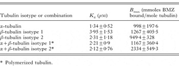
Table 1. Kd and Bmax values (± standard error) calculated from specific binding data for soluble and polymerized tubulin
 You have
Access
- 21
- Cited by