Discordant atrioventricular with concordant ventriculo-arterial connections is an extremely rare cardiac defect. It can occur as an isolated entity, but it is mostly a part of more complex CHDs, especially heterotaxy syndromes. To perform a correct and complete anatomical diagnosis remains challenging, especially in the setting of complex cardiac defects. Haemodynamically, discordant atrioventricular with concordant ventriculo-arterial connections has the same physiology as classical transposition of the great arteries, marked by neonatal cyanosis. As a consequence, survival depends on adequate intra-cardiac – atrial or ventricular septal defect – and extra-cardiac – persistent arterial duct – mixing to allow sufficient oxygenation before surgery. In cases with complex anatomy, the pathophysiology will also depend on associated heart defects such as anomalous pulmonary venous drainage or pulmonary atresia.
In total, five different segmental arrangements taking into account the different possible spatial relationships of the great arteries have been described by Pasquini et al in this rare anomaly, also named “atrioventricular discordance with ventriculo-arterial concordance” or in certain cases “isolated ventricular inversion”.Reference Pasquini, Sanders and Parness 1 , Reference Van Praagh and Van Praagh 2 Reports about this complex cardiac defect remain rare and are mainly case descriptions and small patient series, often lacking information on long-term follow-up.Reference Espino-Vela, De la Cruz, Muñoz-Castellanos, Plaza and Attie 3 – Reference Kannan, Kamath, Rao and Kumar 8 Atrial switch operation by Mustard or Senning technique is the surgical method of choice.Reference Espino-Vela, De la Cruz, Muñoz-Castellanos, Plaza and Attie 3 – Reference Kannan, Kamath, Rao and Kumar 8
We report here a series of eight patients with discordant atrioventricular and concordant ventriculo-arterial connections by discussing the difficulties in correctly describing the anatomy and obtain an accurate pre-operative diagnosis in these complex settings. We also describe the type of surgery conducted based on haemodynamics, complications, and long-term follow-up.
Materials and methods
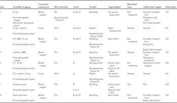
The institutional database was searched for all patients with the diagnosis of discordant atrioventricular with concordant ventriculo-arterial connections seen at the Necker Enfants Malades Hospital from January, 1983 to January, 2013 included. All medical files of the identified patients were studied, including all the available clinical data, chest X-rays, echocardiography, CT scan, or MRI reports, catheter lab reports and images, surgery, or autopsy protocols. All patient files were reviewed by L.H. for segmental analysis. All the available CT scans and MRIs were reviewed by F.R. Details on the available imaging modalities for each patient are shown in Table 1. Attending cardiologists were contacted for reports of the last follow-up visit if the patients were not followed-up in the institution’s outpatient department. Data are presented as median values (ranges) or as numbers (percentages).
Detailed cardiac features including bronchial and abdominal arrangement in all cases.

A=anterior; MA=mitral-aortic continuity; NA=not applicable because of missing data; **No=tiny mitro-aortic discontinuity; P=posterior; R=right; S by S=side by side;
* External shape of the appendages as evaluated by the cardiac surgeon during intervention when information available in surgery report
Definitions
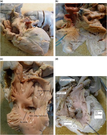
Cardiac morphology of the eight patients was analysed according to the morphological and segmental approach developed by Van Praagh,Reference Van Praagh 9 based on echocardiographic findings, CT scans, MRIs, cardiac catheter lab images, and surgical or post-mortem protocols. The external morphology of the appendages was described by the surgeon during intervention, or by non-invasive imaging studies such as CT scans when available. Their internal morphology – extent of the pectinate muscles – could not be determined except in one cardiac specimen (Fig 1a–d). For this reason, as we will explain in detail further, and because the venoatrial connections determine the haemodynamics, we chose to define the morphologically right atrium as the atrium receiving the inferior caval vein or, in case of interrupted inferior caval vein, the hepatic veins, but also the orifice of the coronary sinus and the attachment of septum primum on the controlateral side of septum secundum, according to Van Praagh.Reference Pasquini, Sanders and Parness 1 , Reference Van Praagh, Kakou-Guikahue, Kim, Becker, Alday and Van Praagh 10 Discordant atrioventricular connections were defined as the morphologically right atrium opening through the mitral valve or the left part of the common atrioventricular valve into the morphologically left ventricle, and the morphologically left atrium opening through the tricuspid or right part of the common atrioventricular valve into the morphologically right ventricle. Concordant ventriculo-arterial connections were defined as the aorta arising from the left ventricle and the pulmonary artery arising from the right ventricle. Heterotaxy syndrome was defined as an abnormal arrangement of the internal thoraco-abdominal organs across the left–right axis of the body, including left or right isomerism, as described by Jacobs et al.Reference Jacobs, Anderson and Weinberg 11
( a ) Patient 8; {I, D, S}: macroscopic view of the heart specimen with dextrocardia (posterior view) showing the left-sided RA receiving the SHV opening to the LV, and the right-sided LA opening to the RV. ( b ) Patient 8; {I, D, S}: macroscopic view of LV opened showing a narrow, stenotic LVOT and left part of the AV valve. ( c ) Patient 8; {I, D, S}: macroscopic view of the left-sided atrium receiving the SCV and all the SHV. The pectinate muscles do not extend to the crux of the heart; one can delimit the ostium primum-type ASD and the underlying AV valve. The left PVs are draining into the left-sided atrium. ( d ) Patient 8; {I, D, S}: macroscopic view of the right-sided atrium. The atrial appendage is triangular in shape and broad-based, but the pectinate muscles do not extend to the crux; one can delimit the ostium primum type ASD and the underlying AV valve. The right PVs are draining into the right-sided atrium. Ao=aorta; ASD=atrial septal defect; AV=atrioventricul; LA=left atrium; LV=left ventricle; LVOT=left ventricular outflow tract; PV=pulmonary vein; RA=right atrium; RV=right ventricle; SCV=superior caval vein; SHV=sub-hepatic veins.

Results
General patient characteristics and cardiac morphology
A total of eight patients with discordant atrioventricular with concordant ventriculo-arterial connections were identified via the institutional database during the study period. There were two male and six female patients; four patients had had a prenatal diagnosis of CHD, but only one had been recognised as having discordant atrioventricular with concordant ventriculo-arterial connections before birth.
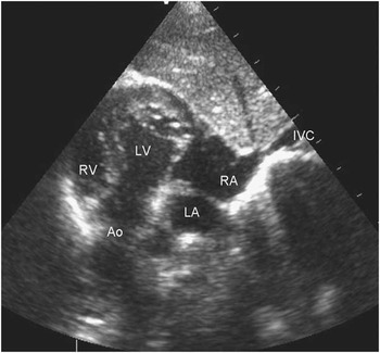
According to the anatomical types of discordant atrioventricular with concordant ventriculo-arterial connections described by Pasquini et al, there were six patients with {I, D, S} segmental arrangement and spiralling great arteries – with inversus or mirror-imaged atria, D-looped ventricles, and solitus or normally related great arteries with the aortic valve posterior and to the right; two patients had parallel great vessels, of whom one had {S, L, D} arrangement – with solitus or normal atria, L-looped ventricles, and parallel great arteries with the aortic valve anterior and to the right (Fig 2) – and the other had {S, L, “A”} arrangement – with solitus or normal atria, L-looped ventricles, concordant ventriculo-arterial connections with parallel great vessels, and the aortic valve strictly anterior to the pulmonary valve (Fig 3).Reference Cavalle-Garrido, Bernasconi, Perrin and Anderson 12 Of the eight patients, five had heterotaxy syndrome, diagnosed on left bronchopulmonary isomerism in four – two of these four patients had leftish external morphology of both appendages – and on midline liver with polysplenia in one. The detailed description of the respective bronchial and abdominal arrangement can be found in Table 1. All patients but two (patient no. 2 and 6) had spiralling great arteries with right and posterior aorta (Supplementary Figures S2–S4), including patient no. 5 who had pulmonary atresia without the main pulmonary trunk, but had a normal relationship of the ascending aorta and the pulmonary bifurcation in the upper mediastinum (Supplementary Figure S4). Patient no. 2 and 6 had parallel great vessels (Supplementary Figures S1A,B and S5); two patients had a very small discontinuity between the mitral and the aortic valve. Patient no. 1 had a long discontinuity (Supplementary Video S1).
Patient 2; {S, L, D}: two-dimensional echocardiographic sub-xiphoid oblique view showing the IVC draining to the RA, which opens to the LV giving rise to the Ao. The ventricles are supero-inferior with a moderate hypoplasia of the RV not fully shown in this view. Ao=aorta; IVC=inferior caval vein; LA=left atrium; LV=left ventricle; RA=right atrium; RV=right ventricle.

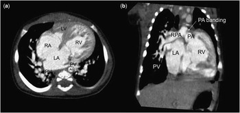
( a ) Patient 6; {S, L, A}: axial CT image showing a four-chamber view of discordant atrioventricular with concordant ventriculo-arterial connections with a large surgically created atrial septal defect and a moderate hypoplastic LV. ( b ) Oblique coronal CT image showing the LA opening through the tricuspid valve into the RV giving rise to the PA, which has been banded. Ao=aorta; LA=left atrium; LV=left ventricle; PA=pulmonary artery; PV=pulmonary vein; RA=right atrium; RPA=right pulmonary artery; RV=right ventricle.

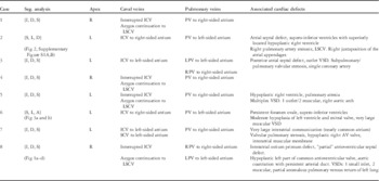
In six patients, discordant atrioventricular with concordant ventriculo-arterial connections were part of a more complex CHD; four of them had variants of obstructive right heart disease: pulmonary atresia with multiple ventricular septal defects and hypoplastic right ventricle (patient no. 5); supero-inferior ventricles with hypoplastic right ventricle and right pulmonary artery stenosis (patient no. 2); an equivalent of tetralogy of Fallot with subpulmonary and valvular pulmonary stenosis and outlet ventricular septal defect (patient no. 3); and a case of severe pulmonary valvular stenosis and hypoplastic tricuspid valve (patient no. 7). Patient no. 6 had supero-inferior ventricles with moderate hypoplasia of the left ventricle and a very large muscular ventricular septal defect making surgical septation impossible (Fig 3a and b). Patient no. 8 had a partial form of atrioventricular septal defect with small left component of the common atrioventricular valve and aortic coarctation (Fig 1a–b). Table 2 summarises the detailed anatomical description of all patients including segmental analysis, systemic and pulmonary venous return, and associated cardiac defects.
Supplementary cardiac features including all associated cardiac defects in the patient series.

ICV=inferior caval vein; L=left; LPV=left pulmonary veins; LSCV=left superior caval vein; PV=pulmonary veins; R=right; RPV=right pulmonary veins; SCV=superior caval vein; Seg.=segmental analysis; VSD=ventricular septal defect
Difficulties in pre-operative diagnosis
Only five patients were correctly diagnosed before surgical intervention by two-dimensional echocardiography. In the remaining patients, anatomical diagnosis was completed or corrected during or after surgery because of an unusual peri-operative clinical course. These three patients all had complex forms of discordant atrioventricular with concordant ventriculo-arterial connections (Table 2).
Patient no. 5 with pulmonary atresia and multiple ventricular septal defects was initially thought to have concordant atrioventricular and ventriculo-arterial connections and had a neonatal Blalock–Taussig–Thomas shunt without complication. During follow-up, anatomical diagnosis was corrected for discordant atrioventricular with concordant ventriculo-arterial connections by echocardiography before final surgical repair – atrial switch, ventricular septal defect closure, and pulmonary artery heterograft – was performed at 8 years of age.
Patient no. 6 with supero-inferior ventricles, a large ventricular septal defect, and parallel great vessels initially had neonatal pulmonary artery banding (Fig 3a and b). The post-operative course was marked by profound cyanosis requiring the urgent creation of a surgical atrial septal defect. Anatomical diagnosis was then corrected to discordant atrioventricular with concordant ventriculo-arterial connections, parallel arterial trunks, and a large ventricular septal defect. She had a Senning procedure three and half months later in order to improve streaming pattern and oxygenation without closure of the ventricular septal defect, which was too large for surgical closure. Pulmonary artery banding has been maintained in consequence.
Patient no. 7 operated in 1983 was correctly diagnosed only after the first surgery in which the right ventricular outflow obstruction was relieved by a transannular pulmonary patch. Mustard operation was performed 2 days later after confirmation of suspected discordant atrioventricular connections by cardiac catheterisation.
General surgical strategy
Median age at first cardiac surgery was 3 weeks (with a range from 1 day to 1.7 years). The first cardiac surgery was a palliative operation like the Blalock–Taussig–Thomas shunt, creation or enlargement of atrial septal defect in four patients. Median age at repair surgery or at last surgical intervention was 1.4 years (with a range from 1.1 months to 8.1 years). The repair surgery finally performed was the atrial switch procedure of Senning or Mustard type in seven out of eight patients. This included one hemi-Senning procedure due to a small right ventricle and a previous partial cavopulmonary connection (patient no. 2). Closure of the ventricular septal defect was performed from the morphological right ventricle in one patient and once via the aortic valve, because the morphological right ventricle was too hypoplastic to allow good surgical access. All but two patients had a biventricular repair: patient no. 6 who had an atrial switch procedure but a ventricular septal defect too large to be closed, and patient no. 8 died in 1984 from heart failure before repair surgery.
Complications and re-interventions
Immediate post-operative complications included two chylothoraces treated medically and one supra-ventricular tachycardia due to Wolff–Parkinson–White syndrome. There was no immediate or late complete heart block after closure of the ventricular septal defect; one patient progressively developed sick sinus syndrome and received a pace maker 18 years after surgical repair.
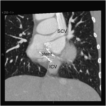
Atrial switch was complicated by atrial baffle obstruction twice. Surgical refection of the pulmonary venous channel was necessary in one patient 6 months after initial repair. In the other patient, inferior caval venous channel obstruction with portal hypertension became apparent 8 years after initial repair and was treated by percutaneous dilatation and stenting with a good immediate and mid-term result (Fig 4). The patient is doing well 6 years after this procedure. There has been no need for re-operation on the right ventricular outflow tract neither in the patient treated by the Senning–Rastelli-type procedure (patient no. 5) nor in the patient with tetralogy of Fallot who was repaired with a transannular patch (patient no. 3).
Patient 3; {I, D, S}: reconstructed sagittal CT image of the stented venous baffle. The metallic stent in the proximal part of the ICV can be seen as a white longitudinal structure in the vessel walls. The stent is well permeable. ICV=inferior caval vein; SCV=superior caval vein.

Follow-up and outcome
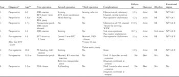
There were six late survivors and two early post-operative deaths, concerning the two patients operated in 1983 and 1984, one 11 days and the other 3 months after surgical intervention. Median follow-up from initial diagnosis to last contact was 4.2 years (with a range from 3.9 to 26.7 years). Median follow-up after complete repair or last surgical intervention was 3.6 years (with a range from 1.7 to 26.3 years). At the last follow-up visit, all the patients were in NYHA Class I or II and in regular sinus rhythm, except for the adult with the pacemaker for sick sinus syndrome. Echocardiographic left ventricular systolic function was normal in all patients. There was no pulmonary or caval venous channel obstruction in the patients who had had the atrial switch procedure. There was only minimal or no regurgitation of the atrioventricular valves; one patient has a moderate aortic regurgitation due to dysplastic aortic valve repaired by triple commissuroplasty at the time of repair surgery. The patient who had been treated by partial cavopulmonary connection due to hypoplastic right ventricle has no clinical or echocardiographic dysfunction. Table 3 summarises the surgical strategy, complications, and outcome of this patient series.
Overview on surgical strategy, post-operative complications and outcome.

ASD=atrial septal defect; BTT=Blalock–Taussig–Thomas shunt; IVC=inferior caval vein; PA=pulmonary artery; PCPC=partial cavopulmonary connection; PA=pulmonary artery; PDA=patent arterial duct; RV=right ventricle; SR=sinus rhythm; VSD=ventricle septum defect; WPW=Wolff–Parkinson–White
* Moment of complete anatomic diagnosis
** Age at first operation: y=years; m=months; d=days
Discussion
In this study, we report a monocentric series of eight patients whose haemodynamics is that of discordant atrioventricular with concordant ventriculo-arterial connections, in which only 60% of patients were correctly diagnosed before surgical repair. Surgical strategy in this series was mainly a biventricular approach, essentially including the atrial switch procedure. Most patients are long-term survivors with an overall encouraging functional long-term outcome.
More than 40 cases of isolated or complex forms of discordant atrioventricular with concordant ventriculo-arterial connections have been reported, including cases with heterotaxy.Reference Pasquini, Sanders and Parness 1 – Reference Kannan, Kamath, Rao and Kumar 8 , Reference Sharma, Marwah, Shah and Maheshwari 13 The challenge of diagnosing this rare cardiac defect, especially if it is part of a complex CHD, has not changed since its first description. The main problem in all historical patients was that the correct anatomical diagnosis was only made at autopsy. Snider et alReference Snider, Enderlein, Teitel, Hirji and Heymann 14 were the first to fully describe the positive and complete pre-operative diagnosis of this CHD by modern two-dimensional echocardiography. More recently, small case series of three to five patients, focussing on the successful surgical management and sometimes long-term follow-up of isolated patients, have been reported.Reference Konstantinov, Lai and Colan 7 , Reference Sharma, Marwah, Shah and Maheshwari 13
It is of notice that the correct diagnosis was missed pre-operatively in three of our patients, including the one with pulmonary atresia, in whom neonatal cyanosis was attributed to the prostaglandin-dependent pulmonary circulation. All of these misdiagnosed patients had a complex intraventricular anatomy increasing the diagnostic difficulty. A thorough and detailed segmental analysis is, therefore, essential for the paediatric cardiologist in order to accurately describe the cardiac anatomy and haemodynamics and avoid surprises for the surgeon. As the surgical strategy will depend above all on haemodynamics, the analysis of venoatrial connections is of utmost importance in these patients. In this complex setting, the anatomical definition of the morphological right and left atrium becomes crucial, especially in the case of inversus or mirror-imaged venoatrial connections, as frequently seen in this series. According to the morphological method developed by Van PraaghReference Van Praagh, Kakou-Guikahue, Kim, Becker, Alday and Van Praagh 10 , cardiac structures should be defined on the basis of their own intrinsic morphology, and features that are themselves variable should not be used to define other variable entities. This is relatively easy for the ventricles and the great vessels, but much more difficult for the atria. In fact, the most constant components of the atria are the atrial appendages, which are easy to distinguish in the normal heart, but are much more difficult in situations where the laterality of the heart is distorted, such as heterotaxy syndrome. Externally, the shape of the appendages is very different: triangular, with a broad junction with the rest of the atrium, for the right atrial appendage, and finger-like, with marked indentations and narrow junction, for the left one; however, the external shape of the atrial appendages has turned out to be highly variable in malformed hearts. Their most reliable anatomical characteristics lie, therefore, in their inner structure – namely, the extent of the pectinate muscles relative to the atrioventricular junctions. In the morphologically right atrium, the pectinate muscles extend to the crux of the heart, whereas in the morphologically left atrium the pectinate muscles are confined within the left atrial appendage.Reference Uemura, Ho, Devine, Kilpatrick and Anderson 15 The problem is that this feature, although anatomically constant, is extremely difficult to assess in the living patient, even with modern imaging techniques such as CT-scan or MRI. In our study, we had to rely only on the external descriptions of the appendages provided by the surgeon or cardiac imaging studies, with the exception of the available heart specimen. This is a source of possible error, as the external shape of the appendages does not always reflect the inner extent of the pectinate muscles,Reference Uemura, Ho, Devine, Kilpatrick and Anderson 15 and the surgeon rarely takes the opportunity to open both atria to check the extent of the pectinate muscles. The illustration of this problem becomes apparent in Table 1.
Owing to these diagnostic deficiencies, we chose to focus on the venoatrial connections, because they determine the haemodynamics and thus the surgical strategy. We will come back later to the issue of atrial morphology and its impact on the interpretation of our patient series in the setting of isomerism of the atrial appendages. According to Van PraaghReference Van Praagh, Kakou-Guikahue, Kim, Becker, Alday and Van Praagh 10 , we chose to define the morphological right atrium by the connection of the inferior caval vein or, in case of interrupted inferior caval vein, the hepatic veins, which of course constitutes a noteworthy exception to the rules of the morphologic method. We are, thus, fully aware that this approach is debatable, especially in heterotaxy patients where the venoatrial connections are often abnormal. In this setting, atrioventricular connections and venoatrial connections are defined by the English school as mixed and quasi-discordant, atrioventricular discordance being possible only if there is usual or mirror-imaged arrangement.
Another key problem of our series lies in the definition and the diagnosis of heterotaxy. Heterotaxy can be defined as an abnormal arrangement of the internal thoraco-abdominal organs across the left–right axis of the body, including left or right isomerism, as described by Jacobs et al.Reference Jacobs, Anderson and Weinberg 11 This means that each patient having evidence of isomerism somewhere within his bodily make-up, including often the absence of the spleen or the presence of multiple spleens, will be classified as having heterotaxy syndrome. Heterotaxy is considered to result from a defect of the laterality of the body, occurring during the earliest stages of cardiac development. In experimental studies, mice knocked-out for the laterality genes demonstrate bilaterally right or left atrial appendages, based on the extent of the pectinate muscles to the crux in case of right isomerism of the atrial appendages or on their confinement inside the appendages in case of left isomerism of the atrial appendages.Reference Bamforth, Braganca and Farthing 16 , Reference Hildreth, Webb and Chaudhry 17 Following these animal models, some have inferred that isomerism of the atrial appendages is the hallmark of heterotaxy.Reference Uemura, Ho, Devine, Kilpatrick and Anderson 15 A constellation of heart defects is usually, but not always, associated with these syndromes, although defects commonly found in the two syndromes can co-exist in the same patient. The diagnosis of left or right isomerism of the atrial appendages would in consequence become extremely probable if one considers such association of lesions and organ arrangements as typical. When we now analyse the associated lesions and specifically the overall abdominal and bronchial arrangement of our patient series as described in Table 1, we then become aware that the constellation of cardiac and extra-cardiac lesions at least for patients 1, 3, 4, 5, and 8 are very much in favour of left isomerism as defined by Anderson et al. Specifically, the left bronchial isomerism observed in four patients of our series is often indicative of left isomerism of the atrial appendages, but this is not an exclusive rule as exceptions have been reported. Notably, abdominal and bronchial arrangement in favour of left isomerism has been described associated with usual or mirror-imaged atrial arrangement.Reference Anderson, Devine, Anderson, Debich and Zuberbuhler 18 In summary, defining the atria either by the internal morphology of the atrial appendages, which we cannot know with certainty, or by their venous connections may change completely the analysis of our patient series. If we consider the patients of this series mentioned above as having left isomeric – and not mirror-imaged – atrial appendages, then the atrioventricular connections would not be considered as discordant any more, but mixed or “quasi-discordant”.Reference Macartney, Zuberbuhler and Anderson 19 This would lead us to interpret our series of eight patients quite differently: patients 1, 3, 5, and 8 who have left bronchopulmonary isomerism would have left isomerism of the atrial appendages with mixed atrioventricular and concordant ventriculo-arterial connections. This diagnosis could also be suspected in patient 4 with mid-line liver and hypoplastic polysplenia, although there is normal bronchial arrangement. In logical consequence, these patients would not have partial anomalous pulmonary venous connection as described in Table 2 for patients 3 and 8, but would be considered as having symmetrical pulmonary vein drainage; three patientsReference Van Praagh and Van Praagh 2 , Reference McElhinney, Reddy, Silverman and Hanley 6 , Reference Konstantinov, Lai and Colan 7 would have truly discordant atrioventricular and concordant ventriculo-arterial connections including one historic patient (no 7) without any information on thoracic and abdominal organs, and patients no 2 and 6 with parallel arterial trunks. The distinction between the hearts with parallel as opposed to spiralling arterial trunks is also extremely important in this setting. Concordant ventriculo-arterial connections with parallel arterial trunks, or anatomically corrected malposition of the great arteries,Reference Van Praagh, Perez-Trevino and Lopez-Cuellar 20 , Reference Anderson, Becker, Losekoot and Gerlis 21 have often been associated with atrioventricular discordant with ventriculo-arterial concordant connections.
Several terms are found in the literature to describe the various anatomical forms of atrio-ventricular discordant with ventriculo-arterial concordant connections and spiralling arterial trunks. The terms “isolated atrial inversion”, “isolated atrial discordance”, “isolated ventricular inversion”, or “isolated ventricular discordance” were used in several articles but appear to be less precise than to describe the lesions in totality.Reference Clarkson, Brandt, Barrat-Boyes and Neutze 22
Our study is naturally limited because of the low number of cases and its retrospective design. Existing literature on discordant atrioventricular with concordant ventriculo-arterial connections remains scarce, making it difficult to draw conclusions concerning long-term outcome with regard to mortality and morbidity in this rare CHD population. Possible outcome in this rare patient group might be extrapolated from long-term follow-up of atrial switch in the setting of transposition of the great arteries. Late survival is known to be high with good functional status in patients with transposition of the great arteries operated by atrial switch procedure.Reference Moons, Gewillig and Sluysmans 23 – Reference Warnes 25 In our series, there was no late mortality with a median follow-up of 4.2 years. Functional status at the last outpatient visit was rather encouraging with all patients being in NYHA Class I and II. Few of the existing surgical reports on patients with discordant atrioventricular with concordant ventriculo-arterial connections describe long-term follow-upReference Konstantinov, Lai and Colan 7 , Reference Sharma, Marwah, Shah and Maheshwari 13 with information often being limited to immediate post-operative survival.
Well-known long-term problems in patients with transposition of the great arteries operated by atrial switch are the failing of the systemic right ventricle, atrial rhythm disturbances, and atrial baffle obstruction or leakage.Reference Moons, Gewillig and Sluysmans 23 – Reference Warnes 25 In the case of discordant atrioventricular with concordant ventriculo-arterial connections, the sub-aortic ventricle is the morphological left ventricle. This leaves arrythmias and channel obstructions, which are both encountered in our our series, as potential major immediate and long term problems. Development of atrial bradyarrhythmia or tachyarrhythmia is known as a typical long-term complication of the atrial switch procedure. Arrhythmia-free survival rate for simple transposition after the Mustard procedure is higher than that after the Senning procedure with, for example, 70.1 versus 54.1% of patients free of arrhythmias, respectively, 20 years after procedure.Reference Moons, Gewillig and Sluysmans 23 Pacemaker implantation was reported in 5.5% of patients in this same study. Patients treated by the Senning procedure seem to develop sinus node dysfunction sooner than patients after Mustard repair.Reference Dos, Teruel and Ferreira 24 Rhythm disturbances occurred post-operatively in two patients of our series, including one girl who had a pacemaker implantation for sinus node dysfunction 18 years after the Senning procedure.
It is of note that none of our patients had post-operative complete heart block, although this has already been reported in patients having discordant atrioventricular with concordant ventriculo-arterial connections and ventricular septal defects who were successfully operated.Reference Ranjit, Wilkinson and Mee 5 , Reference Konstantinov, Lai and Colan 7 , Reference Sharma, Marwah, Shah and Maheshwari 13 , Reference McGrath, Kirklin and Blackstone 26 Conduction bundle pathways are not really documented in discordant atrioventricular with concordant ventriculo-arterial connections even if electrophysiological peri-operative explorations have been performed; however, several anatomical and electrophysiological studies in hearts with corrected transposition of the great arteries in atrial situs inversus {I, D, D} have shown that, in this case, the atrioventricular node and the atrioventricular connection tissue are generally located posteriorly, contrary to corrected transposition of the great arteries in atrial situs solitus {S, L, L}, even if exceptions exist, particularly when there is associated pulmonary stenosis or atresia, leading to a better alignment between the atrial and ventricular septa.Reference Dick, Van Praagh, Rudd, Folkerth and Castaneda 27 – Reference Hosseinpour, McCarthy, Griselli, Sethia and Ho 30 In the presence of discordant atrioventricular connections, the location of the atrioventricular node would, thus, depend on the alignment between the atrial and ventricular septa, normally aligned in atrial situs inversus and malaligned in atrial situs solitus.Reference Thiene, Nava and Rossi 28 Reported cases with post-operative heart block concerned cases of discordant atrioventricular connections in atrial situs solitus.Reference Ranjit, Wilkinson and Mee 5 , Reference Konstantinov, Lai and Colan 7 , Reference McGrath, Kirklin and Blackstone 26 Interestingly, both the patients with ventricular septal defect closure without complications in this series had atrial situs inversus. Moreover, it is generally acknowledged that ventricular septal defect closure from the morphologically right ventricle is to be preferred in order to diminish post-operative heart block.Reference de Leval, Bastos, Stark, Taylor, Macartney and Anderson 31 This was also the preferred approach by our surgeons when technically possible.
Conclusion
Discordant atrioventricular with concordant ventriculo-arterial connections is an extremely rare cardiac defect whose treatment strategy of choice is the atrial switch procedure. Clinical experience is scarce and echocardiographic diagnosis is challenging, especially if discordant atrioventricular with concordant ventriculo-arterial connections are part of a complex CHD. Moreover, discordant atrioventricular with concordant ventriculo-arterial connections are often part of a heterotaxy syndrome. There are ongoing controversies about the definition of atrial morphology and heterotaxy syndrome animating the anatomic discussion of these complex heart defects. Morbidity is closely related to problems well-known from the long-term follow up of patients with transposition of the great arteries treated by the Senning or Mustard procedure 20–40 years ago. Main problems are rhythm disturbances such as supraventricular arrhythmias and complete heart block, the latter being known in the literature but not seen in this series. Post-operative complete heart block after closure of the ventricular septal defect can probably be diminished by a surgical approach from the morphological right ventricle.
Acknowledgements
The authors thank Dr Ivan Bouzguenda from Jacques Cartier Private Hospital, Massy, France, for providing the MRI images performed at his institution concerning one of the patients of the series.
Financial Support
This research received no specific grant from any funding agency, commercial, or not-for-profit sectors.
Conflicts of Interest
None.
Ethical Standards
The authors assert that all procedures contributing to this work comply with the ethical standards of the relevant French guidelines on human experimentation and with the Helsinki Declaration of 1975, as revised in 2008. The need for informed consent was waived by the institutional committee because of French guidelines for retrospective studies.
Supplementary Material
To view Supplementary Materials for this article, please visit http://dx.doi.org/10.1017/S1047951115000736