Implications
This study demonstrated the changes in plasma and milk proteome of lactating dairy cows receiving sequentially increasing doses of α-linolenic acid (LNA) via duodenal infusion, which improved the milk composition by enhancing its LNA content. Haptoglobin in plasma, and albumin, and fragments of β-casein and αs1-casein in milk were altered after the cows received infusion of 400 g/day of LNA. This report displayed a new insight into understanding the effects of duodenal infusion of high doses of LNA on the lactating dairy cows, which can result in metabolic stress within the bovine intestine and in changes in milk composition.
Introduction
Milk is rich in proteins and lipids, and its ingestion may have beneficial health effects for people. Polyunsaturated fatty acids, a type of diverse fatty acids found in milk, may reduce development of some chronic diseases, such as cancer and cardiovascular disease, in humans (Burdge and Calder, Reference Burdge and Calder2006). α-Linolenic acid (LNA), one of the polyunsaturated fatty acids found in milk, is an essential fatty acid that can be converted to fatty acids with longer-chain and more double bonds, principally eicosapentaenoic acid and docosahexaenoic acid in the body. These long-chain fatty acids are important for cell membrane function and are beneficial to the health of humans (Burdge and Calder, Reference Burdge and Calder2006; Mansson, Reference Mansson2008).
In recent years, many researchers have used plant seed supplementations or altered diets to improve the fatty acid profile of milk by enhancing its LNA content (Ward et al., Reference Ward, Wittenberg and Przybylski2002; Bu et al., Reference Bu, Wang, Dhiman and Liu2007; Khiaosa-Ard et al., Reference Khiaosa-Ard, Klevenhusen, Soliva, Kreuzer and Leiber2010; Zachut et al., Reference Zachut, Arieli, Lehrer, Livshitz, Yakoby and Moallem2010). In attempts to study the underlying mechanisms of LNA transport to milk and modification of the fatty acid composition, LNA has been infused post-ruminally to avoid ruminal biohydrogenation (Khas-Erdene et al., Reference Khas-Erdene, Wang, Bu, Wang, Drackley, Liu, Yang, Wei and Zhou2010; Côrtes et al., Reference Côrtes, Kazama, da Silva-Kazama, Benchaar, Zeoula, Santos and Petit2011). Interestingly, we found that increasing the LNA content of milk through post-ruminal infusion was accompanied by reduced oxidative stability of the milk fat (Liu et al., Reference Liu, Wang, Bu, Khas, Liu, Wei, Zhou and Beitz2010) and suppressed immune function of the cows as revealed using the ELISA method (Sun et al., Reference Sun, Wang, Yang, Khas and Liu2010).
To further examine the immunomodulatory effects of post-ruminal infusion of high doses of LNA, we selected a proteomic approach in this study. Proteome analysis generally consists of protein separation by two-dimensional gel electrophoresis (2-DE) and protein identification by mass spectrometry (MS). The availability of protein databases has made it feasible to analyze the protein profiles of various cells, tissues and body fluids (Yamane et al., Reference Yamane, Minamoto, Yamashita, Takamura, Miyamoto-Myoken, Yoshizato, Nabetani, Tsugita and Mishima2003). Use of this method allows us to determine whole protein profiles of body fluids in cattle receiving post-ruminal infusion of high doses of LNA. The proteomic approach allows us to identify any proteins affected by LNA infusion, providing a significant advantage over more targeted approaches such as ELISA. We hypothesized that infusion of high doses of LNA would result in alteration of plasma and milk proteome of lactating dairy cows. Thus, the objective of this study was to investigate the changes of plasma and milk proteome following duodenal infusion of increasing amounts of LNA in lactating dairy cows using 2-DE in combination with matrix-assisted laser desorption/ionization time-of-flight/time-of-flight tandem mass spectrometry (MALDI-TOF-TOF MS).
Material and methods
Animals and experimental treatments
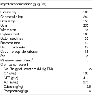
All animal care and procedures were approved by the Animal Care Advisory Committee of the Chinese Academy of Agricultural Sciences. Experimental treatments and sample collection procedures have been previously described (Sun et al., Reference Sun, Wang, Yang, Khas and Liu2010). In brief, four primiparous Chinese Holstein cows (BW 476 ± 6 kg, 100 ± 2 days in milk) were fitted with duodenal cannulas and used in a two-treatment crossover design study. The cows were housed in individual tie stalls, fed with a total mixed ration twice daily at 0700 and 1900 h and milked twice daily at 0500 and 1700 h. The diet ingredients and chemical composition are listed in Table 1. The study lasted for 12 weeks, which included two 5-week experimental periods and a 2-week washout period. During the experimental periods, the two experimental cows received increasing doses, from 0, 100, 200, 300 and 400 g, of LNA via duodenal infusion twice daily for one week at each dose, with each infusion even lasting for 10 h (from 0700 to 1700 h and from 1930 to 0530 h). The two control cows received an equivalent volume of the infusion vehicle with 0 g of LNA. Dry matter intake (DMI) was measured for each cow once daily.
Ingredient and composition of the basal diet on DM basis
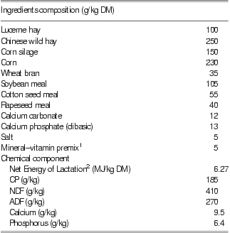
DM = dry matter.
1Based on DM: Fe = 5500 mg; Cu = 4080 mg; Zn = 17 500 mg; Mn = 4980 mg; Se = 110 mg; I = 180 mg; Co = 88.5 mg; vitamin A = 42 000 000 IU; vitamin D3 = 600 000 IU; vitamin E = 10.8 g.
2Calculated according to the China NY/t 34 (2004) guidelines.
Blood and milk samples were collected on the last day of administration of each infusion dose. One aliquot of the milk samples was centrifuged at 3000 × g for 15 min at 4°C to remove fat, and the supernatant was collected. The remaining volume of the milk samples was used to measure somatic cell count (SCC) in milk with Fossomatic 5000 system (Foss Electric, Hillerød, Denmark). Blood samples were centrifuged at 3000 × g at 4°C for 15 min, and the supernatant was collected. Milk and plasma protein samples were stored at −70°C until analysis.
2-D electrophoresis
Protein concentrations were quantified by the Coomassie Brilliant Blue method using bovine serum albumin solutions as reference standards. Milk proteins (300 μg) and plasma proteins (600 μg) from individual cows were diluted in 350 μl of rehydration solution (8 M urea, 4% w/v 3-[(3-cholamido-propyl)-dimethylammonio]-1-propanesulfonate, 40 mM Tris, 65 mM dithiothreitol (DTT), 2% v/v immobilized pH gradient (IPG) buffer) and separated by a Protean IEF Cell (Bio-Rad, Hercules, CA, USA) using first-dimension isoelectric focusing (IEF) with 17 cm, pH 4 to 7 IPG strips according to the manufacturer's instructions. All IEF steps, including passive rehydration for 13 h, 30 min at 250 V, 1 h at 1000 V, gradient from 1000 V to 9000 V in 5 h and 9000 V to the steady state for 8 h, were carried out at 20°C. After IEF, the gel strips were stored at −70°C.
Before the second-dimension separation, the IPG strips were incubated in an equilibration solution containing 6 M urea, 2% w/v SDS, 20% v/v glycerol, 50 mM Tris-HCl, 0.01% w/v bromophenol blue and equilibrated for two 12-min periods with gentle shaking. DTT (2% w/v) was added to the equilibration solution for the first incubation and 2.5% w/v iodoacetamide was added to the solution for the second incubation. After equilibration, the strips were applied to the surface of vertical SDS-PAGE and then embedded in agarose (0.5% w/v agarose in electrode buffer). The gels were run in a Protean II xi electrophoresis instrument (Bio-Rad) at 50 V for 30 min and then at 200 V until the bromophenol blue dye marker had reached the bottom of the gel. Subsequently, the gels were stained with colloidal Coomassie Blue G-250 solution (Candiano et al., Reference Candiano, Bruschi, Musante, Santucci, Ghiggeri, Carnemolla, Orecchia, Zardi and Righetti2004), and the stained gels were scanned using a GS800 calibrated densitometer (Bio-Rad) with 256 gray-scale and 600 dpi parameters. Protein spot detection was performed using PDQuest 8.0.1 software (Bio-Rad).
In this study, there were four biological replications (four cows) and two technical replications (individual cows). Plasma and milk proteins from cows receiving 0 g of LNA served as controls (reference gel) and were then compared with plasma and milk proteins of the cows that received 100, 200, 300 or 400 g of LNA. For intensity and density calculations, volumes were corrected for background and then plotted v. protein amount. Protein spots that exhibited a twofold or more decrease or increase in staining density relative to their levels before LNA infusion were selected.
In-gel digestion
Selected protein spots were manually excised from the Coomassie Blue-stained gels, washed twice with Milli-Q water for 15 min each time and then destained with 50% v/v acetonitrile in 0.1 M ammonium bicarbonate. Subsequently, the gel pieces were dehydrated in acetonitrile and dried in a SpeedVac (Eppendorf, Hamburg, Germany) under vacuum. The gel pieces were digested by overnight incubation at 37°C in a 25 mM ammonium bicarbonate solution containing 0.01% w/v trypsin. The digestion was terminated by the addition of 1% v/v trifluoroacetic acid (TFA).
Mass spectrometric analysis
MS was performed using an Ultraflex MALDI-TOF-TOF MS (Bruker Daltonics, Bremen, Germany) in the reflector mode with an m/z range of 600 to 4000. Extracted peptides were placed on the AnchorChip target and then a matrix solution containing α-cyano-4-hydroxycinnamic acid (4 mg/ml) in 70% v/v acetonitrile with 0.1% v/v TFA was added. The spectra were processed using FlexAnalysis 2.2 software tools (Bruker Daltonics). Protein identification using peptide mass fingerprinting was performed using the MASCOT software to search the NCBI Bos Taurus database (http://www.ncbi.nlm.nih.gov). The search parameters were as follows: monoisotopic mass accuracy <100 ppm, missed cleavages 1, minimum signal-to-noise (S/N) ratio 15 and carbamidomethylation of cysteine as a fixed modification. On the basis of the peptide mass fingerprinting results, the three peptides with the highest accuracy and abundance were subjected to tandem MS analysis (fragment mass tolerance <0.6 Da). Protein probability scores are derived from ion scores (individual ion scores >46 indicate identity or extensive homology (P < 0.05)) that is −10 × Log(P), in which P is the probability that the observed match is a random event.
Western blots
Plasma proteins were separated by 12% w/v SDS-PAGE at room temperature with a constant voltage of 120 V. After electrophoresis, the proteins in the gel were electrotransferred onto a polyvinylidene difluoride (PVDF) membrane. The PVDF membrane was blocked with 5% v/v horse serum in TBST buffer (0.1 M Tris pH 7.9, 0.15 M NaCl, 0.1% v/v Tween 20) at room temperature for 2 h, probed with a primary polyclonal rabbit anti-haptoglobin antibody (prepared by Beijing Protein Institute, Beijing, China) at 37°C for 1 h and then incubated with a horseradish peroxidase-coupled goat-anti rabbit secondary antibody and visualized using diaminobenzidine.
Statistical analysis
The PDQuest 8.0.1 software (Bio-Rad) was used to determine which protein spots on the 2-DE gel images exhibited a twofold or greater change in staining density following infusion of LNA. Staining densities at lower and higher doses of LNA were compared using a nonparametric Mann–Whitney test assuming equal variances using the Statistical Package of Social Science 16.0 (SPSS). Differences were considered significant at P < 0.05.
Results
DMI and SCC
Average daily DMI and SCC in milk are presented in Table 2. In the current study, the magnitude of decrease of DMI was significantly associated with the amount of LNA infused. However, the SCC of milk was significantly increased linearly from the 59 × 103 to 3079 × 103 cells/ml after the cows were infused with 400 g/day of LNA.
DM intake and milk SCC in cows receiving incremental infusions of α-linolenic acid at the duodenum

DM = dry matter; SCC = somatic cell count; LNA = α-linolenic acid; DMI = dry matter intake.
Identification of proteins
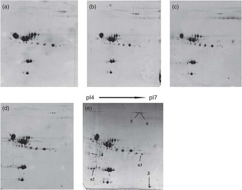
The representative 2-DE gel map of the plasma and milk proteome is shown in Figures 1 and 2, respectively. The gel images, as analyzed by PDQuest 8.0.1 software, showed that the plasma protein expression of cows did not significantly differ irrespective of infusion of LNA. However, three protein spots significantly increased after infusion with 400 g/day of LNA, but not after infusion at the lower doses (0, 100, 200 or 300 g/day). Similarly, milk protein profile was not affected by any of the infusions with LNA, except six protein spots that were increased in staining density by at least twofold after duodenal infusion with 400 g/day of LNA.
Two-dimensional gel maps of the bovine plasma proteome from the cows following duodenal infusion of 0 (a), 100 (b), 200 (c), 300 (d) and 400 (e) g/day α-linolenic acid. Upregulated proteins in cows infused with 400 g/day α-linolenic acid are marked with arrows. Arrow numbers correspond to protein identification in Table 3.

Two-dimensional gel maps of the bovine milk proteome from the cows following duodenal infusion of 0 (a), 100 (b), 200 (c), 300 (d) and 400 (e) g/day α-linolenic acid. Upregulated proteins in cows infused with 400 g/day α-linolenic acid are marked with arrows. Arrow numbers correspond to protein identification in Table 3.

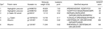
The MALDI-TOF-TOF MS helped identify two protein spots of the same protein from plasma and four protein spots of three different proteins from milk. The other three spots (n1 to n3) did not reach the significance level probably because of the limited number of protein sequences for bovines referenced in the database. These three protein spots were not reported in this paper. The excised protein spots are listed in Table 3. The upregulated protein in the plasma of cows that received duodenal infusion of 400 g/day LNA was identified as haptoglobin. In the milk of these same cows, the upregulated proteins were β-casein A2 variant, αs1-casein and albumin.
Identification of changed proteins in plasma and milk by matrix-assisted laser desorption/ionization time-of-flight/time-of-flight mass spectrometry

*Two spots detected on the gels with different isoelectric points, but similar molecular weights, were identified as the same protein. This is possible because of the presence of genetic variants or post-translational modifications.
Western blot analysis
To confirm the differential expression of plasma proteins, plasma haptoglobin following duodenal infusion of 0, 100, 200, 300 and 400 g/day of LNA was analyzed by Western blotting (Figure 3). The results of this analysis firmly supported our proteomic data; haptoglobin was significantly upregulated after the cows were infused with 400 g/day of LNA.
Western blot analysis of haptoglobin in plasma of cows following infusion of 0, 100, 200, 300 and 400 g/day of α-linolenic acid (LNA).

Discussion
LNA is an essential fatty acid in the human diet and might be protective against cardiovascular and inflammatory diseases. To explore mechanisms for increasing the LNA content in milk, we infused increasing doses, from 0 to 400 g/day, of LNA into the duodenum of dairy cows. This resulted in linear elevation of concentrations of LNA from 0.46% to 24.12% w/w of the total fatty acids in the milk (data not shown). However, as the cows received duodenal infusions of increasing amounts of LNA, their DMI decreased, whereas the milk SCC increased. DMI was also decreased as cows were fed rumen-inert fats or given abomasal infusions of unsaturated fatty acids. Decreases in DMI indicate changes in the metabolism of cows, which were associated with gut hormones, including increased plasma concentrations of glucagon-like peptide-1 (7-36) amide and cholecystokinin, a decreased insulin concentration, as well as with altered plasma metabolites, including increased concentrations of non-esterified fatty acids and decreased concentration of cholesterol (Litherland et al., Reference Litherland, Thire, Beaulieu, Reynolds, Benson and Drackley2005; Relling and Reynolds, Reference Relling and Reynolds2007). In addition, concentrations of prostaglandin E2 also declined, whereas plasma IgG levels rose linearly with increasing duodenal infusion doses of LNA (Sun et al., Reference Sun, Wang, Yang, Khas and Liu2010). By comparing the proteome between plasma and milk, we found that haptoglobin increased in the plasma, whereas β-casein A2 variant, αs1-casein and albumin increased in the milk of cows following the infusion of 400 g/day of LNA for one week. However, plasma haptoglobin was the only plasma protein elevated after the infusion with 400 g/day of LNA. This may be due to the high amounts of plasma albumin and immunoglobulin, which may have prevented detection of changes in minor proteins.
Haptoglobin is an acute phase protein primarily synthesized and secreted by hepatocytes. Its level was elevated in the blood of cows after parturition (Piccinini et al., Reference Piccinini, Binda, Belotti, Casirani and Zecconi2004; Cairoli et al., Reference Cairoli, Battocchio, Veronesi, Brambilla, Conserva, Eberini, Wait and Gianazza2006; Bionaz et al., Reference Bionaz, Trevisi, Calamari, Librandi, Ferrari and Bertoni2007), as well as in cows suffering from mastitis (Moyes et al., Reference Moyes, Drackley, Salak-Johnson, Morin, Hope and Loor2009), respiratory disease and stress (Aich et al., Reference Aich, Jalal, Czuba, Schatte, Herzog, Olson, Ross, Potter, Babiuk and Griebel2007; Cooke and Bohnert, Reference Cooke and Bohnert2011). It may serve as a non-specific host immune response to inflammation or stress. Recently, Araujo et al. (Reference Araujo, Cooke, Hansen, Staples and Arthington2010) showed that haptoglobin concentrations in beef calves were reduced by supplementation with rumen-protected polyunsaturated fatty acid (2.5% w/w as-fed basis) after transport and feedlot entry, regardless of how the supplement was fed before or after transportation. In contrast, our results suggest that haptoglobin concentrations increased in plasma because of intestinal metabolic stress, which could result in host non-specific immune response and higher milk SCC in cows infused with 400 g/day of LNA. Of particular note, the haptoglobin concentrations in the cows with high milk SCC were significantly higher than those in plasma of the healthy cows (Eckersall et al., Reference Eckersall, Young, McComb, Hogarth, Safi, Weber, McDonald, Nolan and Fitzpatrick2001; Pyörälä, Reference Pyörälä2003). These results are consistent with previous findings that transition cows supplemented with Ca salts of safflower oil had greater plasma haptoglobin concentrations than cows supplemented with Ca salts of palm oil (Silvestre et al., Reference Silvestre, Carvalho, Crawford, Santos, Staples, Jenkins and Thatcher2011). Dietary supplementation with polyunsaturated fatty acids has immunosuppressive effects through antigen presentation, T cell responses and fatty acid-derived inflammatory mediator production (Shaikh and Edidin, Reference Shaikh and Edidin2007; Yaqoob and Calder, Reference Yaqoob and Calder2007). Our proteomic approach did not detect haptoglobin in milk, probably because of the presence of large amounts of caseins. Thus, it is important to note that caution must be used when cows receive high doses of LNA by duodenal infusion to ensure that their health status is not compromised.
β-Casein A2 variant, αs1-casein and albumin in milk were also elevated in cows following duodenal infusion of 400 g/day of LNA, but not of lower doses (0, 100, 200 and 300 g/day). Albumin is a plasma protein whose elevation in milk indicates disintegration of the blood–milk barrier (Vangroenweghe et al., Reference Vangroenweghe, Duchateau and Burvenich2004). The observed elevation in milk albumin after high-dose post-ruminal infusion of LNA was consistent with changes in the protein composition of milk seen during naturally and experimentally induced mastitis (Hogarth et al., Reference Hogarth, Fitzpatrick, Nolan, Young, Pitt and Eckersall2004; Boehmer et al., Reference Boehmer, Bannerman, Shefcheck and Ward2008; Boehmer et al., Reference Boehmer, Ward, Peters, Shefcheck, McFarland and Bannerman2010; Ibeagha-Awemu et al., Reference Ibeagha-Awemu, Ibeagha, Messier and Zhao2010). In the current study, the elevated albumin and SCC in milk were accompanied by disintegration of the blood–milk barrier, suggesting a metabolic stress of mammary gland owing to the LNA infusion.
The elevated β-casein A2 variant and αs1-casein levels detected in the milk following high-dose LNA infusion correlates with enhanced proteolytic activity of endogenous enzymes toward β-casein precursor and α-casein (Napoli et al., Reference Napoli, Aiello, Di Donna, Prendushi and Sindona2007). Milk endogenous proteolysis has two main origins: plasmin activity and milk somatic cell proteases. Plasmin is primarily responsible for the observed proteolysis in milk with a low SCC, whereas cathepsins and elastase are implicated in proteolysis of milk with an elevated cell count (Wedholm et al., Reference Wedholm, Moller, Lindmark-Mansson, Rasmussen, Andren and Larsen2008). Increases in milk SCC (from 55 × 103 to 1382 × 103 cells/ml) have been correlated with increased plasmin, cathepsin D and cysteine protease activity (Somers et al., Reference Somers, O'Brien, Meaney and Kelly2003), and with concomitant increases in proteolysis in milk, leading to the formation of a lower molecular weight casein fraction (Larsen et al., Reference Larsen, Hinz, Jorgensen, Moller, Wellnitz, Bruckmaier and Kelly2010). In our study, fragments of β-casein and αs1-casein likely originated from increased caseinolysis in the milk of cows that received post-ruminal infusion of LNA at 400 g/day.
Conclusion
Our results show that the plasma and milk proteome is not affected by duodenal LNA infusion. However, an acute phase protein haptoglobin was elevated after post-ruminal infusion of LNA at 400 g/day. In addition, milk levels of albumin, fragments of β-casein and αs1-casein were increased, with a concomitant change in milk composition after high-dose infusion. Thus, increasing the amount of LNA in milk fat by high-dose post-ruminal LNA infusion may lead to adverse effects on the health and milk quality of lactating dairy cows. Future research is needed to explore the underlying mechanisms of LNA transport to milk fat and to comprehensively investigate the impact of post-ruminal infusion of LNA on lactating dairy cows.
Acknowledgments
The project was supported by the National Key Basic Research Program of China (Project No.2011CB100805).