

Background
Delirium is a severe and complex neuropsychiatric disorder associated with poor short-term outcome (Ely et al. Reference Ely, Gautam, Margolin, Francis, May, Speroff, Truman, Dittus, Bernard and Inouye2001). High prevalence rates in elderly and critically ill patients up to 80% have been described (Girard et al. Reference Girard, Pandharipande and Ely2008). Despite growing recognition of the importance of early diagnosis, delirium is regularly overlooked or poorly managed (Ely et al. Reference Ely, Stephens, Jackson, Thomason, Truman, Gordon, Dittus and Bernard2004). Several studies have identified numerous predisposing and precipitating factors (Girard et al. Reference Girard, Pandharipande and Ely2008). Refeeding syndrome is a well known cause of delirium and occurs after nutrition is started following a period of starvation. A variety of clinical symptoms following severe electrolyte and fluid shifts associated with metabolic abnormalities have been described. Due to the complexity of presentation and broad range of aetiologies, refeeding syndrome as a cause of delirium may be overlooked (Caplan, Reference Caplan2008). The literature is inconclusive regarding the prevalence of refeeding syndrome, although it has been described as being present in up to 34% of critically ill patients (Marik & Bedigian, Reference Marik and Bedigian1996). In this report the case of a 53-year-old man who experienced a severe delirium due to refeeding syndrome is presented. It illustrates the importance of the recognition of this poorly identified iatrogenic cause of neurological disturbance.
Case report
A 53-year-old man with unknown medical history was admitted to the emergency department of our hospital. He looked dishevelled and presented with psychomotor agitation, marked deficits in attention and irritability. He complained of right abdominal pain and severe headaches but was unable to provide details about the duration and severity. Physical examination revealed several haematoma's on his back, extremities, and head. Neurological examination did not reveal further pathology.
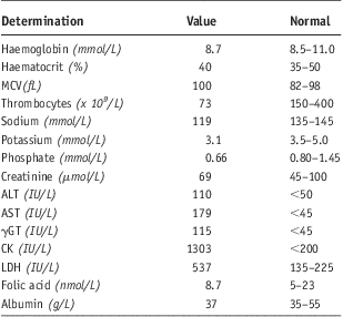
Blood analysis (Table 1) revealed a normal haemoglobin level with an increased macrocytic volume, a mild thrombocytopenia, and electrolyte derangements. Plasma creatinine level was normal. Liver biochemistry tests showed mild derangements of transaminases and elevated gamma glutamyl transpeptidase (γGT). Vitamin levels (i.e. B1, B6, and B12) were normal, only folic acid level was slightly decreased. Elevated levels of creatine phosphokinase and lactic dehydrogenase, indicated concomitant rhabdomyolysis.
Laboratory values of blood analysis
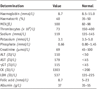
MCV – Mean corpuscular volume; ALT – alanine aminotransferase; AST – aspartate aminotransferase; γGT – gamma glutamyl transpeptidase; CK – creatine phosphokinase; LDH – lactic dehydrogenase
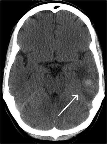
Because of his facial injuries and clouded mental state, diagnostic imaging was indicated as traumatic intracranial pathology was suspected. Non-contrast brain CT-scan (Fig. 1) and CT-angiography were performed. The non-contrast CT-scan revealed an ill defined lesion, sized 1.5 cm. The lesion was located in the left temporal area at the junction of white and grey matter and surrounded with vasogenic oedema, indicating a pathological process or intracranial haematoma. The CT-angiography scan showed the hypo-attenuated lesion most likely indicated a haematoma. The diagnosis of post-stroke delirium was considered as cognitive impairment and delirium are frequent consequences of stroke (Haring, Reference Haring2002; McManus et al. Reference McManus, Pathansali, Stewart, Macdonald and Jackson2007).
Axial CT-scan of the brain. The lesion located in the left temporal at the junction of white and grey matter, surrounded with vasogenic oedema, most likely indicating an intracranial hematoma (white arrow).

On the day of admission his consciousness improved rapidly, allowing us to obtain more relevant anamnestic information. The patient mentioned a period of alcohol abuse two years ago and claimed he used alcohol only occasionally since then. During the last years he had built up a successful career in finance and did not suffer from any major mental illnesses. However, recently a close family member had died and he had experienced severe mood disorders, i.e. depressed mood with fluctuations without manic episodes that were accompanied by a loss of appetite. His mental and physical state had not limited his ability to work until the day of admission. Strikingly, thereafter his appetite improved.
On the third day after admission a second episode of acute deterioration of his mental state occurred. He became agitated, suspicious, and showed inappropriate behaviour. Initially, this acute cognitive dysfunction was explained as a fluctuating course of post-stroke delirium. The possibility of a reoccurrence of the intracranial haemorrhage was considered. Blood analysis revealed that his serum phosphate had fallen to 0.1 mmol/l. Because of his severe delirious state and hypophosphataemia, he was transferred to the medical intensive care unit. The diagnosis of refeeding syndrome as the possible cause of the delirium was made.
The delirium was managed with intravenous haloperidol, which is described as the gold standard. Although optimal dose regimen has not been determined in clinical trials, current guidelines advise managing hyperactive delirium with 2 mg haloperidol followed by repeated doses every 15 minutes while agitation persists (Jacobi et al. Reference Jacobi, Fraser and Coursin2002). During acute management of delirium with intravenous haloperidol our patient was monitored for adverse effects including extrapyramidal effects, malignant hyperthermia and anticholinergic effects. One of the most important risks is prolongation of the corrected QT interval (QTc), which is associated with an increased risk of arrhythmias, including torsade de pointes.
In addition, his treatment included a new dietary regimen comprising a restricted caloric intake and supplementation of phosphate was initiated (0.5 mmol/kg over 12 hours). His mental status improved rapidly (<24 hours). Normal phosphate plasma concentration (0.801.45 mmol/l) was reached within 12 hours of initiating phosphate supplementation. Thereafter, no further episodes of severe confusion or disorientation occurred. Electroencephalography (EEG) was performed and demonstrated no abnormalities. The patient remained in the hospital for a total of 14 days.
Discussion
Delirium is defined as an acute disturbance of consciousness and cognition which fluctuates over time (American Psychiatric Association, 2000). Incidence rates of up to 70% in medical intensive care units have been described (Pandharipande et al. Reference Pandharipande, Cotton, Shintani, Thompson, Pun, Morris, Dittus and Ely2008) and elderly and/or critically ill patients often develop this complex, critical condition during the first days of hospital admission. Various studies have shown delirium to be associated with poor short term outcome and noted the importance of early recognition, diagnosis, and treatment. Numerous predisposing and precipitating factors have been described: e.g. intracranial pathology, malnutrition, metabolic disturbances, medication effects, alcohol or drug withdrawal or intoxication, and infection (Girard et al. Reference Girard, Pandharipande and Ely2008). In general, possible causal agents are identified during physical examination, investigations or from medical history. Stroke is a known precipitating factor for the development of delirium (Ferro et al. Reference Ferro, Caeiro and Verdelho2002). Whereas specific anatomical site and extent of brain injury, presence of cerebral oedema or cerebral hypoperfusion may be of influence. Although difficult to diagnose, post-stroke delirium prevalence ranges from 13 to 48% (McManus et al. Reference McManus, Pathansali, Stewart, Macdonald and Jackson2007).
In this case laboratory investigation revealed a severe hyponatraemia (119 mmol/l) most likely due to excessive extra-renal sodium wasting or malnutrition. Acute hyponatraemia is known to cause varying neurological sequelae. It is probable that hyponatraemia developed progressively in this patient due to malnutrition and therefore mild clinical manifestation may be expected. The patient showed a second episode of neurological deterioration on the third day of admission and recurrence of the intracranial haemorrhage or a fluctuating course of post-stroke delirium were considered. Although both are characterized by fluctuation of mental state, other causes of delirium should be considered (e.g. delirium tremens, as this classically presents on day three after cessation of alcohol intake).
Remarkably, blood analysis showed a severe hypophosphataemia (0.1 mmol/l). Normally phosphate plasma concentrations vary between 0.80 and 1.45 mmol/l. Hypophosphataemia usually becomes symptomatic in severe cases (< 0.32 mmol/l) (Brunelli & Goldfarb, Reference Brunelli and Goldfarb2007). Causes of hypophosphataemia can be described using three mechanisms: reduced intestinal absorption, and/or increased urinary excretion, and/or internal shifts of phosphate (Gaasbeek & Meinders, Reference Gaasbeek and Meinders2005). The refeeding syndrome is a major cause of hypophosphataemia, as 34% of critically ill patients experience refeeding syndrome (Marik & Bedigian, Reference Marik and Bedigian1996). Clinical manifestations include neurological, hematological or musculoskeletal disorders, or cardiovascular and respiratory failure (Caplan, Reference Caplan2008). Because patients frequently have multiple other potential causes for delirium, refeeding syndrome may be overlooked, given the frequent presence of infection, delirogenic medication or neuropathology.
After a new dietary regimen and aggressive phosphate suppletion was initiated, the mental state of our patient improved dramatically. Therefore, we concluded that the second episode of acute cognitive impairment was a symptom of refeeding syndrome. Moreover, at admission the patient's initial plasma phosphate level was only slightly decreased, asymptomatic, and could not explain the psychiatric disturbance at presentation whereas the observed serious intracranial pathology could.
Patients with alcohol dependence are at risk of developing hypophosphataemia because of low dietary intake of phosphate and vitamin D, chronic diarrhoea and alcohol-induced renal tubular impairment (Knochel, Reference Knochel1980). Blood analysis revealed elevated creatine phosphokinase (up to 30 000) indicating rhabdomyolysis, which is a common consequence of (severe) phosphate deficiency and/or pre-existing myopathy (Knochel et al. Reference Knochel, Barcenas, Cotton, Fuller, Haller and Carter1978; Singhal et al. Reference Singhal, Kumar, Desroches, Gibbons and Mattana1992). Patients with chronic alcohol dependency are prone to develop clinically significant rhabdomyolysis as muscle cells are pre-injured by the alcohol and phosphate depleted. In this case report we cannot fully exclude influences of alcohol abuse in the recent past. The increased creatine phosphokinase might also be the result of muscle injury following trauma, or confinement in a static position after stroke.
This case report demonstrated intensive liaison between psychiatry and medicine. This connection is of particular importance for psychiatrists, who are often involved in the management of delirium in the medical intensive care unit, as several causative factors of delirium are often intertwined and concurrently present. In this case a consultant psychiatrist was intensively involved during the patient's stay in the medical intensive care unit. This resulted in early recognition of the presenting symptomatology and optimal drug (haloperidol) dosage regimen. Therefore, in order to improve patient care, the importance of including psychiatrists in the treating medical team needs to be emphasized.
Complementary diagnostics (blood analysis, imaging studies) are essential for identification of underlying physical conditions. Furthermore, the cause of delirium can change during management which illustrates its complexity. For instance, post-stroke delirium can induce malnutrition, as the reinstitution of nutrition is potentially followed by the refeeding syndrome, which is characterized by further deterioration of mental state. The management of delirium can be challenging from both the medical and psychiatric perspective as disturbances of consciousness and cognition hinders appropriate treatment.
Conclusions
This case report illustrates the point that clinicians in both psychiatric and general intensive care must be aware of the numerous causes of delirium, which can concurrently be present. However, refeeding syndrome may be overlooked when more alarming symptoms are also present.
This case also highlights the intensive nature of interventions in an individual who was displaying challenging psychiatric symptomatology. It demonstrates ‘a credible argument that Psychiatric Intensive Care is much better defined as a set of interventions aimed at acute disturbance and crisis, than it is as a place in which these interventions are traditionally enacted’ (Dix, Reference Dix2007).
Acknowledgements
The assistance of Dr Steve Dye in the preparation of this article is greatly appreciated.