Introduction
Foreign body aspiration remains an important cause of morbidity and mortality in children in developing countries, especially in lower socioeconomic groups. Serious complications from aspirated foreign bodies, such as severe airway obstruction and death, can occur in younger children because of the small calibre of their airways.Reference Kosucu, Ahmetoglu, Koramaz, Orhan, Ozdemir and Dinc1 Long-standing foreign bodies can cause tissue reaction, oedema and secondary infection.
Because of the high risk associated with overlooked foreign body aspiration, bronchoscopy is often performed for definitive diagnosis and treatment, even when there is little suspicion or a doubtful history.Reference Kosucu, Ahmetoglu, Koramaz, Orhan, Ozdemir and Dinc1 Rigid bronchoscopy remains the ‘gold standard’ in managing these cases, for diagnostic as well as therapeutic purposes. Its outcome depends on many factors. However, it is a potentially dangerous procedure that relies on good visualisation and precise instruments such as foreign body grasping forceps and optically guided forceps. Furthermore, the foreign body must be removed as quickly as possible after visualisation. Repeated removal attempts are likely to result in increased complications. Serious complications can occur, such as pneumothorax, pneumonia, respiratory distress, cardiac arrest, tracheal laceration and subglottic oedema. Therefore, the availability of a noninvasive, harmless technique to locate the foreign body pre-operatively would greatly minimise subsequent morbidity and mortality. Virtual bronchoscopy is such a technique, and is particularly useful in locating nonradiopaque foreign bodies missed on plain radiography.
This study aimed to compare the usefulness of virtual bronchoscopy with actual rigid bronchoscopy in the detection of paediatric tracheobronchial foreign bodies not detected on plain chest radiography. To the best of our knowledge, very few published studies have compared and evaluated the diagnostic parameters and assessed the usefulness of virtual bronchoscopy in managing tracheobronchial foreign bodies.
Materials and methods
This was a prospective, cross-sectional, comparative study conducted in a tertiary referral hospital in a developing country, over the course of one year.
The medical ethics committee of our institute approved the study protocol.
We included patients attending the ENT out-patient clinic or admitted to our hospital (to the ENT department, including referrals from other departments). All patients gave written, informed consent for chest radiography, computed tomography (CT) scanning and rigid bronchoscopy.
Our inclusion criteria were: (1) a history suggestive of foreign body inhalation; and (2) clinical features of lower airway foreign body, such as sudden onset of coughing, difficulty in breathing, wheezing and decreased air entry into the lungs.
Our exclusion criteria were: (1) contraindications to rigid bronchoscopy (including unstable neck, severely ankylosed cervical spine, temporomandibular joint pathology and unstable cardiovascular status likely to worsen during endoscopy), unless there was clear evidence of life-threatening foreign body aspiration; and (2) chest radiography features suggestive of tracheobronchial obstruction (such as widening of rib spaces, consolidation, collapse or bronchiectasis).
Forty consecutive cases of suspected tracheobronchial foreign body aspiration were initially included in the study. After detailed history-taking and physical examination, all these patients underwent chest radiography and screening for foreign bodies. Twenty cases had either a radiopaque foreign body or features suggestive of tracheobronchial obstruction on chest radiography, and were excluded from the study. The remaining 20 cases had a normal chest radiograph, and thus underwent CT virtual bronchoscopy to locate the foreign body. All patients underwent rigid bronchoscopic evaluation performed by an otolaryngologist blinded to the CT virtual bronchoscopy findings, within 24 hours, to confirm the presence or absence of a foreign body.
Written consent for bronchoscopy and tracheostomy was obtained from all the patients and their attendants.
Computed tomography and virtual bronchoscopy
Helical CT scans (GE Medical Systems, Wilmington, New York, USA) and consecutive virtual bronchoscopy reformations were obtained in the 20 cases described above, prior to actual rigid bronchoscopy. Scanning extended from the level of the thoracic inlet to the superior portions of the lung bases. No intravenous contrast was used. Scanning was performed during spontaneous breathing in all patients, under sedation with 0.3 mg/kg intravenous diazepam.
The following scanning parameters were used: slice width, 2 mm; slice collimation, 2 mm; feed rotation, 6 mm; rotation time, 1 second; pitch, 1.5; tube current, 80–100 mA, 120 kV; and scanning time, 20–30 seconds.
The cross-sectional images were transferred to an attached workstation and analysed using Dicom compatible software. Navigation was possible up to the level of the segmental bronchi. All axial images were evaluated for the presence or absence of foreign bodies, using standard lung and soft tissue settings (window 1400 and level −450, and window 300 and level 50, respectively). Any additional parenchymal and mediastinal abnormalities were noted. Interactive navigation through the tracheobronchial tree was performed using the computer monitor and mouse. The generation of virtual bronchoscopic images was performed in three stages, as described by Sorantin et al. Reference Sorantin, Geiger, Lindbichler, Eber and Schimpl2 The virtual images for each patient were saved as digital files. The axial images and virtual bronchoscopic images were evaluated together, and reported by an experienced radiologist.
Rigid bronchoscopy
Bronchoscopy was performed in all patients by an otolaryngologist, using a rigid paediatric bronchoscope (Karl Storz GmbH, Tuttlingen, Germany) with the patient under general anaesthesia. A rigid, ventilating paediatric bronchoscope was initially inserted with the help of a Macintosh laryngoscope under direct vision, and advanced past the vocal folds. First, the unaffected bronchus was examined. Then, the foreign body was visualised on the affected side. The foreign body was extracted using optical grasping forceps. The bronchoscope was reinserted to look for any retained fragments, to suction out secretions, and to examine the severity of tissue reaction and oedema.
Outcome measures
We assessed two outcome measures: (1) the presence or absence of foreign body in the bronchus, as revealed by CT virtual bronchoscopy and rigid bronchoscopy; and (2) the accuracy and efficacy of CT virtual bronchoscopy in detecting an airway foreign body, in comparison with rigid bronchoscopy.
Results and observations
A history of foreign body inhalation was present in 14 cases. At least one clinical sign was positive in 12 cases. Various types of foreign bodies were encountered during rigid bronchoscopy: peanut (n = 7), tamarind seed (two), betel nut (one), jowar seed (one) and Bengal gram (seed of pulses) (two). In seven cases, no foreign body was seen.
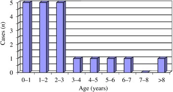
Patients' ages ranged from six months to 11 years (Figure 1), with a mean age of 3.3 years.
Age distribution of paediatric patients with suspected aspirated foreign body.

There were seven (35 per cent) boys and 13 (65 per cent) girls, giving a male:female ratio of 1:1.8.
Computed tomography virtual bronchoscopic analysis of airway foreign body
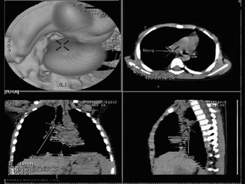
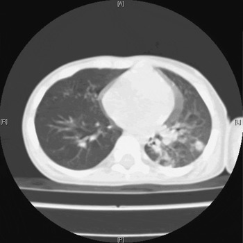
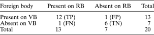
In 12 cases, foreign bodies detected by conventional bronchoscopy were also revealed on virtual bronchoscopy. Figure 2 depicts a foreign body in the right main bronchus of one such case. In a separate case, a foreign body detected by virtual bronchoscopy was found, on rigid bronchoscopy, to be a mucous plug. In another case, virtual bronchoscopy missed a foreign body in the bronchus. In six cases, no foreign body was detected either by virtual bronchoscopy or rigid bronchoscopy (Table I). A chest CT revealed hyperaeration of the ipsilateral lung in nine cases, atelectasis in five, compensatory emphysema in four, lobar collapse in one (Figure 3) and consolidation in two. One case had no abnormal findings. A 2 × 2 contingency table was prepared to enable calculation of the various statistical parameters for virtual bronchoscopic analysis of airway foreign body (Table I). These were as follows: sensitivity, 92.3 per cent; specificity, 85.7 per cent; validity, 90 per cent; positive likelihood ratio, 6.45; negative likelihood ratio, 0.089; positive predictive value, 92.3 per cent; negative predictive value, 85.7 per cent; and relative risk, 6.46.
Virtual bronchoscopic images showing a foreign body in the right main bronchus. Navg = Navigation

Computed tomography scan showing left lower lobe collapse due to a foreign body in the lobar bronchus. A = anterior; P = posterior; R = right; L = left

2 × 2 contingency table for virtual bronchoscopic analysis of airway foreign body, compared with rigid bronchoscopy

Data represent patient numbers. RB = rigid bronchoscopy; VB = virtual bronchoscopy; TP = true positive; FP = false positive; FN = false negative; TN = true negative
Discussion
Foreign body aspiration is a very serious and life-endangering emergency which is quite frequently encountered in small children. Its sequelae range from choking and fatal asphyxiation to (in the case of a forgotten episode) symptoms mimicking chronic and recurrent chest infection, with a spectrum of severity in between.
Virtual bronchoscopy is a term used to describe a variety of software-based, three-dimensional visualisation formats created from noninvasive medical imaging methods such as CT and magnetic resonance imaging, with the goal of creating views similar to a minimally invasive bronchoscopy procedure.Reference Bauer and Steiner3 Rigid bronchoscopy is an invasive procedure and has its own risks. Chest radiography is the preferred initial investigation in cases of tracheobronchial foreign body aspiration. Unfortunately, almost 90 per cent of foreign bodies are radiolucent, and the chest radiograph is normal in up to 30 per cent of children with aspirated foreign bodies.Reference Svedstrom, Puhakka and Kero4 Compared with rigid bronchoscopy, virtual bronchoscopy has some advantages: it is noninvasive, and can visualise areas inaccessible to the rigid bronchoscope.
In developing countries, many poorer patients are unaware of the relevant symptoms and do not give a proper history of foreign body aspiration. Hence, many cases present late with symptoms of chest infection. These patients are very often treated medically in paediatric wards for unresolving chest infection, for many days. It is only then that suspicion of foreign body aspiration arises, and a request for bronchoscopy is made. In many of these cases, the chest radiography is not pathognomonic of foreign body aspiration. We have found virtual bronchoscopy to be particularly useful in such cases, to confirm the presence of a foreign body. The risk of rigid bronchoscopy in these cases is higher, as the chest is already compromised due to infection. Thus, performing rigid bronchoscopy on every case of suspected foreign body aspiration can be avoided. Virtual bronchoscopy is also useful to determine the exact size, shape and type of foreign body. Hence, virtual bronchoscopy serves as a guide to the bronchoscopist and reduces the time taken for the procedure, as well as its morbidity and mortality. Yet another advantage of CT virtual bronchoscopy is that it enables simultaneous visualisation of inner and outer structures of the tracheobronchial tree, thus showing any pathology in the surrounding region. Virtual bronchoscopy is also capable of going beyond stenoses, whereas rigid or flexible bronchoscopy does not have this advantage.Reference Sodhi, Saxena, Singh, Rao and Khandelwal5 Multidetector CT technology has enabled the visualisation of segmental and subsegmental bronchi, thereby visualising possible foreign bodies which the rigid bronchoscope may not be able to reach.
One of the main drawbacks of virtual bronchoscopy is that it cannot be used as a therapeutic tool. Also, virtual bronchoscopy may sometimes be unable to distinguish soft tissue or a mucous plug from a foreign body, especially if it is of vegetable origin. In one of our cases, a long-standing, minute, vegetative foreign body was missed by virtual bronchoscopy, probably due to inadequate slice thickness (i.e. partial volume reduction). This has previously been described by Kocaoglu et al. Reference Kocaoglu, Bulakbasi, Soylu, Demirbag, Tayfun and Somuncu6 Another disadvantage of virtual bronchoscopy, compared with rigid bronchoscopy, is that it cannot reveal the colour, morphology and vascularity of the mucosa.Reference Summers, Shaw and Shelhamer7
• This study aimed to evaluate computed tomography (CT) virtual bronchoscopy for the detection of paediatric tracheobronchial foreign bodies not evident on plain chest radiography, compared with rigid bronchoscopy
• Results for CT virtual bronchoscopy of children with suspected foreign body were compared with those for rigid bronchoscopy
• Computed tomography virtual bronchoscopy is particularly useful to detect occult tracheobronchial foreign bodies missed on chest radiography
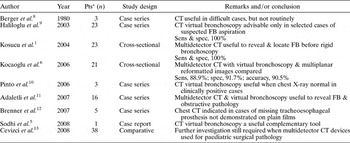
Various authors have investigated the utility of CT virtual bronchoscopy in detecting tracheobronchial foreign bodies, and a few have published statistical parameters (Table II).
Previous studies of CT virtual bronchoscopy for tracheobronchial foreign body aspiration

*Cases undergoing both rigid bronchoscopy and computed tomography (CT) virtual bronchoscopy. Pts = patients; FB = foreign body; sens = sensitivity; spec = specificity
Conclusion
In the presence of a positive clinical diagnosis and negative chest radiography, CT virtual bronchoscopy must be considered in all cases of tracheobronchial foreign body aspiration, in order to avoid needless rigid bronchoscopy. Computed tomography virtual bronchoscopy is particularly useful in screening cases of occult foreign body aspiration, as it has a high sensitivity, specificity and validity. Our study also suggests that, in cases of tracheobronchial foreign body aspiration, virtual bronchoscopy has an important, complementary diagnostic role prior to rigid bronchoscopy.