Introduction
Chronic otitis media (COM) accounts for 65–330 million individuals globally, 60 per cent of whom suffer from significant hearing impairment.1 COM accounts for 28 000 deaths and a disease burden of over 2 million.1 In India, pooled estimated prevalence of ear diseases is 11.66 per cent among children.Reference Bhatia, Chauhan, Kaur, Rana, Pradhan and Singh2
Meatoplasty is an integral part of canal wall down (CWD) tympanomastoidectomy in the surgical management of squamous chronic otitis media. The creation of a dry mastoid cavity primarily depends on surgical technique being the lowering of the facial ridge to the level of the facial nerve and development of a large external auditory meatus.Reference Kveton, ME III and Gulya3 To date, the consensus for reconstruction of the external opening (meatoplasty) for the newly created single mastoid cavity and external auditory canal is that it is to be ‘adequate or optimal’ to effectively maintain its functions. It has to be large enough to provide a dry, self-cleaning ear with adequate ventilation, unfavourable for microbial growth, debris accumulation and recurrent disease, as well as simultaneous in-office surveillance. At the same time, it should not be too large to cause symptoms, especially giddiness, if air directly contacts the medial wall of the cavity, or the opening appears to be unnatural and/or cosmetically unacceptable. Nevertheless, the challenge for the operating surgeon is the ‘on-table’ decision for a meatoplasty which would be ‘optimal or adequate on healing’.
A range of techniques and variations have been reported in literature since the original descriptions in 1893 by Stacke and Shwartze.Reference Sellars4, Reference Roulleau5 Korner’s meatoplasty has been described for both post-aural and endaural incisions.Reference Raut6 For the post-auricular approach the most popular meatoplasty is Portmann’s 3 flap techniqueReference Portmann7, though others have also been reportedReference Raut6, Reference Wormald and Van Hasselt8-Reference Patil, Ahmed and Patel14 In the past decade, newer techniques of large meatoplasty in endaural approach CWD mastoidectomy with modifications of transposition post-auricular flapReference Elsamanody, Topsakal, Grolman and Tange15, a posterior skin flap with removal of the conchal cartilageReference Bansal and Singh16 and mastoid obliteration with Palva’s flap following endaural incision, meatoplasty with skin unsutured and cavity packed for two weeks have also been suggested.Reference Memari, Delarestaghi, Mir, GolMohammadi and Koushki17 However, the latter article reported change in the shape of the ear among 16.1 per cent and external canal stenosis in 6.5 per cent with no mention of achieving dry ear.Reference Memari, Delarestaghi, Mir, GolMohammadi and Koushki17
Numerous continuously evolving techniques of meatoplasty in CWD tympanomastoidectomy suggest a lack of a technique that is standardised, surgeon-friendly and reproducible with consistently successful outcome, as well as applicable for effective ongoing specialist training programmes. Our randomised controlled trial was thus initiated.
Materials and method
Aim and objective
The primary objective of our study was to identify on-table ‘optimal’ meatal opening size that predicts post-operative mastoid cavity healing. The secondary objective was to develop a technique that can be standard as well as measurable, achievable, acceptable, replicable and time-bound (SMAART) as compared to the standard Portmann 3 flap meatoplasty.
Methodology
This study was carried out in the Department of Otorhinolaryngology in a tertiary academic hospital over eighteen months from January 2019 to June 2020, among patients diagnosed with a squamous COM undergoing CWD tympanomastoidectomy. It was a randomised double-blind controlled trial. The study was initiated following clearance from the Institutional Review Board and Ethics Committee (IEC No. ECR/400/Inst/PY/2013/RR-16). Informed written consent was obtained from the patients/guardians prior to enrolling in the study. The study was registered under National CTRI (Clinical Trial Registry – India) Number: CTRI/2019/12/022503. All consecutive adult patients with squamous chronic otitis media undergoing modified radical mastoidectomy with tympanoplasty (CWD tympanomastoidectomy) were included in the study. Those with serious medical comorbidities were excluded.
Parameters studied
• Volume of the tympanomastoid cavity
• Intra-operatively on-table
• Post-operatively after one and three months
• Size of meatoplasty opening
• Intra-operatively on-table
• Post-operatively after one and three months
• Time taken for creation of meatoplasty
• Complications associated with meatoplasty
• Healing Measures
• Dry cavity
• Absence of wax
• Absence of giddiness
• Cosmetically acceptable
Sample size
Numerous existing techniques suggested for meatoplasty reported their benefits. Considering that surgical outcome is satisfactory in the majority, the outcome of interest in our study was taken as time and cost in terms of efficiency. Therefore, our calculation of sample size is based on time. We considered the average time for creation of meatoplasty as 45 and 20 minutes for Portmann’s 3 flap technique and the new 2 flap technique respectively, the difference between means of the 2 groups as 10, including the standard deviations of the former and the latter as 15 and 20 based on previous studies. With a confidence interval of 95 per cent, power of 80 per cent and an alpha error of 5 per cent, the present study obtained a sample size of 35.
Participants were assessed clinically with a detailed history and ENT examination. These were noted in case report forms. All routine blood investigations, X-rays of the mastoid and para-nasal sinuses and pure tone audiometry (PTA) were done. After obtaining written informed consent, pre-anaesthetic clearance was obtained, and patients were randomised into two groups using www.randomizer.org, one being an accepted popular meatoplasty technique Portmann 3 flap and the other the new SMAART 2 flap technique. Following completion of CWD mastoidectomy and tympanoplasty, the patients were randomised to either of the two techniques mentioned below.
Portmann 3 flap meatoplasty
A lateral circumferential incision was made from the 1 to the 6 o’clock position, 10 mm lateral to the upper drum. The three flaps (lateral, superior and inferior) were created. An upper lateral radial incision was made from the upper part of the circumferential incision to the spine of the helix, and a similar lower incision was done from the inferior edge of the circumferential incision towards the concha. A finger was placed through the canal, exposing the lateral flap, which was thinned out. The visible edge of the conchal cartilage was excised, and the laterally based flap was turned around and fixed to the posterior aspect to form the lateral covering of the cavity. The ear canal skin was then divided at the 9 o’clock position down to the drum, creating the superior and inferior flaps. These flaps were then thinned out and sutured.Reference Tos18
SMAART 2 flap meatoplasty
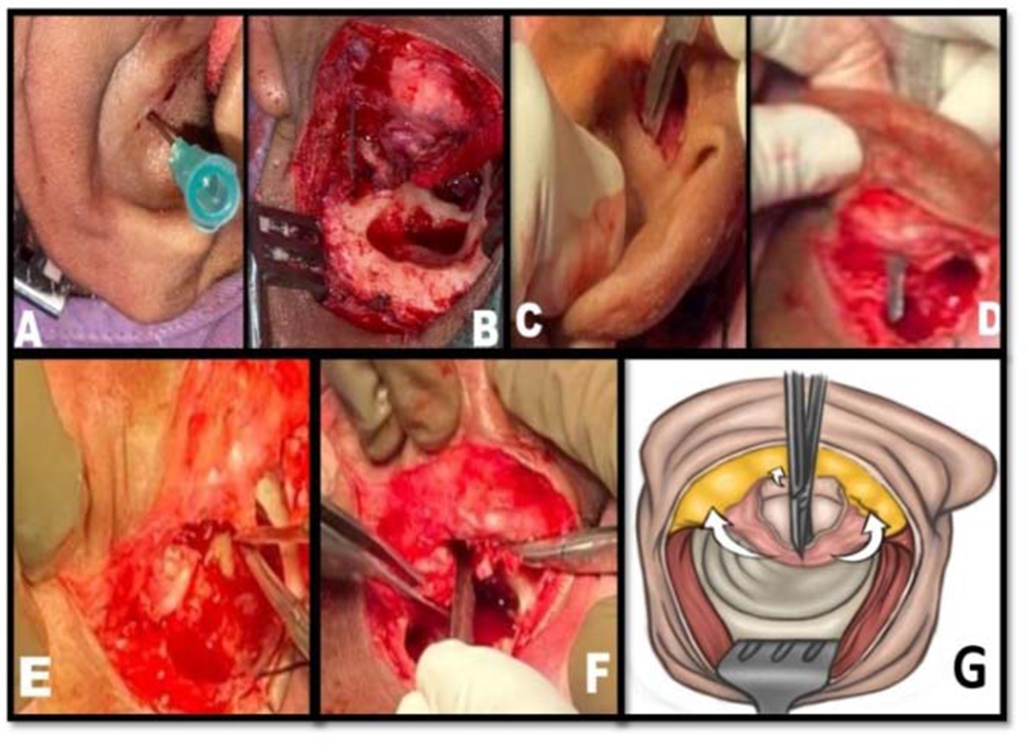
A lateral circumferential incision from the 12 to the 6 o’clock position was made on the outer elevated posterior canal wall skin, just within the medial margin of the conchal cartilage. From the medial end of the deep meatal incision made earlier, around the 9 o’clock position, a horizontal incision was extended laterally to bisect the flap created by the above lateral circumferential incision to the level of the root of the helix. The latter was marked by a needle (Figures 1A and B). A horizontal incision was made from the medial end of the raised flap to the level of the needle (Figures 1C and D). Thus two flaps, a superior and inferior were created (Figures 1E, F and G). The flaps were thinned out if there was excess soft tissue.
SMAART 2 flap meatoplasty technique. Figure 1A–F. Intraoperative pictures of the new SMAART 2 flap meatoplasty technique. Figure 1G. Schematic representation of Figures 1E and F

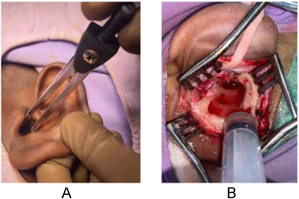
The graft was then placed over the mastoid and tympanic cavities, and the mastoid cavity volume was measured using saline instilled with a syringe (Figure 2A). The external meatal opening size was measured using callipers and a standard ruler (Figure 2B). All patients were on antibiotics post-operatively for 10 to 14 days, along with analgesics initially. The mastoid bandage was changed on the second and fourth post-operative days. The sutures were removed on the seventh post-operative day. Patients were reviewed two weeks after discharge and followed up for a period of three months and/or until the cavity became dry.
Intra-op diameter and volume of meatoplasty. Figure 2A. Intra-op diameter of meatoplasty. Figure 2B. Intra-op volume of meatoplasty.

The visible medial edge of the conchal cartilage was also trimmed, so that it was not exposed. The superior flap was then sutured superiorly, and the inferior one sutured inferiorly.
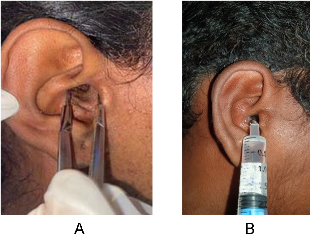
Measurements of the pre-operative external auditory canal volume and meatal opening size, as well as immediate post-op on-table, after one and three months, and mastoid cavity volume and meatal opening size were noted. The surface area of meatoplasty at each time was calculated using the formula A = πr2 (where A is the surface area and r is the radius). The size of the meatoplasty and volume of the cavity was also measured during each review (Figures 3A and B). The cavity and meatoplasty were assessed by a team not involved in the surgery.
Post-op diameter and volume of meatoplasty. Figure 3A. Post-op diameter of meatoplasty. Figure 3B. Post-op volume of meatoplasty using syringe post-operatively.

Results
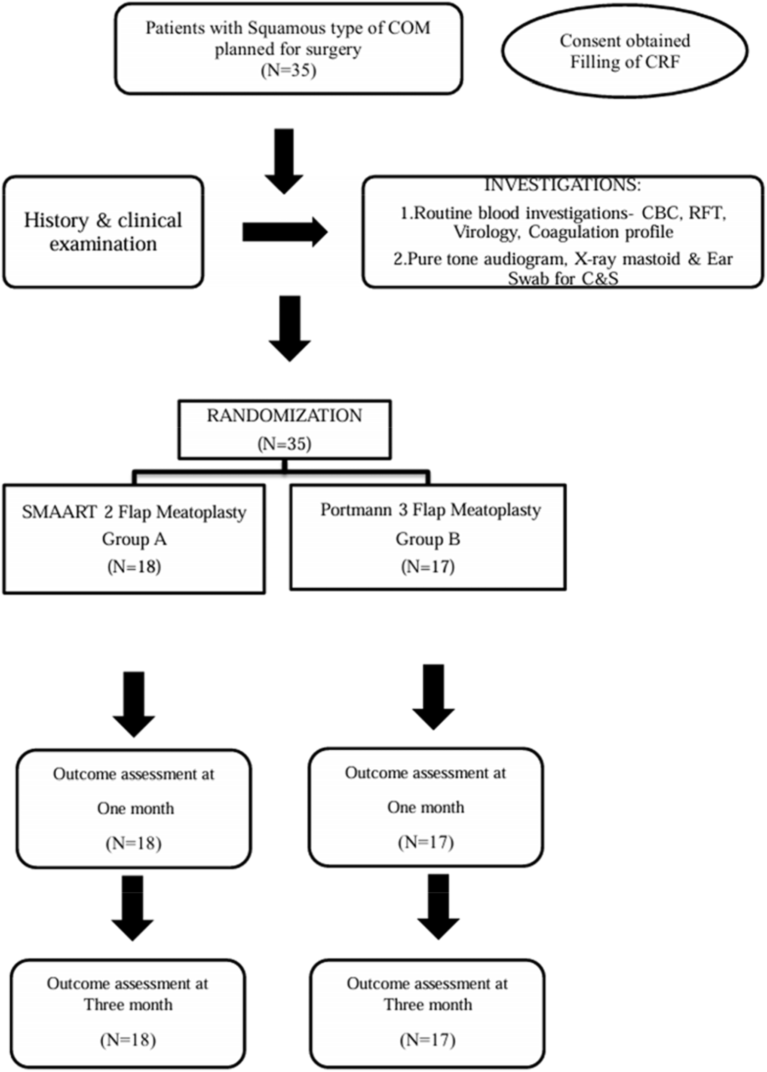
There were 35 patients in this study, 18 in the new SMAART 2 flap (Group A) and 17 in the Portmann 3 flap (Group B) meatoplasty. There were no drop-outs in the study. Among these 35 patients, 22 were males and 13 were females. The new 2 flap group had 12 male (66 per cent) and 6 female (34 per cent) patients, while the 3 flap group had 10 male (58 per cent) and 7 female (42 per cent) patients.
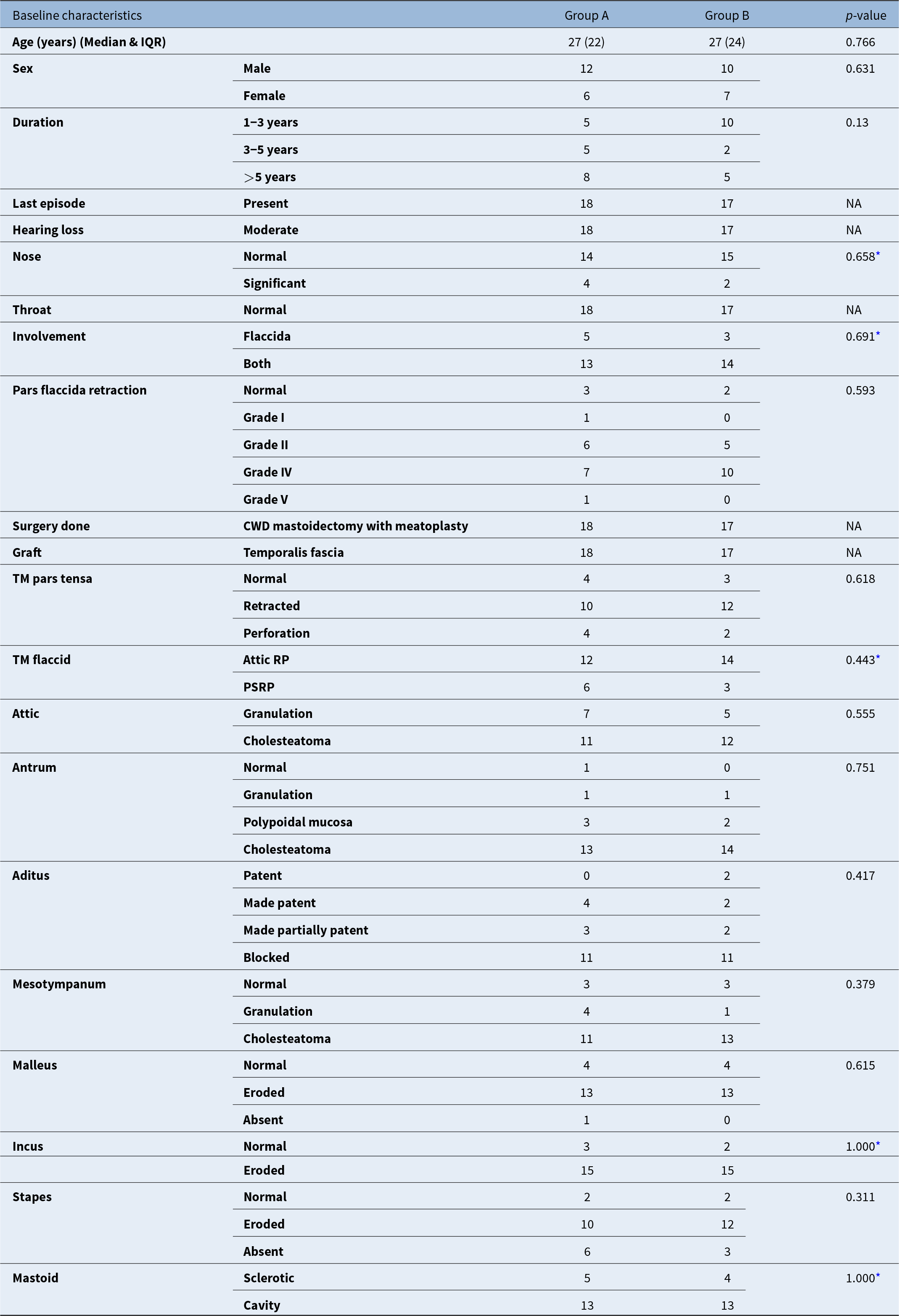
The baseline characteristics were similar in both groups (Table 1).
“Baseline characteristics of both groups; IQR = inter-quartile range; TM = tympanic membrane; RP = retraction pocket; PSRP = posterosuperior retraction pocket”

* Mann–Whitney U test
†chi-square test
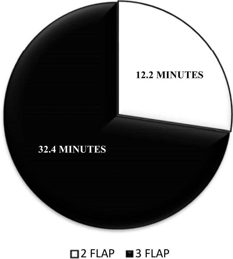
The mean time taken to complete meatoplasty in the 2 flap group was 12 minutes and 31 seconds, whereas it was 32 minutes and 5 seconds in the 3 flap group (Figure 5), which was significant (p-value < 0.001).
At the end of one month, ear discharge was present in 20 per cent and 6.25 per cent in 2 flap and 3 flap meatoplasty, respectively. However, at the end of three months, all had dry ears in both groups (Table 2).
Ear discharge of post-op mastoid cavity at one and three months

With regard to otorrhoea, the mean volume and mean meatoplasty size were compared immediately post-operatively on-table, at one month and at three months among patients with ear discharge present and absent post-operation. It was noted that the size of the mastoid cavity was twice that of meatoplasty among those who did not have discharge, unlike those with discharge who had more than twice the cavity size (Table 3).
Comparison of mean volume and mean meatoplasty size on-table and at one month for patients correlating with discharge

* Size of cavity more than twice that of meatoplasty in those with persistent discharge
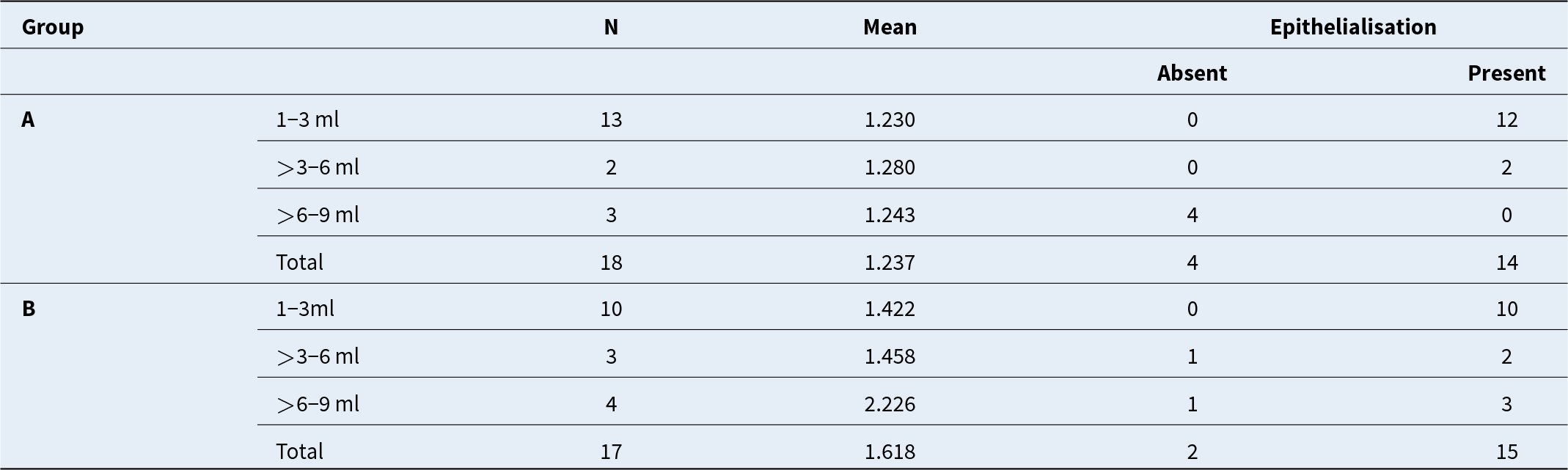
By one month, epithelialisation was complete among 78 per cent of patients and 41 per cent of patients with cavity size of 6 ml or less in 2 flap and 3 flap meatoplasty, respectively (Table 4).
Epithelialisation at one month with respect to volume

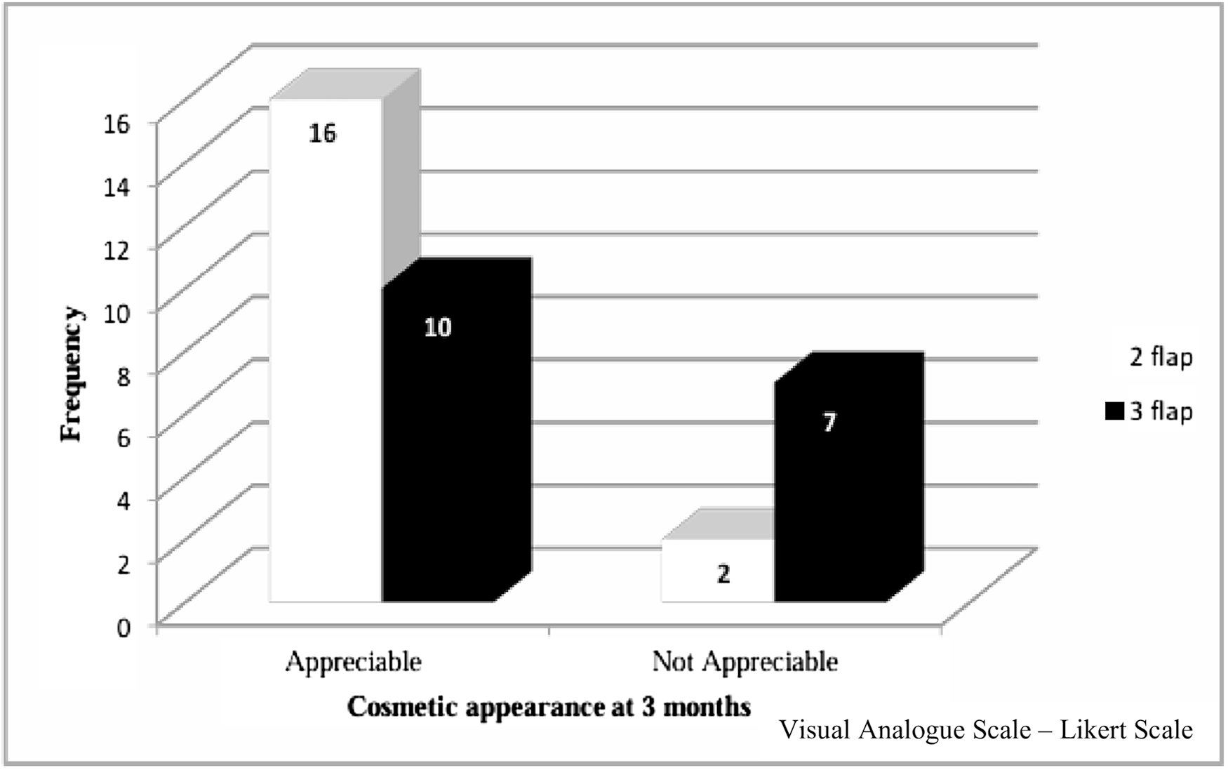
At three-month follow-up when assessed using a Likert scale, 16 patients from the 2 flap group and 10 patients from the 3 flap group had cosmetically acceptable outcomes. This was statistically significant (p-value 0.048, Figure 6).
Discussion
Meatoplasty is a crucial but often overlooked component of CWD mastoidectomy. In a landmark article, Sade et al. described that four factors such as an open Eustachian tube, a large cavity, a high facial ridge and a small meatoplasty were additive. If all were present, the cavity had a 100 per cent chance of being wet.Reference Sadé, Weinberg, Berco, Brown and Halevy19 Meatoplasty should be performed so as to achieve an adequate size that is needed to provide surface volume ratio for aeration, epithelial stability and good post-operative visualisation of the mastoid cavity.
The reported rates of post-operative otorrhoea in conventional meatoplasty range from 4 to 28 per cent.Reference Kveton, ME III and Gulya3 Studies in the recent past have addressed various types of meatoplasty and mastoid cavity size, obliteration or avoiding exposed bone, including epithelialisation issues in CWD tympanomastoidectomy. Good success rates have been reported in a comparative study, following either inferiorly based or division in middle-flap technique meatoplasty without incision or excision of a piece of cartilage from the pinna. Persistent granulations, discharge or stenosis of less than eight per cent were noted in both groups.Reference Vadiya, Makwana, Mehta and Khetani20 Post-operative otorrhoea was reported from a period of two to six months post-operatively in 14.3 per cent of patients in a prospective study, where one-cut meatoplasty technique was used.Reference Hovis, Carlson, Sweeney and Haynes21 Using a posterior fixation meatoplasty flap technique, a large external meatus and healed cavity with a mean follow-up of twenty-six months was reported in all ears in a retrospective chart review.Reference Choi, Song, Jang and Chang22 Concurrent mastoid obliteration and meatoplasty, using an anteriorly based musculo-periosteal flap and a horizontal skin incision on the concha, reported in a retrospective study that 86 per cent and 78 per cent of patients with cholesteatoma and adhesive otitis media achieved a dry and self-cleaning mastoid, respectively. These authors also noted complete control of infection duration ranging from four to twenty-four weeks, with mean complete epithelialisation time being 11.1 ± 4.6 weeks.Reference Kim, Oh, Choi, Park and Park23
Placing a large graft on the mastoid cavity has also facilitated rapid epithelialisation and healing in another relatively recent prospective study.Reference Tansuker, Ş, Özyılmaz, Cengiz, ŞE, Oktay and Korkut24
Previously, two prospective studies of CWD tympanomastoidectomy for squamous otitis media have reported on the post-operative size of the mastoid cavity and the diameter of the reconstructed external meatal opening.Reference Wormald and Van Hasselt8, Reference Lakra25 The first study reported a 90 per cent success rate for a dry cavity and graft-take when the mean volume of the cavity was 2.6 ml, and the post-operative on-table mean diameter of the meatoplasty was 1.01 cm.Reference Wormald and Van Hasselt8 The latter study, a comparative one between different techniques, reported the mean volume of the mastoid cavity created on-table was 4.37 ml with a marginal difference in Korner’s, Siebenman’s, Mawson’s and Bhatia et al.’s techniques at the end of three months. The overall mean reduction in the volume of the mastoid cavity was found to be less in Siebenman’s and Mawson’s techniques (0.68 ml and 0.67 ml) in contrast to Korner’s and Bhatia et al.’s, which was more (0.89 ml and 1.0 ml). They achieved a mean meatoplasty size of 1.59 cm in terms of diameter, which for Korner’s technique was 1.6 cm; Siebenman’s technique 1.57 cm; Mawson’s technique 1.57 cm; and for Bhatia et al.’s technique, it was found to be 1.64 cm.Reference Lakra25
Unlike previously reported studies, ours was a randomised double-blind control trial of two meatoplasty techniques: the standard Portmann 3 flap with a new SMAART 2 flap. Our study had also analysed the outcome of healing in relation to the volume of the exteriorised mastoid cavity to the size of the post-op meatoplasty external canal opening, measurements being noted immediately on-table, as well as post-op at one and three months. The mean post-operative on-table overall mastoid cavity volume was found to be 3.4 ml (new 2 flap and Portmann 3 flap groups being 2.8 ml and 3.9 ml, respectively). However, the mean reduction in the volume of the mastoid cavity at the end of three months was only 0.44 ml (new 2 flap 0.36 ml versus Portmann 3 flap 0.52 ml). The mean on-table post-operative meatoplasty size of 1.65 cm by diameter was noted (new 2 flap group 1.42 cm and Portmann 3 flap group 1.79 cm).
CONSORT.

It was noted that at post-operative one month, cavities with a mean volume of 2.852 ml and mean meatoplasty size of 1.440 cm2 did not have discharge, whereas cavities with a mean volume of 4.077 ml and a mean size of 1.517 cm2 had discharge. All cavities in both groups were dry by the end of three months (Table 2). It appears that if the ratio of ‘cavity volume to meatoplasty size’ of 2:1 is maintained immediately on-table, the post-operative cavities are less likely to develop otorrhoea, and healing is ensured. This has not been previously reported.
Considering epithelialisation, the authors in an earlier report had noted that the mean time for control of infection and complete epithelialisation following mastoid obliteration with meatoplasty was 11.1 ± 4.6 weeks.Reference Kim, Oh, Choi, Park and Park23 Our present study revealed that both groups had well-epithelialised cavities by the end of three months. Early epithelialisation at post-operative one month was noted more among those with the new 2 flap than Portmann 3 flap technique (77.7 per cent versus 58.5 per cent). We further assessed epithelialisation, considering the volume of the mastoid cavity in three categories of 1–3 ml, greater than 3–6 ml and greater than 6 ml with both techniques. Those with a volume greater than 6 ml did not have epithelialisation in the new 2 flap group at the end of one month, unlike those in the Portmann 3 flap meatoplasty technique where cavity epithelialisation was present even in cavities more than 6ml (Table 4). This, too, has not been previously reported.
Unlike other studies, the time taken for creation of meatoplasty was also noted in our study. The new 2 flap technique took an average of 12.2 minutes, whereas the Portmann 3 flap was completed in 32.4 minutes (Figure 5). This was statistically significant (p-value < 0.001) and has not been reported earlier.
Time taken for meatoplasty.

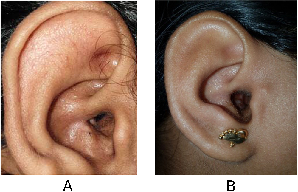
Regarding cosmetic acceptance, an observational case-series study reported that 16 per cent of patients had a change in the shape of their ears.Reference Memari, Delarestaghi, Mir, GolMohammadi and Koushki17 Though a large external meatus with a healed mastoid cavity has also been reported by another earlier report, its cosmetic acceptance by patients had not been assessed.Reference Choi, Song, Jang and Chang22 In our study, 25 per cent of patients noted that the external meatal opening was too large and were not happy with the cosmetic outcome of their ears. More than two-thirds of them were in the Portmann 3 flap meatoplasty group (Figure 6). This was found to be statistically significant post-operatively both at one month (p-value = 0.019) and at three months (p-value = 0.048). This has also not been reported previously.
Cosmetic appearance at three months.

Meatoplasty in patient who underwent new SMAART 2 flap technique (7A) and standard Portmann 3 flap meatoplasty technique (7B). Figure 7A. New 2 flap SMAART Figure. 7B. Standard Portmann 3 flap meatoplasty.

Conclusion and clinical significance
In our double-blind randomised control trial of 35 participants assigned to new SMAART 2 flap and standard Portmann 3 flap meatoplasty technique, it was noted that to achieve a dry ear on healing, the ratio of immediate on-table, as well as post-operative one- and three-month volume of the tympanomastoid cavity to the size of the meatal opening was 2:1. At post-operative one month, among cavity size of 6 ml or less, epithelialisation was complete in 78 per cent and 41 per cent with 2 flap and 3 flap meatoplasty, respectively. In order to attain a healed mastoid cavity in CWD tympanomastoidectomy, in addition to the already widely accepted principles for a well-saucerised and exteriorised tympanomastoid bowl to the external auditory canal, its volume should be proportional to the size of meatoplasty to a ratio of 2:1. The meatoplasty is then termed ‘optimal’. The SMAART 2 flap meatoplasty was significantly less time-consuming, with better cosmetic acceptance as compared to Portmann 3 flap meatoplasty. It is the choice of meatoplasty when the volume of the post-operative tympanomastoid cavity is or can be obliterated to less than 6 ml.
Financial support
This research received no specific gran from any fundraising agency, commercial or not-for-profit sectors.
Competing interests
The authors declare none.
Ethical statement and informed consent
The study was conducted in accordance with the Declaration of Helsinki and was approved by the Institutional Review Board and Ethics Committee of the Pondicherry Institute of Medical Sciences. Informed consent was obtained from patients/guardians prior to enrolment.