Arginine–vasopressin is an endogenous peptide hormone that is increasingly used for treating hypotension in paediatric cardiac patients.Reference Rosenzweig, Starc and Chen 1 – Reference Mastropietro, Davalos, Seshadri, Walters and Delius 4 Recent reports have shown that some children have absolute or relative vasopressin deficiency after cardiopulmonary bypass and, if hypotensive, exhibit a favourable haemodynamic response to low-dose, exogenous vasopressin.Reference Mastropietro, Rossi and Clark 5 Most children after cardiopulmonary bypass, however, have markedly elevated vasopressin levels, and administration of exogenous vasopressin may be ineffective or lead to excessive vasoconstriction with potentially deleterious effects in cardiac output and organ perfusion.Reference Morrison, Simone, Conway, Tumulty, Johnson and Cardarelli 6 , Reference Indrambarya, Boyd, Wang, McConechy and Walley 7 Furthermore, there are concerns that increased afterload may be detrimental in the presence of impaired ventricular systolic function. Despite these theoretical physiological concerns, routine use of arginine–vasopressin in postoperative paediatric cardiac patients has been recommended.Reference Mastropietro 8 There are, however, limited data on outcomes in relation to ventricular function and incomplete description of clinical predictors of response to arginine–vasopressin therapy.Reference Mastropietro, Davalos, Seshadri, Walters and Delius 4 Of note, reported mortality in paediatric cardiac patients treated with vasopressin is very high, ranging from 18 to 24%.Reference Rosenzweig, Starc and Chen 1 – Reference Mastropietro, Davalos, Seshadri, Walters and Delius 4
The objectives of this study were to determine the clinical and haemodynamic effect of arginine–vasopressin therapy in relation to ventricular dysfunction and explore clinical and echocardiographic associations. We hypothesised that paediatric cardiac patients with ventricular dysfunction following cardiac surgery have impaired haemodynamic response to arginine–vasopressin therapy compared with patients with normal ventricular function.
Materials and methods
Patients
This study was approved by the Institutional Review Board of Cincinnati Children’s Hospital Medical Center. The requirement for parental consent was waived because of the retrospective nature of the study. We retrospectively reviewed the medical records of all patients who met the following inclusion criteria:
-
∙ age ⩽12 months at the time of cardiac surgery (neonates/infants);
-
∙ underwent cardiopulmonary bypass for corrective or palliative cardiac surgery between January 2010 and January 2014;
-
∙ treated with intravenous arginine–vasopressin within 48 hours of completion of cardiopulmonary bypass;
-
∙ had an echocardiogram, either transthoracic or transoesophageal, assessing ventricular function within 12 hours before initiation of vasopressin therapy.
We excluded patients who met the following exclusion criteria:
-
∙ received arginine–vasopressin within 24 hours before cardiopulmonary bypass;
-
∙ had incomplete relevant data on primary end points – that is, blood pressure and catecholamine score.
We identified study subjects by reviewing electronic medical and pharmacy records, as well as surgical and echocardiographic databases. Following initial electronic screening and cross-check through different databases, research personnel confirmed eligibility of each subject by reviewing the medical records of all potential study subjects. We used standardised data collection templates such as case report forms, and trained research personnel extracted all data. Data collection was initially piloted and reviewed by the principal investigator to ensure accuracy.
We evaluated the response to arginine–vasopressin therapy over the first 12 hours after initiation. The primary end points were systolic blood pressure 1 hour after titration of arginine–vasopressin to the “target” dose and catecholamine score for the entire period following titration to the “target” dose. The “target” arginine–vasopressin dose was defined as the dose after upward titration of arginine–vasopressin was completed; in other words, it was followed by the same or lower dose for all subsequent hourly measurements. Catecholamine score was calculated by adding 100 points for every microgram per kilogram per minute (mcg/kg/minute) of epinephrine or norepinephrine on the basis of previous reports in the literature.Reference Gaies, Gurney and Yen 9
Patients with increased systolic blood pressure by 10% without concomitant increase in the catecholamine score or with maintained systolic blood pressure with decreased catecholamine score were considered “responders”. Patients with increased systolic blood pressure by <10% and/or with an increased catecholamine score were deemed “non-responders”. For the primary end points, we assessed response 1 hour after the “target” arginine–vasopressin dose was achieved. In addition, we also evaluated the response linearly over the entire study period of 12 hours.
Demographic, perioperative, including surgical complexity and operations performed, and postoperative variables were recorded for all patients. We recorded haemodynamic data including blood pressure, heart rate, catecholamine score, lactate levels, urine output, near-infrared spectroscopy values, and central venous oxygen saturation, if available, at baseline and hourly for 12 hours. We also recorded fluid balance, plasma sodium levels, use of inhaled nitric oxide or postoperative steroids, cortisol levels, if available, and complications such as arrhythmias, temporary pacing, necrotising enterocolitis, use of mechanical circulatory support, delayed sternal closure, and infections. We recorded the following outcome data: duration of mechanical ventilation, length of ICU stay, and in-hospital mortality.
Operative and postoperative management
Our study group included patients who underwent a variety of cardiac operations performed by four different surgeons, and therefore surgical management was diverse. Anaesthesia and perfusion management, however, were consistent for similar cases. All patients received 30 mg/kg methylprednisolone when cardiopulmonary bypass was initiated. Cardiopulmonary bypass was performed using a Jostra-HL20 Heart-Lung machine system with a roller arterial pump (Maquet Cardiopulmonary, Solna, Sweden) and a 3-T heater–cooler unit (Sorin Group GmBH, Munchen, Germany). The target flow rate during CBP was 2.4–3.0 L/minute/m2 at normothermia, and acid–base status was managed with alpha-stat strategy. Dilution ultrafiltration was performed during bypass. Target haematocrit during cardiopulmonary bypass was 28%. According to institutional protocol during the study period, modified ultrafiltration was not used. Instead, haemo-concentrated pump blood was returned to each patient after separation from cardiopulmonary bypass according to blood loss and haemodynamic needs with a target haematocrit of 30–40%. All neonates and many infants in our study had a planned peritoneal drain placed in the operative room as part of standard care at our institution. Almost all patients had a transoesophageal echocardiogram in the operating room after completion of cardiopulmonary bypass.
Postoperative haemodynamic management in our cardiac ICU for the first 48 postoperative hours is uniform and directed towards achieving certain haemodynamic goals: target blood pressure (mean 45–55 mmHg for neonates, >55 mmHg for infants) with concomitant improvement of end-organ perfusion (arteriovenous oxygen saturations difference <30%, urine output >1 ml/kg/hour, decreasing blood lactate). We generally aim for central venous pressure of 8–12 cm H2O and a heart rate below 170 bpm. Near-infrared spectroscopy monitoring is used routinely at two sites (cerebral and somatic), although near-infrared spectroscopy values are not yet part of a standardised management protocol. Trends are, however, noted and acted upon at the discretion of the attending clinical team, although specific goals are not set. We aim to avoid excessive fluid administration and usually prefer vasoactive agent titration to achieve haemodynamic goals. Almost all of our patients return from the operating room on milrinone, but very few receive either a load or a dose higher than 0.5 mcg/kg/minute. Milrinone is not usually discontinued before initiation of vasopressin. Dopamine is not used. Norepinephrine and phenylephrine are used only as “rescue” vasoconstrictors if arginine–vasopressin is deemed to have failed. Arginine–vasopressin is usually initiated at a dose 0.3–0.5 milli-units per kilogram per minute (mU/kg/minute) and titrated (up to 2 mU/kg/minute) to achieve haemodynamic targets. Weaning is initiated once several hours of haemodynamic stability has been achieved, and epinephrine is decreased to low dose (usually ⩽0.05 mcg/kg/minute).
All patients are fluid restricted to 50 and 75% maintenance for the first and second postoperative days, respectively. All patients receive either diuretics or peritoneal dialysis during the first postoperative night if there is haemodynamic stability and if urine output is <1 ml/kg/hour for 4 hours. Peritoneal drains, if not used for dialysis, are placed for gravity drainage. Some of our study patients received furosemide or peritoneal dialysis as part of a randomised control trial taking place concurrently. For patients not enrolled in that study, we had a low threshold to use peritoneal dialysis if the patient had anasarca or if there was difficulty in achieving the target fluid balance.
We tend to avoid escalation to high doses of catecholamine (>0.05 mcg/kg/minute) and we have a low threshold to adding arginine–vasopressin to achieve haemodynamic stabilisation, especially in cases of apparent clinical vasodilation. Postoperative strategy and haemodynamic goals are discussed in detail and agreed upon between ICU, cardiac surgery, and anaesthesiology teams on arrival to the ICU from the operating room in a standardised “sterile cockpit” handoff, which takes places on all occasions in the presence of attending physicians from all involved teams. Overnight adjustments according to the evolving clinical situation are led by an in-house cardiac ICU attending.
Data analysis
Measures of central tendency, variability, and association were calculated for all variables in the study. Frequencies, means (standard deviations), as well as medians (interquartile ranges) were used to describe the distribution of data. The bivariate associations between the demographic or clinical variables and the outcome variables were tested using either the χ2 test or the Student t-test to assess statistical significance. Mixed modelling was used to evaluate trends for end points measured serially.
We performed two related types of analyses. The first analysis assessed baseline differences among responders and non-responders, addressing the question of whether response or non-response could be evaluated sooner, potentially changing intervention in the future. The second analysis evaluated the clinical course of those responding or not responding to vasopressin therapy, adjusting for baseline differences that would provide greater insight into the risks versus benefits of therapy. An alpha level of <0.05 was used to determine significance, and all data were analysed using SAS™ 9.3 (SAS Institute, Cary, North Carolina, United States of America).
Results
During the 48-month study period (January 2010–January 2014), 609 neonates and infants underwent cardiac surgery with cardiopulmonary bypass at our institution. A total of 118 patients received arginine–vasopressin (Vasostrict, Par Pharmaceutical Inc., Spring Valley, New York, United States of America) within 48 hours of surgery, and 48 of them had an echocardiogram within the 12 hours before initiation of therapy. Among all, twelve patients were excluded for receiving arginine–vasopressin on the day of surgery before cardiopulmonary bypass or for missing data on primary end points. The remaining 36 patients were included in the final analysis.
There were 17 males (47%) and 19 females (53%) in the study group. The median age at the time of surgery was 10.4 weeks (interquartile range=1.1–26.7). There were 10 (28%) patients with risk adjustment for congenital heart surgery – one with category 2, 10 (28%) with category 3, 12 (33%) with category 4, and four (11%) with category 6. Patient diagnoses were as follows: eight (22%) single-ventricle anatomy, five (14%) aortic arch abnormalities, four (11%) atrioventricular septal defect, three (8%) double-outlet right ventricle, and three (8%) tetralogy of Fallot. A total of 13 patients (36%) had “other” primary cardiac diagnoses. Most frequently performed operations were aortic arch reconstruction in five (14%) patients and atrioventricular septal defect repair and Norwood palliation with four patients (11%).
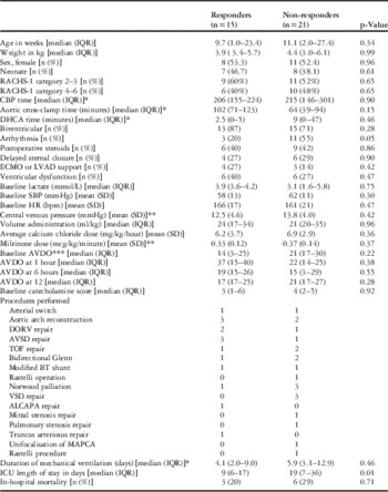
According to our blood pressure-based definitions, 15 patients (42%) responded favourably to arginine–vasopressin and 21 (58%) did not. There were no statistical differences between responders and non-responders with respect to gender, age at the time of surgery, preoperative or postoperative steroid use, surgical complexity, or cardiopulmonary bypass or aortic cross-clamp times.
Baseline haemodynamic measurements, before initiation of arginine–vasopressin, did not differ significantly between the two groups, specifically blood pressure, central venous pressure, heart rate, blood lactate, and near-infrared spectroscopy. Furthermore, there was no difference in central venous pressure values, volume, and calcium administration among “responders” versus “non-responders” during the study period. Arteriovenous difference of oxygen – defined as the difference between the systemic oxygen saturation as measured by a pulse oximeter and the average of cerebral and somatic infrared spectroscopy value – remained statistically indifferent in both groups from baseline throughout the entire study period of 12 hours. The mean time to titration to “target” arginine–vasopressin was 1.9 hours (standard deviation=3.1) for the “responders” and 3.1 hours (standard deviation=4.2) for the “non-responders”, with a mean “target” dose of 1.07 mU/kg/minute (standard deviation=0.71) and 1.01 mU/kg/minute (standard deviation=0.71), respectively. Table 1 shows a summary of patient characteristics, as well as comparisons between vasopressin responders and non-responders.
Patient characteristics and comparison based on vasopressin response.

RACHS-1=risk adjustment for congenital heart surgery; CBP=cardiopulmonary bypass; DHCA=deep hypothermic circulatory arrest; ECMO=extracorporeal membrane oxygenation; LVAD=left ventricular assist device; DORV=double-outlet right ventricle; AVSD=atrioventricular septal defect; TOF=tetralogy of Fallot; BT=Blalock–Taussig; VSD=ventricular septal defect; ALCAPA=anomalous left coronary artery from the pulmonary artery; MAPCA=major aortopulmonary collaterals
* CBP, aortic cross-clamp, DHCA time are in minutes, duration of mechanical ventilation is in days
** Milrinone and CVP values at 1 hour after titration of AVP to the “target” dose
*** AVDO (arteriovenous difference of oxygen)=systemic oxygen saturation (pulse oximetry)−average cerebral and somatic near-infrared spectroscopy value (NIRS)
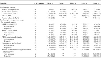
The average blood pressure for the entire cohort of patients increased and the catecholamine score decreased over the study period of 12 hours. Keeping the same definition of response but assessing the response not at 1 hour after the “target” dose but linearly over time by comparing every hour of treatment with the baseline before initiation of arginine–vasopressin, we found that only 46% of patients responded to therapy at 1 hour, 54% at 6 hours, and 42% at 12 hours. A summary of clinical response for the entire cohort as well as a comparison between the two groups at five time points are given in Table 2.
Measurements of clinical outcomes at five time points for the entire cohort.

NIRS=near-infrared spectroscopy
* Systolic blood pressure in mmHg
** Data were not available at this specific time point
*** P/F: ratio of arterial oxygen partial pressure to fractional inspired oxygen
**** NIRS and arterial-central venous oxygen saturation difference values are in %
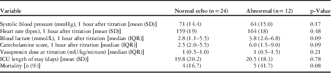
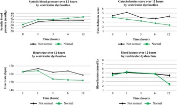
Of 36 patients, 12 (33%) had abnormal baseline ventricular function by echocardiography. Blood pressure response did not correlate with echocardiographic assessment of ventricular function. Comparing the groups with normal and abnormal ventricular function, there was no statistically significant difference in systolic blood pressure, catecholamine score, heart rate, blood lactate levels, or mortality. Interestingly enough, patients with abnormal ventricular function received higher baseline, average hourly, and cumulative doses of calcium chloride. The cumulative dose (mean/standard deviation) for the entire 12 hours for “normal versus abnormal” ventricular function was 58 (32) mg/kg versus 98.6 (33.5) mg/kg (p=0.002), and the average calcium dose was 5.2 (2.7) mg/kg/hour versus 8.6 (2.9) mg/kg/hour (p=0.002); however, the total dose of calcium after “target” vasopressin dose was similar [“normal versus abnormal”=32.7 (33.3) mg/kg versus 31.5 (24) mg/kg (p=0.3)] in both groups. Clinical response by ventricular function is summarised in Table 3.
Clinical assessments by ventricular function.

IQR=interquartile range
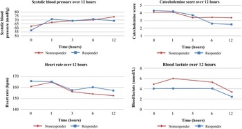
Figure 1 shows the blood pressure, heart rate, catecholamine score, and lactate response over time between the responders and the non-responders. Figure 2 shows the blood pressure, heart rate, catecholamine score, and lactate levels over time between patients with normal and abnormal ventricular function.
Comparison of “responders” with “non-responders” for blood pressure, catecholamine score, heart rate, and blood lactate levels over time. Data are shown as mean values. Blood pressure is measured in mmHg and blood lactate in mmol/L.

Comparison of patients with normal ventricular function with patients with ventricular dysfunction for blood pressure, catecholamine score, heart rate, and blood lactate levels over time. Data are shown as mean values. Blood pressure is measured in mmHg and blood lactate in mmol/L.

From the entire cohort, nine patients died before hospital discharge (in-hospital mortality 25%). There was no statistical difference in mortality between “responders” (20%, three patients) and “non-responders” (30%, six patients), nor between those with normal (17%, four patients) and abnormal (42%, five patients) ventricular function. “Responders”, however, had significantly shorter ICU length of stay (9 days versus 21.5 days, p=0.02) and less arrhythmias [three patients (20%) versus 11 patients (55%), p=0.05].
Discussion
This study is the first to investigate the relationship between clinical response to arginine–vasopressin therapy and ventricular function in neonates and infants after cardiac surgery. We found that only half of the patients responded to arginine–vasopressin with increasing blood pressure or decreasing catecholamine requirements, and moreover that this response was not associated with ventricular dysfunction. Furthermore, there was no change in response rate with duration of therapy, suggesting that alternative strategies should be used if an early response to therapy is not observed.
Only half of our patients showed a response to arginine–vasopressin therapy. Although somewhat disappointing, our data are in keeping with previous reports. In 2012, Mastropietro et al similarly reported a response of only 50% in a population of children after cardiac surgery.Reference Mastropietro, Davalos, Seshadri, Walters and Delius 4 These investigators used a similar definition of response, but did not assess response in relation to ventricular function. We found that some children with ventricular dysfunction still responded favourably, whereas others with preserved ventricular function did not. Our data do not allow us to explain this finding. The explanation is probably hidden in the multiple mechanisms of action of arginine–vasopressin. Our understanding of the entire spectrum of its pharmacological action is still evolving. We speculate, however, on the basis of previous findings that arginine–vasopressin can cause both vasodilation and vasoconstriction in different vascular beds.Reference Asfar and Radermacher 10 Several arginine–vasopressin receptors have been identified with different, and sometimes opposing, actions. Among them, vascular V1 receptors mediate systemic, and possibly coronary, vasoconstriction, whereas renal V2 receptors are located in the renal collecting tubule and are responsible for free water re-absorption and urine concentration.Reference Walker, Childs and Adams 11 , Reference Fenton, Brønd, Nielsen and Praetorius 12 Oxytocin receptors, which may also respond to arginine–vasopressin stimulation, are located in all cardiac chambers and in the pulmonary vasculature, with roles in atrial natriuretic peptide production and pulmonary vasodilation, respectively.Reference Holmes, Patel, Russell and Walley 13 Finally, purinergic receptors, present in the heart and coronary arteries, also bind arginine–vasopressin, leading to nitric oxide synthase production and vasodilation.Reference Diéguez, Martínez, Fernández, Climént, García-Villalón and Monge 14 The direct action of vasopressin on the myocardium and coronaries, however, remains unclear. Arginine–vasopressin may have a positive inotropic effect and may increase responsiveness of catecholamine receptors.Reference Noguera, Medina and Segarra 15 Improved cardiac function has been attributed to direct inotropy and to improved coronary perfusion either due to coronary vasodilation or due to improved perfusion pressure.Reference Lindner, Prengel and Pfenninger 16 On the contrary, other investigators have reported decreased cardiac function with arginine–vasopressin therapy, particularly at high doses.Reference Wilson, Brackett, Archer and Hinshaw 17 It has also been suggested that arginine–vasopressin may exhibit different effects in various physiological and disease states – hypoxia versus ischaemia-reperfusion versus sepsis – likely through alteration in different receptor expression.Reference Indrambarya, Boyd, Wang, McConechy and Walley 7 , Reference Asfar and Radermacher 10 , Reference Sellke and Quillen 18 It is likely that the action of arginine–vasopressin on the myocardium is dose-dependent with cardio-protective and positive inotropic effects at lower doses but coronary vasoconstriction and negative effects at very high doses.Reference Nazari, Sadr, Faghihi, Imani and Moghimian 19 , Reference Boyle and Segel 20 The effect on the myocardium also relates to the ratio of different myocardial receptors – V1 versus oxytocin versus purinergic – which can be altered in different disease states.Reference Indrambarya, Boyd, Wang, McConechy and Walley 7 In addition, it has been suggested that response to treatment might be related to pre-treatment levels. Patients with relative arginine–vasopressin deficiency might respond more favourably to exogenous administration compared with patients with elevated pre-treatment levels.Reference Mastropietro, Rossi and Clark 5
The retrospective nature of our study necessarily precludes mechanistic analysis of response, but our results clearly show that baseline ventricular dysfunction does not preclude favourable haemodynamic response to arginine–vasopressin therapy. Furthermore, our groups of “responders” and “non-responders” did not differ significantly in any of the factors tested. In particular, they had similar demographics, surgical complexity, cardiopulmonary bypass times, fluid resuscitation, markers of end-organ perfusion, and mortality. The average baseline blood pressure was similar in both groups, but this cannot assure us that both groups were similar in the nature of circulatory failure they experienced. Hypotension in children following cardiac surgery can be due to cardiogenic shock, vasodilatory shock with preserved cardiac function, or a mixture of both; however, ventricular dysfunction, baseline urine output, blood lactate, and near-infrared spectroscopy values did not differ significantly between the two groups. Although most clinicians agree that arginine–vasopressin therapy may be beneficial in cases of vasodilatory circulatory failure, concern has been expressed with regard to the use of arginine–vasopressin in patients with ventricular dysfunction.Reference Dünser and Hasibeder 21 This concern stems from early experimental studies that used very high arginine–vasopressin doses and reported negative inotropy and evidence for myocardial ischaemia.Reference Wilson, Brackett, Archer and Hinshaw 17 In addition, the first clinical report in a paediatric cardiac population by Rosenzweig et alReference Rosenzweig, Starc and Chen 1 contributed further to this concern. Their study described the rescue use of arginine–vasopressin in 11 children with catecholamine-resistant hypotension. Although this cohort showed uniform increase in blood pressure with arginine–vasopressin therapy, the two children with severe ventricular dysfunction constituted the entire mortality. This finding was not replicated in our study, as ventricular dysfunction was associated with neither blood pressure response nor mortality in our own cohort.
Our findings are consistent with recent experimental animal studies with low-dose vasopressin. In an experimental rat model, it was found that arginine–vasopressin increases cytosolic calcium and is associated with positive inotropy at low doses.Reference Xu and Gopalakrishnan 22 It should be noted that in the same experiment a dose-dependent effect was shown with suggestion of a negative inotropic effect at higher doses.Reference Xu and Gopalakrishnan 22 An alternative explanation for the negative effects on the heart at high-doses through marked afterload increase rather than direct negative inotropy has also been suggested.Reference Gassanov, Jankowski and Danalache 23 Similar to experimental evidence, clinical reports on the use of arginine–vasopressin in adults support the notion of its positive cardiac effect at low doses. In heart failure, arginine–vasopressin was found to increase systemic vascular resistance without decreasing cardiac index, suggesting a mechanism of positive inotropy.Reference Gold, Cullinane and Chen 24 Supportive of this notion is our finding that calcium administration decreased in patients with abnormal ventricular function after the arginine–vasopressin “target” dose was achieved. In 41 adults with postcardiotomy shock, arginine–vasopressin was devoid of adverse effects on the heart.Reference Dünser, Mayr and Stallinger 25 In particular, arginine–vasopressin increased systemic vascular resistance and left ventricular stroke work index without affecting cardiac or stroke volume indices, resulting in increased blood pressure. The relatively low mean “target” vasopressin dose of 1 mU/kg/minute in our cohort may explain the lack of untoward effects in cases of ventricular dysfunction. Although our findings are reassuring for the use of arginine–vasopressin in the setting of ventricular dysfunction, our study sample was small and likely underpowered to detect small differences.
Given that we did not establish statistically significant discriminators between “responders” and “non-responders”, among the factors that we tested, we questioned our definition of response and examined the blood pressure response by hour. We discovered that roughly half of our patients were “responders” at each point in time, and moreover some patients vacillated between the “responder” and “non-responder” groups. Although our study was not designed to address this, a plausible explanation for this observed response to arginine–vasopressin treatment can be searched in the mechanism of “homoeometric autoregulation” of the myocardium or the so-called Anrep effect, which constitutes a powerful physiological mechanism of adaptation to acute afterload changes.Reference Burton, Kaufman, Goot and da Cruz 26 , Reference Cingolani, Pérez, Cingolani and Ennis 27 It is conceivable that the Anrep effect is responsible for the tolerance of moderately increased afterload and lack of clinically deleterious effects in cases of ventricular dysfunction and cardiogenic shock demonstrated in our own and previous studies of arginine–vasopressin.Reference Lechner, Hofer, Mair, Moosbauer, Sames-Dolzer and Tulzer 2 , Reference Alten, Borasino, Toms, Law, Moellinger and Dabal 3 , Reference Villa-Abrille, Caldiz and Ennis 28
Reflecting the complexity of the mechanisms of action of arginine–vasopressin, the controversy continues beyond the cardiac effects and surrounds its effect on splanchnic perfusion.Reference Burton, Kaufman, Goot and da Cruz 26 Clinical studies have reported intestinal hypo-perfusion in septic patients with catecholamine-resistant shock.Reference Asfar, Radermacher and Hauser 29 Animal studies, however, have demonstrated improved splanchnic perfusion when low-dose arginine–vasopressin is used.Reference Van Haren, Rozendaal and van der Hoeven 30 In keeping with previous reports in the paediatric cardiac population, none of our patients experienced necrotising enterocolitis or other clinical sequela of splanchnic hypoperfusion.Reference Lechner, Hofer, Mair, Moosbauer, Sames-Dolzer and Tulzer 2
Urine output was maintained or increased after initiation of arginine–vasopressin in our cohort. Although arginine–vasopressin has antidiuretic action via V2 receptors at the renal collecting tubule, it also constricts afferent glomerular arterioles, resulting in increased filtration fraction and perhaps urinary output.Reference Fenton, Brønd, Nielsen and Praetorius 12 , Reference Pelletier, LaBossiere and Dicken 31 – Reference Edwards and Grantham 33
Despite the relatively small patient number, we were able to demonstrate some clinical responses that may be instructive. The “responder” group had shorter ICU stay, although there was no difference in baseline haemodynamics or surgical complexity. Furthermore, the “responders” had statistically lower incidence of arrhythmias. A speculative explanation of this finding relates to less catecholamine administration to patients who responded favourably to arginine–vasopressin. It has, also, been suggested that the use of vasopressin is associated with less arrhythmias in septic adults, but the very small number of patients with arrhythmias in our cohort did not allow for solid clinical conclusions to be drawn.Reference Patel, Chittock, Russell and Walley 34 Similarly, given the retrospective, non-controlled nature of our study, causation cannot be inferred between arginine–vasopressin use and improved recovery from surgery. Both these associations should be tested in prospective, randomised studies.
Our study examined arguably the most vulnerable group of postoperative paediatric cardiac patients, neonates, and infants with circulatory failure, treated with multiple inotropic and vasoactive agents. It is, therefore, of no surprise that both mortality and need for mechanical circulatory support were high in our cohort. Our high mortality indeed corresponds to previous reports.Reference Rosenzweig, Starc and Chen 1 – Reference Mastropietro, Davalos, Seshadri, Walters and Delius 4 It is relatively reassuring that arginine–vasopressin therapy in the group of patients with ventricular dysfunction did not worsen these outcomes; however, the size and heterogeneity of our group do not allow generalisation of our findings.
Our study has several limitations. First, we acknowledge the limitations of the predominant use of the intra-operative transoesophageal echocardiogram to assess ventricular function. Ideally, an immediate pre-therapy assessment would have been performed, but this was not available given the retrospective nature of our analysis. In the present study, however, we excluded patients in whom assessment of ventricular function had not been performed within 12 hours before initiation of therapy. In future prospective studies, assessment of ventricular function before and after arginine–vasopressin treatment and, more importantly, measurement of cardiac output and systemic vascular resistance should be addressed in a systematic, controlled manner. A further limitation is that this was a retrospective, single-centre study. Although the largest so far reported in this population, our sample size was insufficient to detect small differences. We recognise the need for a larger study, probably through multi-institutional collaboration. Finally, our study is limited by not measuring arginine–vasopressin levels before initiation of treatment. It has been suggested that response to treatment might be at least partly related to pre-initiation levels.Reference Mastropietro, Rossi and Clark 5 To address this knowledge gap, we are currently conducting a prospective study with measurement of arginine vasopressin levels.
In conclusion, we found that only half of infants with haemodynamic instability after cardiac surgery showed increased blood pressure in response to administration of arginine–vasopressin. Ventricular function does not predict this response. Prospective systematic comparison of arginine–vasopressin with other vasoactive agents and/or inotropes with meaningful clinical end points is warranted in this population.
Acknowledgements
The authors acknowledge the staff of Heart Institute Research Core who provided regulatory, data, and statistical support for this study.
Financial Support
This study received intramural funding from the Heart Institute Research Core at Cincinnati Children’s Hospital Medical Center.
Conflicts of Interest
None.
Ethical Standards
This study complies with the Helsinki Declaration and was approved by the local Institutional Review Board.