Introduction
Supracricoid laryngectomy with cricohyoidoepiglottopexy is an organ-preserving surgical procedure used to treat laryngeal cancer. It was first described by Majer and Rieder in 1959.Reference Majer and Rieder1 The procedure allows the patient to breathe through the natural airway, and also restores the vocal and swallowing functions.Reference Laccourreye, Laccourreye, Weinstein, Menard and Brasnu2–Reference Naudo, Laccourreye, Weinstein, Hans, Laccourreye and Brasnu4
Because of the increasing attention paid to cancer patients' quality of life, supracricoid laryngectomy with cricohyoidoepiglottopexy is gaining importance in laryngeal cancer treatment.Reference Nakayama, Seino, Hayashi, Miyamoto, Takeda and Masaki5, Reference Nakayama, Okamoto, Miyamoto, Takeda, Yokobori and Masaki6 However, in view of the fact that this procedure resects the vocal folds, it is unclear how the sound source and airway morphology are involved in phonation via the post-operative neoglottis. Since the vibration of the neoglottis is irregular in the laryngeal region following this procedure, thorough observation of the neoglottis via videostroboscopy (the usual technique for observing vocal fold vibration) is difficult. Based on such observation, the sound source has been assumed to be derived from interaction between the remaining arytenoid and the epiglottis.Reference Weinstein, Laccourreye, Ruiz, Dooley, Chalian and Mirza7
However, more recently a new, ultra-high-speed digital imaging system has been developed to analyse neoglottal vibration.Reference Granqvist and Lindestad8 This imaging modality has indicated that the sound source may be mucosal vibration of the remaining arytenoid region.Reference Saito, Araki, Ogawa and Shiotani9
However, vibratory analysis of the neoglottis is generally based only on findings from a lateral-viewing scope, which observes the neoglottis from above. No reports have been published describing three-dimensional observation of the positional relationship between the neoglottis and the thickened arytenoid, or of the arytenoid cartilage motion or airway morphology. Three-dimensional observation of the neoglottis would seem to be indispensable in facilitating further improvement in post-operative phonetic function, as the latter is directly related to neoglottal morphology.
Multidetector helical computed tomography (CT) scanning is relatively easy to perform during short phonation (or breath-holding), because of the short scanning time involved. Nevertheless, there appear to be no previous reports of three-dimensional reconstruction of post-surgical neoglottal structural and/or functional imaging data, following supracricoid laryngectomy with cricohyoidoepiglottopexy.
The current study was designed to examine the larynx using multidetector-row CT, during the post-operative follow-up period. Following supracricoid laryngectomy with cricohyoidoepiglottopexy, three-dimensional models of the larynx were reconstructed from CT data obtained from two patients.
Patients and methods
Case one
A 66-year-old man with T3 N0 M0 glottic squamous cell carcinoma was referred to us for supracricoid laryngectomy with cricohyoidoepiglottopexy. The patient received 34 Gy radiation before surgery.
Supracricoid laryngectomy with cricohyoidoepiglottopexy was performed in 2003. The laryngeal surface of the left arytenoid was resected en bloc, leaving one portion of the arytenoid cartilage and the corniculate cartilage. A neoglottis was reconstructed according to the standard technique, using a three-string pexis.
Although the patient suffered post-operative wound infection, he acquired a communicative voice and satisfactory swallowing function within two months of surgery.
Case two
A 66-year-old woman with rT2 N0 M0 glottic squamous cell carcinoma was referred to us for supracricoid laryngectomy with cricohyoidoepiglottopexy.
The patient had received 66 Gy radiation for her initial T1b N0 M0 lesion four years before referral. However, an irregularly shaped tumour had recurred and invaded the anterior commissure and anterior half of the bilateral vocal folds.
Supracricoid laryngectomy with cricohyoidoepiglottopexy was performed in 2005. Upon removal of the main tumour, including the thyroid cartilage, the entire right arytenoid was preserved but the anterior half of the left arytenoid was resected. A neoglottis was reconstructed according to the standard technique, using a three-string pexis.
The patient's immediate post-operative course was uneventful, and she acquired a communicative voice and satisfactory swallowing function within two months of surgery.
Imaging methods
We used a 16-channel multidetector-row CT system (LightSpeed Ultra 16, GE Healthcare, Buckinghamshire, England). Computed tomography scanning was performed under the following conditions: an X-ray tube voltage of 120 kV; an X-ray tube current of 250 mA; a slice thickness of 1.25 mm; and a slice time of 0.6 seconds.
Non-enhanced CT scanning was performed from the oropharynx down to the level of the cricoid cartilage for approximately 5 to 6 seconds, both at inspiration and at phonation. If the patient was unable to continue phonating, they were instructed to hold their breath to form a closed neoglottic portion.
Computed tomography images were processed as a Digital Imaging and Communications in Medicine (DICOM) file and analysed using DICOM Viewer and Intage Realia and Volume Player software (KGT, Tokyo, Japan), installed on a Windows personal computer. In order to evaluate neoglottal morphology, the region from the upper end of the epiglottis to the lower end of the cricoid cartilage was designated in each case. Using the volume-rendering method, 0.3-mm-thick coronal multiplanar reconstruction images of the cricoid and arytenoid cartilages were used to create a three-dimensional image of the airway. The airway was visualised using the contrast in CT values between air and airway mucosa.
Computed tomography was performed seven years and four months after surgery in case one, and two years and 11 months after surgery in case two.
Before the start of this study, we obtained informed consent from both patients, after giving sufficient explanation about the content and necessity of the present study, and with careful consideration of the ethical issues. This study was approved by Ethical Committee B (B ethics 09-20) of Kitasato University School of Medicine.
Results
Cricoid and arytenoid visualisation
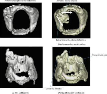
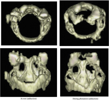
Ossification of the cricoid and arytenoid cartilages was well visualised on both patients' three-dimensional images. The muscular and vocal processes and the cricoarytenoid joint on the side of the resected arytenoid cartilage were also identified. No corniculate cartilage was visualised.
In case one, the left arytenoid cartilage was not visualised. Slightly excessive adduction of the right arytenoid cartilage was observed, but no dislocation had occurred (Figure 1).
Three-dimensional images for case one. The thyroarytenoid and cricothyroid intrinsic laryngeal muscles have been separated at surgery. The left arytenoid cartilage is not visualised. Slightly excessive adduction of the right arytenoid cartilage is observed, but no dislocation has occurred.

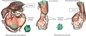
In case two, bilateral arytenoid cartilage motion was present, with both adduction and abduction. There were no findings indicative of dislocation (Figure 2).
Three-dimensional images for case two. The thyroarytenoid and cricothyroid intrinsic laryngeal muscles have been separated at surgery. Bilateral arytenoid cartilage motion is favourable, with both adduction and abduction. There are no findings suggestive of dislocation.

Airway visualisation
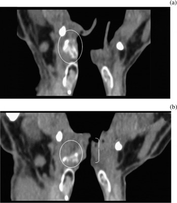
In case one, coronal multiplanar reconstruction images indicated that airway constriction was achieved by right arytenoid adduction. The airway was almost straight in the midline because the mucosa was rotated inward during phonation, even on the side of the resected cartilage (Figure 3).
Coronal multiplanar reconstruction images for case one: (a) at rest (abduction), and (b) during phonation (adduction). The airway is almost straight in the midline (bracket) because the mucosa is rotated inward during phonation, even on the side of the resected cartilage. Circles indicate the right arytenoid cartilage. Window level (WL) = 30; Window width (WW) = 350

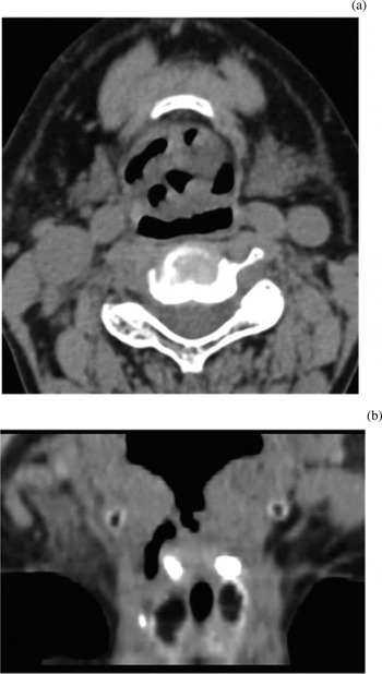
However, in case two it was difficult to identify an obvious airway and structure (Figure 4); therefore, we could not determine the form of the neoglottis. In contrast, three-dimensional images made it possible to visualise both the inside of the arytenoid region and the outside of the right arytenoid (Figure 5).
(a) Axial and (b) coronal multiplanar reconstruction images for case two. It is difficult to identify an obvious airway and structure. Window level (WL) = 30; Window width (WW) = 350

Three-dimensional images of the airway, cricoid cartilage and arytenoid cartilage of case two, showing the inside of the arytenoid region and the outside of the right arytenoid.

Discussion
Arytenoid cartilage movement
In both presented patients, three-dimensional imaging of the laryngeal cartilages showed favourable arytenoid cartilage motion. No post-operative dislocation or fixation had occurred. In both cases, the thyroarytenoid and cricothyroid intrinsic laryngeal muscles had been separated by the surgical procedure. However, arytenoid cartilage motion appeared to be maintained by the remaining lateral cricoarytenoid muscle, transverse arytenoid muscle, oblique arytenoid muscle and posterior cricoarytenoid muscle. Electromyographic studies of intrinsic laryngeal muscles have shown that the lateral and posterior cricoarytenoid muscles move alternately, associated with arytenoid cartilage motion.Reference Nakayama, Hirose, Okamoto, Miyamoto, Yokobori and Takeda10
Airway observation
Three-dimensional neoglottal imaging indicated that our two patients' post-operative airways differed. The neoglottal airway seemed to alter rapidly, from a wide space at the subglottic level to a narrow space at the neoglottis, an arrangement which favoured phonation. Following supracricoid laryngectomy, various arytenoid mucosa vibratory patterns have been reported, and various causes proposed for the observed airway shape and narrowed length.Reference Saito, Araki, Ogawa and Shiotani9 In our first case, high-speed digital imaging of the neoglottal laryngotopography showed mucosal vibrations at the interface between the adducted right arytenoid and the remaining left arytenoid. In our second case, high-speed digital imaging showed mucosal vibrations at the interface between the right arytenoid and the right edge of the epiglottis.Reference Hayashi, Hirose, Tayama, Imagawa, Nakayama and Ueta11 These results reflect the morphological characteristics of the post-operative airway, as viewed using three-dimensional images. The sudden narrowing above the wide subglottic space causes rapid airflow, which can be considered to induce vibration of the arytenoids.
Three-dimensional image construction
Advances in computer technology have enabled the construction of cross-sectional and three-dimensional images, and the creation of virtual endoscopy. Using DICOM files, three-dimensional imaging software is able to construct three-dimensional and multiplanar reconstruction images.
During CT investigation, it is preferable that the radiologist guides the patient on speech method and timing. In order to obtain reliable images during phonation or breath-holding, it is preferable that the patient self-operates the CT scanner if possible.
• Three-dimensional neoglottal image construction was performed for two patients who had undergone supracricoid laryngectomy with cricohyoidoepiglottopexy
• The mobility of the arytenoids was well preserved
• During phonation, one patient had a single stream airway and the other a combination of several streams (which may constitute a sound source)
• Following such surgery, the construction and analysis of three-dimensional images may be useful in analysing neoglottal morphology and function
Our study findings suggest that, following supracricoid laryngectomy with cricohyoidoepiglottopexy, the acquisition of three-dimensional radiological images may be useful in analysing the morphology and function of the post-operative neoglottis. Such images provide useful information which may facilitate future modifications of laryngeal resection and reconstruction techniques, with the aim of improving post-operative phonetic function.