Implications
This study investigated the effects of a novel plant extract product that was added to the drinking water for weaned piglets. Different feeding regimens were tested, and the effects of the extract on growth performance, gut health and protection against Escherichia coli challenge were observed. The results showed that the plant extract may represent a useful additive for improving gut health and microbial ecology and reducing the severity of an E. coli challenge. The results may have a significant impact on nutritional management in conventional farms. Use of the plant extract could enable piglets to better withstand the infections that are often associated with weaning.
Introduction
Phytobiotics is a term used to describe plant-derived natural bioactive compounds that promote livestock health and well-being and improve livestock growth and production efficiency (Wu and Wu, Reference Wu and Wu2012). Although the mechanisms of phytobiotics are not entirely understood, their benefits to the overall health of animals have been noted. Phytobiotics represent a source of various chemical and bioactive compounds, such as terpenes, phenols, glycosides, saccharides, aldehydes, esters and alcohols. When plant extracts (PEs) have been used in swine nutrition, both improvements and reductions in productivity have been observed; however, this has also been the case for common growth promoters (Manzanilla et al., Reference Manzanilla, Nofrarías, Anguita, Castillo, Perez, Martín-Orúe, Kamel and Gasa2006; Nofrarías et al., Reference Nofrarías, Manzanilla, Pujols, Gibert, Majó, Segalés and Gasa2006; Jin et al., Reference Jin, Yang, Choi, Shinde, Yoon, Hahn, Lim, Park, Hahm, Joo and Chae2008). The beneficial effects of phytobiotics may be explained by the activation of feed intake, the secretion of digestive enzymes, immune stimulation, intestinal microflora modulation, anti-bacterial effects and anti-inflammatory properties (Ultee et al., Reference Ultee, Bennik and Moezelaar2002; Windisch et al., Reference Windisch, Schedle, Plitzner and Kroismayr2008).
Increased susceptibility to enterotoxigenic Escherichia coli infections and acute diarrhoea are common problems in newly weaned animals. Experimental models for E. coli K88 challenge have been used to evaluate the role of feed additives for weaning pigs in modulating the gastrointestinal microbial response (Bhandari et al., Reference Bhandari, Xu, Nyachoti, Giesting and Krause2008; Kiarie et al., Reference Kiarie, Bhandari, Scott, Krause and Nyachoti2011). Feed restriction, as part of farm strategy, has been applied in post-weaning piglets to decrease the proliferation of bacteria such as haemolytic E. coli and to reduce the incidence of diarrhoea, and it also has been associated with a transitory decrease in growth compared with piglets fed ad libitum (AD) (Pastorelli et al., Reference Pastorelli, Le Floc’h, Merlot, Meunier-Salaün, van Milgen and Montagne2012). Thus, the current study was conducted to investigate the effects of a novel PE product on the growth performance and gut health (microbial counts, intestinal morphology and enzyme activities) of weaned piglets undergoing an experimental E. coli challenge and feed restriction.
Material and methods
The experimental protocol was reviewed and approved by the Animal Care and Use Committee of the University of Milan (Protocol No. Gra.Piglet.09.10).
Animals and treatments
The experiment was carried out at the facility for Animal Production Research and Teaching Centre of the Polo Veterinario, Università degli Studi di Milano (Lodi, Italy). At weaning (24 days), a total of 144 crossbred (Stambo HBI×Dalland) piglets (6.50±0.35 kg) of the same age and litter origin were randomly assigned to treatment groups. Animals were housed in two identical rooms, equipped with 24 pens each, in an environmentally regulated, isolated stable. A combination of daylight and artificial light was used. Ventilation was achieved by using variable-speed fans. The starting temperature of 28ºC was adjusted weekly to reach a final temperature of 24ºC. Piglets were housed in pens with a slatted floor (3 piglets/pen, 1.20×1.00 m). Each pen was equipped with two standard nursery pig bite-style nipple drinkers or stainless steel nursery push-lever bowl drinkers and a self-feeder. Piglets were raised for 35 days in eight different groups with a 2×2×2 factorial design comparing different treatments (water without product (CT) or 8 μl/kg per day PE in the drinking water (PE)), feeding regimens (AD or restricted (RE)) and oral E. coli challenges (sham (−) or infected (+)). There were six pens per treatment group, and the different combinatorial treatments are designated as follows: (1) CTAD−; (2) CTAD+; (3) CTRE−; (4) CTRE+; (5) PEAD−; (6) PEAD+; (7) PERE−; and (8) PERE+. On day 9 of the trial, the piglets housed in one of the two rooms were orally injected with 4 ml of a solution containing 109 cfu of E. coli 0149: F4 (K88)-positive strain. The K88-positive strain, isolated from pigs with colibacillosis, also expressed heat-labile and heat-stable B toxins, and further prepared as described by Bosi et al. (Reference Bosi, Casini, Finamore, Gremokolini, Merialdi, Trevisi, Nobili and Mengheri2004).
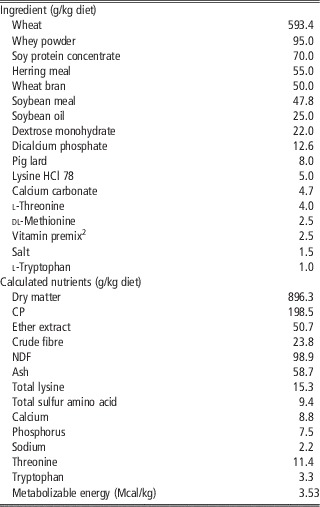
The PE product (GrazixTM, LiveLeaf Inc., San Carlos, CA, USA) is an oral supplement produced by using the LiveXtractTM process on green tea leaves (Camellia sinensis) and pomegranate rinds (Punica granatum). The PE product was administered from 2000 to 0800 h through the drinking water. A graduated tank filled with treated water was linked to the nursery push-lever bowl drinker in each pen. The PE product was diluted with water daily and manually supplemented in the water tank to maintain the correct dosage according to the total animal weight per pen (8 μl/kg per day). This adjustment was carried out at 2000 h, and the provision of water was managed to ensure that the piglets consumed all of the water. Following the E. coli challenge (day 9), PE supplementation was increased; the E. coli-challenged piglets received 200 μl/kg per day of the PE product on days 8, 9 and 12, and they received 400 μl/kg per day on days 10 and 11. The diet was formulated to be isonutritive, exceeding the protein requirement recommended by NRC (1998) for pigs (Table 1), and it did not include any antibiotic growth promoters or antibiotic growth promoter alternatives. In the RE feeding groups, piglets were allowed access to food from 0800 to 2000 h. Feeding troughs for the RE groups were removed at 2000 h, weighed and stored. Each morning, the feeding troughs were filled, weighed and placed in the pens at 0800 h.
Composition and calculated nutrient content of the basal dietFootnote 1

1 Diets were not supplemented with antibiotics.
2 Vitamin-mineral premix contents per kg final feed: Vitamin A: 10 500 IU; Vitamin D3: 2500 IU; Vitamin E: 15 mg; Vitamin B1: 1.5 mg; Vitamin B2: 3.8 mg; Vitamin B12: 0.025 mg; Vitamin B6: 1.6 mg; calcium pantotenate: 12 mg; nicotinic acid: 15 mg; biotin: 0.15 mg; folic acid: 0.5 mg; Vitamin K3: 3 mg; Fe: 100 mg; Cu: 6 mg; Co: 0.75 mg; Zn: 150 mg; Mn: 65 mg; I: 0.75 mg; Se: 0.4 mg; ethoxyquin: 150 mg.
Experimental observations and measurements
All piglets were weighed at weaning (day 0) and subsequently every week until the end of the trial. Piglet feed consumption was measured on a daily basis in the RE regimen groups, and it was measured weekly in the AD regimen groups. The average daily gain (ADG), average daily feed intake (ADFI) and gain to feed ratio values (G : F) were calculated for each pen. Water consumption was measured daily as the amount moved from the graduated tank to the nursery push-lever bowl drinker. The occurrence and severity of diarrhoea were monitored weekly. After the E. coli challenge, the faecal score daily monitored daily for 1 week in the challenged animals. The severity of diarrhoea was characterised by using a 5-point faecal consistency scoring system: 1=hard, dry pellet; 2=firm, formed stool; 3=soft, moist stool that retains shape; 4=soft, unformed stool; 5=watery liquid that can be poured. Liquid consistency (score 4 to 5) was considered indicative of diarrhoea. Pooled faecal samples from each pen (~20 g), were collected on days 0, 14 and 35, placed in small sterile containers, and immediately sent to the laboratory for microbiological analysis. One gram of the fresh sample was diluted with 99 ml sterile physiological NaCl solution. Following homogenisation on a vortex, 1 ml of the suspension was mixed with 9 ml NaCl solution. Serial dilutions (1 : 10) for culturing were prepared down to 10−9. From each diluted sample, 0.1 ml was plated on the appropriate medium for enumeration of microbial populations. Three replicates were carried out for each sample. Plates for the enumeration of Enterobacteriaceae (using Violet-Red Bile Dextrose agar) were incubated aerobically at 37°C for 24 h. E. coli was grown in tryptic soy agar at 37°C. Lactobacillus faecal content was determined using MRS agar (Lactobacillus agar) with an incubation time of 72 h at 37°C (10% CO2), and the Clostridia procedure used an incubation time of 48 h at 37°C with tryptose sulphite cycloserine agar. The microbial counts were expressed as log10 cfu/g.
At the end of the trial, 24 animals from the RE groups (3 piglets per treatment per room) were selected as the most representative of pen performance in terms of weight gain and health, and these animals were slaughtered. The animals were stunned electrically and bled after ~16 h of starvation. Immediately after slaughter, the gastrointestinal tract was removed from each animal, and the distal ileum (2 cm before its opening into the caecum) was collected and promptly fixed in neutral buffered formalin for 24 h at 4°C. The specimens were then dehydrated in a graded ethanol series, cleared with xylene and embedded in paraffin. After dewaxing and rehydration, microtome sections (4 µm thick) were stained with haematoxylin and eosin and examined to either assess the ileum micro-anatomical structure or perform histometry. Histometric assays considered the following parameters for each section: villus height (V) (five villi measured per section), crypt depth (C) (five crypts measured per section), the villus height to crypt depth ratio (V : C ratio), number of lymphatic follicles (counted in 3 fields per section at 400× and expressed as n/mm2 of mucosa) and the area of the lymphatic follicles and their compartments (cortex, medulla, corona; 5 follicles per section). Other ileum sections were processed by immunohistochemical analysis to identify mucosal macrophages with a macrophage monoclonal antibody (1 : 400, ab22506, Abcam) after antigen retrieval with K-protease (20 μg/ml of buffer solution). For each section, the number of immuno-positive mucosal cells was counted in eight fields at 400× and subsequently expressed as n/mm2 of mucosa.
The ileal mucosa was also used to evaluate myeloperoxidase (MPO), nitric oxide (NO) and inducible nitric oxide synthase (iNOS) by using commercial kits from the Nanjing Jiancheng Bioengineering Institute (Nanjing, China). Three 5 cm segments from the ileum were collected, opened longitudinally and cleaned with phosphate buffered saline (PBS). Intestinal mucosa samples were collected by scraping the wall with a glass slide. Nine ml of 4°C PBS was added to 1 g of intestinal mucosa, followed by homogenisation. The homogenates were centrifuged (4000×g for 5 min at 4°C), and supernatant fluid was used according to the manufacturer’s instructions.
Statistical analysis
Data were analysed as a completely randomised block design by ANOVA, as implemented in the MIXED procedure of SAS v. 9.2 (SAS Institute Inc., Cary, NC, USA). For growth performance, faecal scores and microbial counts, a model for a 2×2×2 factorial design was applied. The model statement included the effects of treatment (CT or PE), feeding regime (AD or RE), E. coli challenge (− or +), and interactions among those factors, as suggested by Song et al. (Reference Song, Liu, Soares, Che, Osuna, Maddox and Pettigrew2012). The pen represented the experimental unit for these parameters. Faecal scores recorded for the challenged piglets within 7 days of challenge were analysed as a generalised randomised block design with repeated measurements over time, and the pen was the experimental unit. An ANOVA mixed model with a 2×2 factorial design was used for gut histomorphology and mucosal inflammatory parameters. The applied model included the treatment effect (CT or PE), the E. coli challenge effect (− or +) and a treatment×challenge interaction. The individual piglets were the experimental units for intestinal histometric and mucosal inflammatory parameters. Treatment differences were assessed by using the least squares means with a Tukey adjustment. Treatment effects were considered significant at P⩽0.05, whereas a trend for a treatment effect was noted for P⩽0.10.
Results
Growth performance
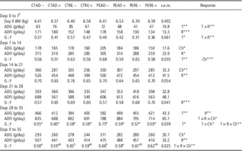
The effects of PE and feeding regimen on growth performance in the E. coli-challenged and non-challenged piglets are shown in Table 2. The piglets that were given supplemental PE showed enhanced ADG during the last week of the trial (P=0.03) and had an increased G : F ratio during the second and last weeks (P=0.02 and 0.10, respectively). Reduced ADG and G : F ratio values were observed in the PE treatment from day 0 to day 7 (P=0.04 and 0.06, respectively). An effect of the feeding regimen was observed on ADFI for the overall experimental trial, with a higher feed intake in the piglets that were fed AD during the 1st week (P<0.01), 2nd week (P=0.06) and 3rd week (P=0.05), and during the overall study period (P=0.05). However, the piglets with RE feeding had higher G : F ratios than those in the AD feeding group(s) from day 21 to day 28 (P=0.01). The E. coli challenge decreased the ADG and G : F ratio during days 7 to 14, the week of the challenge (P=0.08 and <0.01, respectively), and it reduced the ADG from day 14 to day 21 and from day 0 to day 35 (P=0.03 and 0.06, respectively). The treatment×regimen interactions between ADG and G : F ratio from day 0 to day 7 (P=0.04 and 0.02, respectively) indicate that PE had different effects on those performance in the AD and RE regimen animals. The results indicate that G : F ratio was also affected by treatment, feeding regimen, and challenge from day 28 to day 35 and day 0 to day 35 (treatment×regimen×challenge, P=0.05 and 0.02, respectively). The interactions during the last week and for the overall period can be attributed to the differences between the CTAD− (0.55 and 0.58, respectively) and PEAD− (0.75 and 0.66, respectively) groups, but no differences were found in others piglets. In addition, our results indicate that the G : F ratio tended to be affected by treatment and challenge (treatment×challenge, P=0.08), and ADFI tended to be affected by treatment, regimen and challenge (treatment×regimen×challenge, P=0.09) during the last week of the trial. For the PE group, piglets fed AD increased their water consumption compared with piglets with RE feeding (258 ml/piglet per day v. 148 ml/piglet per day; P<0.01, data not shown). No difference was observed in the PE-supplemented water consumption between E. coli-challenged and non-challenged piglets.
Effects of plant extract supplementation on growth performance of Escherichia coli-challenged (+) and non-challenged (−) piglets fed ad libitum or restricted dietsFootnote 1
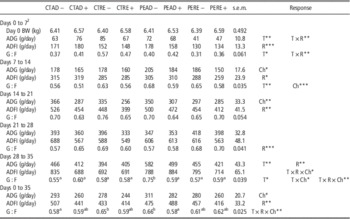
CT=water without supplementation; PE=8 μl/kg per day PE in drinking water; T=treatment (CT v. PE); AD=ad libitum regimen; RE=restricted regimen; R=regimen (AD v. RE); +/−=presence or absence of E. coli challenge; ADG=average daily gain; ADFI=average daily feed intake; Ch=challenge (sham (−) v. infected (+)); T×R=interaction between treatment and regimen; T×Ch=interaction between treatment and challenge; R×Ch=interaction between regimen and challenge; T×R×Ch=interaction between treatment, regimen and challenge.
a,bMeans listed in the same row with different superscripts are significantly different (P<0.05).
1 n=48 (6 pens/treatment). Piglets were weaned at 24 days of age, and half of the piglets were orally challenged with E. coli on day 9 of the trial.
2 Day of the trial.
*P⩽0.10, **P⩽0.05, ***P⩽0.01.
Faecal score and faecal microbial population
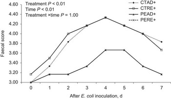
E. coli challenge increased the faecal scores of the CT piglets, resulting in average values above four points (index of diarrhoea occurrence) up to 6 days after challenge (Figure 1). Within 1 week post-infection, there was a treatment (P<0.01) and time effect (P<0.01), but no treatment×time interaction for faecal score. Piglets supplemented with PE had improved faecal consistency, irrespective of their feeding regimen, compared with CT. PE supplementation decreased the faecal score of PEAD and PERE piglets compared with the CTRE group on the 2nd day post-challenge (P=0.05). On the 3rd day post-challenge, the CT piglets had a higher faecal score than the PE piglets, irrespective of the feeding regimen (P=0.05). In addition, diarrhoea occurrence was lower in the PE piglets than in the CT animals (26% v. 62%, data not shown). The effects of PE supplementation and feeding regimen on faecal score in the E. coli-challenged and non-challenged piglets are shown in Table 3. PE supplementation decreased the faecal score values on days 14 (P=0.02), 21 (P<0.01), 28 (P<0.01) and 35 (P<0.01). E. coli challenge significantly affected the faecal score on days 14 (P<0.01), 21 (P<0.01), 28 (P<0.01) and 35 (P<0.01). However, faecal score was not affected by the feeding regimen. In addition, treatment×challenge interactions were observed on days 28 (P<0.01) and 35 (P<0.01), indicating that PE supplementation had different effects on challenged and non-challenged piglets.
Effect of plant extract (PE) on faecal score in piglets with different feeding regimens 7 days after Escherichia coli challenge. CTAD+=no supplement in drinking water (CT), ad libitum regimen (AD), challenge with E. coli (+); CTRE+=no supplement in drinking water (CT), restricted regimen (RE), challenge with E. coli (+); PEAD+=8 μl/kg per day PE in drinking water (PE), ad libitum regimen (AD), challenge with E. coli (+); PERE+=8 μl/kg per day PE in drinking water (PE), restricted regimen (RE), challenge with E. coli (+). Error bars are omitted for presentation purposes. Pooled s.e.m.=0.30. Faecal score were recorded by using a 5-point scoring system: 1=hard; 2=firm; 3=soft (moist stool); 4=soft (unformed stool); 5=watery faeces. Diarrhoea: liquid consistency (score 4 to 5). Day 9 of the trial is shown as day 0 post-inoculation with E. coli.

Effects of plant extract (PE) supplementation on the faecal scoresFootnote 1 of Escherichia coli-challenged (+) and non-challenged (−) piglets fed ad libitum or restricted dietsFootnote 2

CT=water without supplementation; PE=8 μl/kg per day PE in drinking water; T=treatment (CT v. PE); AD=ad libitum regimen; RE=restricted regimen; R=regimen (AD v. RE); +/−=presence or absence of E. coli challenge; Ch=challenge (sham (−) v. infected (+)); T×R=interaction between treatment and regimen; T×Ch=interaction between treatment and challenge; R×Ch=interaction between regimen and challenge; T×R×Ch=interaction between treatment, regimen and challenge.
1 Faecal scores were recorded using a 5-point scoring system: 1=hard; 2=firm; 3=soft (moist stool); 4=soft (unformed stool); 5=watery faeces. Diarrhoea: liquid consistency (score 4 to 5).
2 n=48 (6 pens/treatment). Piglets were weaned at 24 days of age, and half of the piglets were orally challenged with E. coli on day 9 of the trial.
3 Day of the trial.
**P⩽0.05, ***P⩽0.01.
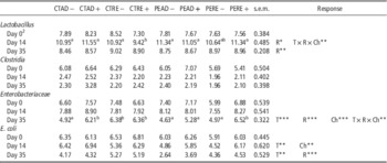
The effects of PE and feeding regimen on faecal microbial populations in the E. coli-challenged and non-challenged piglets are shown in Table 4. No effect was observed on the Clostridia population (P>0.05). PE supplementation decreased the faecal E. coli levels on days 14 and 35 (P=0.05 and 0.02, respectively) and reduced the Enterobacteriaceae (P<0.01) microbial population on day 35. The challenge increased the faecal E. coli (P=0.03) and Enterobacteriaceae populations (P<0.01) on days 14 and 35, respectively. The faecal microbial population was influenced by the feeding regimen, with an increase of faecal Lactobacillus, Enterobacteriaceae and E. coli populations in piglets fed the RE diet on day 35 (P=0.02, <0.01 and <0.01, respectively). In addition, RE fed piglets increased faecal Lactobacillus, Enterobacteriaceae and E. coli population in comparison with piglets fed AD on day 35 (P=0.02, <0.01 and <0.01, respectively), whereas a slight decrease was observed in Lactobacillus population in piglets with RE feeding compared with animals fed AD on day 14 (P=0.07). The treatment×regimen×challenge interactions for the Lactobacillus counts on day 14 (P=0.03) indicate that PE supplementation increased the population in challenged piglets with RE diets but had no effect on other groups. The Enterobacteriaceae population was also affected by PE, regimen and challenge on day 35 (treatment×regimen×challenge, P=0.02). The interaction was because of the differences between CTAD+ and PEAD+ (6.21 v. 5.28 log10 cfu/g), and between CTRE− and PERE− (6.38 v. 4.97 log10 cfu/g), but there were no differences in other groups.
Effects of plant extract (PE) supplementation on faecal microbiological counts (log10 cfu/g) in Escherichia coli-challenged (+) and non-challenged (−) piglets fed ad libitum or restricted dietsFootnote 1

CT=water without supplementation; PE=8 μl/kg per day PE in drinking water; T=treatment (CT v. PE); AD=ad libitum regimen; RE=restricted regimen; R=regimen (AD v. RE); +/−=presence or absence of E. coli challenge; Ch=challenge (sham (−) v. infected (+)); T×R=interaction between treatment and regimen; T×Ch=interaction between treatment and challenge; R×Ch=interaction between regimen and challenge; T×R×Ch=interaction between treatment, regimen and challenge.
a,bMeans listed in the same row with different superscripts are significantly different (P<0.05).
1 n=48 (6 pens/treatment). Piglets were weaned at 24 days of age, and half of the piglets were orally challenged with E. coli on day 9 of the trial.
2 day of the trial.
*P⩽0.10, **P⩽0.05, ***P⩽0.01.
Ileum histology and histometry
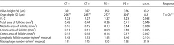
The effects of PE on ileum histological and histometric parameters were examined only for the RE feeding groups, considering both the challenged and non-challenged piglets (Supplementary Figures S1, S2; Table 5). In the ilea of E. coli-challenged animals without PE supplementation, signs of chronic enteritis were observed, such as irregular and flattened enterocytes at the villus tips, confluent villi at the apical zones, and hyperaemia, oedema and inflammatory cell infiltration in the lamina propria. The intestinal structure of E. coli-challenged pigs that had consumed the PE was normal in both the epithelium and the lamina propria. There was no PE effect (P>0.05) or challenge effect (P>0.05) on villus height, crypt depth, V : C ratio, total area of follicles, medulla area of follicles, corona area of follicles, cortex area of follicles, lymphatic follicle number or macrophage numbers in the intestinal mucosa. However, crypt depth in the ileum was affected by PE administration and challenge (treatment×challenge, P=0.03), which was because of a difference between non-challenged piglets (n=277) and challenged piglets (n=304) for those receiving the PE treatment, but not for those in the CT groups.
Effects of plant extract (PE) supplementation on ileum histometric parameters in Escherichia coli-challenged (+) and non-challenged (−) piglets fed a restricted dietFootnote 1

CT=water without supplementation; PE=8 μl/kg per day PE in drinking water; T=treatment (CT v. PE); +/−=presence or absence of E. coli challenge; Ch=challenge (sham (−) v. infected (+)); T×Ch=interaction between treatment and challenge.
a,bMeans listed in the same row with different superscripts are significantly different (P<0.05).
1 n=24 (6 piglets/treatment). Piglets were weaned at 24 days of age, and half of the piglets were orally challenged with E. coli on day 9 of the trial; 24 piglets with the restricted regimen (6 piglets/treatment) were slaughtered at the end of the trial.
**P⩽0.05.
Intestinal inflammatory parameters
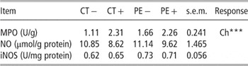
The effects of PE on intestinal inflammatory parameters in E. coli-challenged and non-challenged piglets that were fed a RE diet are shown in Table 6. In the E. coli-challenged animals that were fed a RE diet, MPO activity in the ileum was increased (P<0.01) compared with non-challenged piglets, whereas NO and iNOS were not affected (P>0.05). PE treatment had no effect on these inflammatory parameters (P>0.05).
Effects of plant extract (PE) supplementation on intestinal mucosa inflammatory parameters in Escherichia coli-challenged (+) and non-challenged (−) piglets fed a restricted dietFootnote 1

MPO=myeloperoxidase; NO=nitric oxide; iNOS=inducible nitric oxide synthase; CT=water without supplementation; PE=8 μl/kg per day PE in drinking water; T=treatment (CT v. PE); +/−=presence or absence, respectively, of challenge with E. coli; Ch=challenge (sham (−) v. infected (+)); T×Ch=interaction between treatment and challenge.
1 n=24 (6 piglets/treatment). Piglets were weaned at 24 days of age, and half of the piglets were orally challenged with E. coli on day 9 of the trial; 24 piglets with the restricted regimen (6 piglets/treatment) were slaughtered at the end of the trial.
***P⩽0.01.
Discussion
The objectives of the present study were to determine whether a novel PE product added to the drinking water of weaned piglets under different feeding regimens would affect growth performance and gut health and to determine whether this supplement could protect piglets against an E. coli challenge. In our study, PE did not affect growth performance during the overall period of the trial, but the PE piglets did show an enhanced ADG during the last week of the trial period, and they showed improvements in feed efficiency during the 2nd week and the last week. These results suggest that PE may allow piglets to better respond to stress in the post-weaning period, when impaired piglet performance is most likely to occur. Previous studies regarding the effects of PEs on growth performance in piglets have had inconsistent results. Increased feed intake, ADG and feed to gain ratio were reported by several authors (Kim et al., Reference Kim, Hong, Kwon, Min, Lee and Shon2004; Lien et al., Reference Lien, Horng and Wu2007), but other groups observed no effects on piglet growth performance (Nofrarías et al., Reference Nofrarías, Manzanilla, Pujols, Gibert, Majó, Segalés and Gasa2006; Liu et al., Reference Liu, Song, Che, Almeida, Lee, Bravo, Maddox and Pettigrew2013). These contradictory results may be explained by the PEs used, the compositions of the extracts, the dietary doses tested, the route of administration or the different experimental approaches used to test the effects of the substances (Windisch et al., Reference Windisch, Schedle, Plitzner and Kroismayr2008).
Infections and post-weaning diarrhoea are major causes of mortality and morbidity worldwide, and they are estimated to account for as much as 50% of economic losses in the production of weaned pigs (Cutler et al., Reference Cutler, Lonergan, Cornick, Johnson and Stahl2007). In the current study, E. coli challenge significantly impaired piglet performance, resulting in reduced ADG and G : F ratio in the week after the challenge and, consequently, a reduced ADG in the following week. These results are in agreement with those of other authors (Liu et al., Reference Liu, Piao, Thacker, Zeng, Li, Wang and Kim2010; Song et al., Reference Song, Liu, Soares, Che, Osuna, Maddox and Pettigrew2012; Liu et al., Reference Liu, Song, Che, Almeida, Lee, Bravo, Maddox and Pettigrew2013). In our study, PE supplementation improved G : F only in non-challenged piglets with an AD feeding regimen; no further beneficial effect on growth performance was found during the enteric E. coli challenge. The E. coli challenge was successful in causing a mild infection. Increased faecal consistency and diarrhoea frequency were found up to 7 days after the challenge. The increases in faecal consistency scores are typical of an E. coli challenge model (Kiarie et al., Reference Kiarie, Bhandari, Scott, Krause and Nyachoti2011; Nyachoti et al., Reference Nyachoti, Kiarie, Bhandari, Zhang and Krause2011). The present experiment clearly shows a reduction in the faecal consistency scores and diarrhoea frequency owing to supplementation of PE to weaned pigs subjected to the E. coli challenge. The effect is particularly strong within 1 week of the E. coli challenge. These findings are in agreement with one recent study that showed an anti-diarrhoeal effect in piglets fed PE (Liu et al., Reference Liu, Song, Che, Almeida, Lee, Bravo, Maddox and Pettigrew2013). The effect on faecal consistency may be related to an improved gastrointestinal microbial ecosystem in the PE-supplemented piglets. In the current study, we focused on Lactobacillus, Clostridia, total Enterobacteriaeae and E. coli because these were found to be important for intestinal health in swine (Pluske et al., Reference Pluske, Pethick, Hopwood and Hampson2002). A positive effect was observed after PE supplementation; the Enterobacteriaceae and E. coli populations were reduced. However, PE did not affect the Clostridia population, which may be because of the low basal level of Clostridia in the intestines of piglets after weaning (Risley et al., Reference Risley, Kornegay, Lindemann, Wood and Eigel1992). In addition, PE supplementation increased the Lactobacillus population in the challenged piglets that were fed a RE diet on day 14 of the trial. The mechanisms by which PEs may exert positive effects on gut populations are not yet clearly understood, although in vitro studies have demonstrated that the anti-microbial activities of herbs and spices can provide protection against important pathogens (Burt, Reference Burt2004; Özer et al., Reference Özer, Sökmen, Güllüce, Adigüzel, Sahin, Sökmen, Kilic and Baris2007). Some reports describe the effects of green tea and pomegranate peel extracts. Tea polyphenols have been shown to exert anti-bacterial activities against human and animal disease-causing bacteria, phytopathogenic bacteria and food-borne bacteria (Sakanaka et al., Reference Sakanaka, Juneja and Taniguchi2000), although the potency of the polyphenols is dependent on the bacterial species (Su et al., Reference Su, Henriksson, Nilsson and Mitchell2008; Archana and Abraham, Reference Archana and Abraham2011). Recently, another hypothesis about the possible mechanism of action of polyphenols was reported by Wu and Wu (Reference Wu and Wu2012), who demonstrated that the same PE mix used in our study was capable of binding lipopolysaccharide, the major component of bacterial endotoxins. In vivo studies regarding the effects of PEs on gut microbes are less common. In humans, Goto et al. (Reference Goto, Kanaya, Ishigami and Hara1999) found that consumption of green tea selectively promoted the growth of Bifidobacterium and Lactobacillus in the gut walls of elderly residents in a long-term care facility. Hagmüller et al. (Reference Hagmüller, Jugl-Chizzola, Zitterl-Eglseer, Gabler, Spergser, Chizzola and Franz2006) did not find any significant differences in the shedding of haemolysing E. coli in weanling piglets fed Thymi herba (Thymus vulgaris, rubbed).
The decrease in ADFI and ADG as a consequence of feed restriction observed in this study has been previously reported (Lovatto et al., Reference Lovatto, Sauvant, Noblet, Dubois and van Milgen2006; Pastorelli et al., Reference Pastorelli, Le Floc’h, Merlot, Meunier-Salaün, van Milgen and Montagne2012). However, the RE feeding regimen tended to improve feed efficiency. Our results showed that RE feeding led to reduced food intake during most periods of the trial, but it increased the G : F ratio compared with the AD group from day 21 to day 28. Moreover, there was a significant treatment×regimen interaction for ADG and G : F ratio during the 1st week after weaning, and lower ADG and G : F ratio values were observed in the PE treatment groups. These unexpected results, which were owing to reductions in ADG and G : F ratio in the RE piglets receiving PE compared with the piglets given water without PE supplementation, may indicate that this PE product cannot increase growth performance for piglets with a RE feeding regimen in the short post-weaning period. Similar results have been found by Daza et al. (Reference Daza, Rodríguez, Ovejero and López Bote2003), who reported that pigs fed a RE diet reduced their feed intake but had a feed conversion ratio similar to that of pigs fed AD. Rantzer et al. (Reference Rantzer, Svendsen and Westrom1996) reported that, during the period of feeding restriction, the RE piglets had a lower diarrhoea score than those fed AD. The faecal scores of animals in this study were not influenced by feeding regimen, which indicates that feeding restriction did not impair their health status. Some previous studies observed that feed restriction resulted in a reduced health risk index (mortality+morbidity rates) in weaned rabbits compared with animals fed AD (Gidenne et al., Reference Gidenne, Combes, Feugier, Jehl, Arveux, Boisot, Briens, Corrent, Fortune, Montessuy and Verdelhan2009), which could be explained by a decrease in the flow of nutrients to the caecum that would reduce the proliferation of pathogenic bacteria. Gidenne and Feugier (Reference Gidenne and Feugier2009) found that a 20% reduction in feeding increased the caecal concentration of volatile fatty acids and acidified caecal content while enhancing bacterial fibrolytic activity. In the present study, feed restriction increased the faecal populations of Lactobacillus, Enterobacteriaceae and E. coli on day 35, which is different from the observations of piglets fed a RE diet by Rantzer et al. (Reference Rantzer, Svendsen and Westrom1996). Lázaro et al. (Reference Lázaro, Latorre, Medel, Gracia and Mateos2004) reported that feed restriction and enzyme supplementation reduced the magnitude of problems in broiler chicks, especially jejunum viscosity problems, owing to the presence of rye in feed. In addition, feed restriction implemented by decreasing the time for feeding would be aimed at precisely evaluating the utilisation of the product in the water and correlating the product effects with gut histomorphology.
Intestinal growth and development are critical for the optimal performance of piglets. After weaning, there are marked changes in the histology and biochemistry of the small intestine, including villous atrophy and crypt hyperplasia, which may cause decreased digestive and absorptive capacities and contribute to post-weaning diarrhoea (Pluske et al., Reference Pluske, Hampson and Williams1997). In our study, E. coli challenge induced morphological changes in the ileum, which showed signs of chronic enteritis such as irregular and flattened enterocytes at the villus tips and confluent villi at the apical zones, as well as hyperaemia, oedema and inflammatory cell infiltration in the lamina propria. The intestinal structure of E. coli-challenged pigs given PE supplementation was normal in both the epithelium and the lamina propria. Morphological changes in the gastrointestinal tissues occurring after phytogenic feed supplementation may provide further information about the possible benefits for gut health. The available literature does not provide a consistent picture. In our study, PE supplementation had no effect on villus height, crypt depth or V/C ratio, which is in agreement with previous observations of piglets supplemented with different PEs (Manzanilla et al., Reference Manzanilla, Nofrarías, Anguita, Castillo, Perez, Martín-Orúe, Kamel and Gasa2006; Nofrarías et al., Reference Nofrarías, Manzanilla, Pujols, Gibert, Majó, Segalés and Gasa2006; Sehm et al., Reference Sehm, Lindermayer, Dummer, Treutter and Pfaffl2007). In contrast, other authors observed an increase in villus height in the duodenum, jejunum and ileum, and a decrease in the crypt depth in response to dietary supplementation of Acanthopanax senticosus extract (Fang et al., Reference Fang, Yan, Kong, Ruan, Liu, Huang, Li, Geng, Yang, Zhang, Li, Gong, Wu, Fan, Liu, Hou and Yin2009). The interaction between treatment and challenge indicate that PE supplementation kept the V : C ratio of E. coli-challenged animals at a normal level by increasing villus height and crypt depth. PEs have been reported by several authors to affect the immune system of the intestine. Nofrarías et al. (Reference Nofrarías, Manzanilla, Pujols, Gibert, Majó, Segalés and Gasa2006) and Manzanilla et al. (Reference Manzanilla, Nofrarías, Anguita, Castillo, Perez, Martín-Orúe, Kamel and Gasa2006) found reduced counts of intraepithelial lymphocytes in the proximal jejunum after supplementation with carvacrol, cinnamaldehyde and capsicum oleoresin. Sehm et al. (Reference Sehm, Lindermayer, Dummer, Treutter and Pfaffl2007), after administration of apple pomace or red-wine pomace, observed enlargement of the ileum and the incorporation of Peyer’s patches. These results may indicate an activating and/or modulating effect of PEs on the gut immune system, which may increase an animal’s ability to cope with stress and reduce the effects of E. coli challenge. Dietary supplementation with PEs may modulate immune functions (Liu et al., Reference Liu, Song, Che, Almeida, Lee, Bravo, Maddox and Pettigrew2013) to reduce inflammation in the small-intestinal mucosa, which often occurs in weanling piglets. In the present study, challenge with E. coli caused an increase in MPO, indicating the involvement of the inflammatory response. Similarly, Steadman et al. (Reference Steadman, Topley, Jenner, Davies and Williams1988) showed that human polymorphonuclear leukocytes challenged with defined strains of E. coli could release significant amounts of MPO. However, in the present study, the administration of PEs had no effect on gut inflammatory parameters.
Conclusions
Our results show that the use of a PE product based on green tea leaves (C. sinensis) and pomegranate rinds (P. granatum) improved gut health and microbial ecology in weanling piglets, thus reducing the severity of an E. coli challenge. This may have important implications for nutritional management on conventional farms, where specific PE supplementation could enable piglets to resist the infections that are often associated with weaning.
Acknowledgements
This project was funded by LiveLeaf Bioscience, San Carlos, CA, USA. The authors gratefully acknowledge Dr Thomas Lawson and Ted Belleza of LiveLeaf Bioscience for their kind support and technical assistance.