From the diagnostic point of view, as well as from the stance of surgical correction, the associated malformations seen in the setting of isomeric atrial appendages present the most challenging and complex problems. The morphological spectrum is bewildering, and understanding is not helped by ongoing controversies concerning nomenclature.1, 2 All are agreed, nonetheless, that a segmental approach is desirable both for clinical diagnosis and for pathological description. Isomerism within the heart is usually associated with visceral heterotaxy, the latter term now being accepted for description of abnormal arrangements of the abdominal organs and the lungs in the setting of symmetrical rather than lateralised bodily arrangement.3 In right isomerism,4 the liver is usually midline, the spleen is almost always absent, and there is usually a malrotation of the bowel. Both lungs typically have the morphology of the right lung, with three lobes and bilateral short eparterial bronchuses. In left isomerism,4 multiple spleens are expected to be found on the opposite side to the liver, albeit that the liver often extends to both sides of the abdomen. Both lungs usually have left-sided morphology, with two lobes and bilateral long hyparterial bronchuses. Within the heart, right isomerism is associated with multiple anomalies of the systemic and pulmonary venous connections, major abnormalities of the atrioventricular connections, and usually with pulmonary stenosis or atresia. Although the associated defects found in the setting of left isomerism are typically less severe, major abnormalities can still be found, and death during fetal life is frequent because of the severe bradycardia which often occurs as a consequence of complete heart block.5 Many patients with isomerism of the atrial appendages, in particular those with right isomerism, are not correctable surgically and life expectancy is limited. Survival has now been improved by developments in the palliative surgery required in the newborn period which, when followed by open heart surgery later in childhood, permit a Fontan-type circulation to be constructed in the majority of patients. The overall constellations of anomalies associated with isomerism account for approximately 1 in every 50 patients undergoing open cardiac surgery6 and thus make up a significant caseload in many surgical centres.
With this in mind, we sought to review our knowledge of the disposition of the specialised conducting system in patients with these very complex hearts. Our hope was that this information would not only help to reduce the risk of surgical damage to the conduction tissue, but would also increase our understanding of the disorders of the cardiac rhythm which frequently complicate the clinical course of patients with visceral heterotaxy.
Materials and methods
We were able to assemble 35 hearts obtained from patients with isomeric atrial appendages, including 14 hearts from fetuses, in which we had studied the disposition of the conduction tissues by serial histological sectioning. The material was obtained from the Royal Liverpool Children's Hospital NHS Trust; the Royal Brompton London and Harefield NHS Trust, Guy's & St. Thomas' NHS Trust, London; the Princess Diana Children's Hospital, Birmingham; and the Yorkshire Heart Centre, Leeds. We have previously published accounts of the conduction tissues in 27 of the cases.5, 7 For the purposes of this review, we have re-examined our previous findings in the light of our increased understanding of the implications of isomerism.2 In addition, we have prepared specifically a further 8 hearts.
Hitherto, the morphology of the appendages had been determined generally by recognition of their external shapes, and on the basis of the width of their junctions with the smooth-walled portion of the atrium. We know now that a better arbiter of isomerism within the heart is the internal extent of the pectinate musculature.2 This was the criterion used for inclusion of all the cases. We were able to separate the material into two groups, according to the presence of right or left isomerism, then into subdivisions by the modes of atrioventricular connection, and further, when appropriate, by the topology of the ventricular mass (Table 1). After recording details of the morphology of each heart, we removed blocks of tissue to establish the location of the specialised conduction system. The blocks were processed by the paraffin method. Sections were subsequently cut and stained to demonstrate muscle. Since the stain is not specific for the specialised conduction tissue, the method is based upon the ability to follow the specialised myocardium from section to section in serially sectioned blocks.8 In each heart, we identified the location of the sinus and atrioventricular nodes, and then followed the downstream course of the conduction tissue from the atrioventricular node, relating this to the complex morphology of each individual case. When necessary, the microscopic images were reconstructed to produce three-dimensional models.
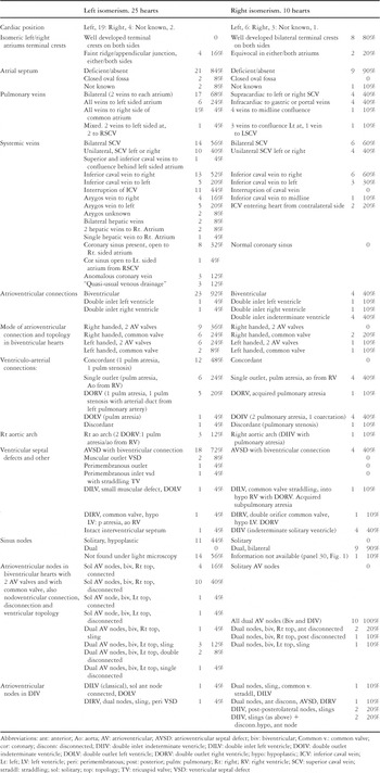
Table 1. Summary of characteristics of the 35 specimens showing comparisons between isomerism of the left and right atrial appendages.

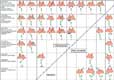
The cartoons in Figure 1 show details of the morphology and the conducting system in individual hearts. We have shown the types of isomerism, the arrangement of the atrial septum, the connections of the pulmonary, systemic and hepatic veins, the orifice of the coronary sinus or its equivalent coronary vein, and the conduction tissues. The colour key to the cartoons is shown in Figure 2. We have shown the sites of the sinus node, or both nodes if this structure is duplicated, and the disposition of the atrioventricular nodes and bundles, in the insets below the main diagrams. The plane of the diagrams is essentially coronal.

Figure 1. The figure summarizes the findings in 35 hearts with isomerism of the atrial appendages, in schematic form. It shows frontal views of each specimen, to include the atriums and their appendages, the status of the atrial septum, the superior, inferior and coronary veins. Interruption of the inferior caval vein is first seen in Figure 1, panel 1. The approximate locations of the sinus nodes are given for each case in individual panels. The atrioventricular conduction system in each heart is drawn below the atriums. Discontinuation of the atrioventricular conduction axis is first seen in Figure 1, panel 4. The key to the colour coding is given in Figure 2. Also included is the purple colouring given to hepatic or portal veins which connect with the heart only in some of those hearts with left isomerism of the atrial appendages.
Figure 1. The single atrioventricular node as shown, for example, in Figure 1, panel 1, is always in the posterior (inferior position) as in the normal (Fig. 2). The exception is seen in Figure 1, panel 24, where the single atrioventricular node, in double inlet left ventricle, lies in an anterolateral position. Where there are 2 atrioventricular nodes, the second node lies anteriorly (superiorly), providing the substrate for a sling (or a potential sling) of conduction tissue, along the crest of the ventricular septum (for example Fig. 1, panels 10 and 12). In double inlet to a solitary and indeterminate ventricle, where it is not possible to demonstrate a ventricular septum (Fig. 1, panels 32–35), the two nodes, connected by a sling, are orientated between an inferior and an inferolateral position, the sling also gives off a single bundle branch. A third, hypoplastic but disconnected node was seen superiorly in two of these cases (Fig. 1, panels 32 and 35). An accessory atrioventricular connection was found in one case which was known to have Wolf-Parkinson-White Syndrome (Fig. 1, panel 2).
Figure 1. Abbreviations: AV: atrioventricular; AVSD: atrioventricular septal defect; DILV: double inlet left ventricle; DIRV: double inlet right ventricle; NK: not known; VSD: ventricular septal defect.

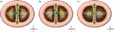
Figure 2. A diagram of the frontal view of the right and left atriums in the setting of usual atrial arrangement. A colour coding of the systemic venous connections and the conduction tissue is given as follows:
Figure 2. Superior and inferior caval veins, red; orifice of the coronary sinus or, as with isomerism, abnormal coronary vein, yellow; cardiac or, as in isomerism, extracardiac connections of the pulmonary veins, blue; hepatic or portal venous connections, purple.
Figure 2. Sinus node, green circle; atrioventricular nodes, green ellipse below atriums; non-branching and branching bundle, continuous green line (occasionally interrupted where dissociation was present); left bundle branch, green triangle.
We have compared subgroups, where possible, according to the presence of right or left isomerism, the modes of atrioventricular connection, and the ventricular topology (Table 1). Comparable specimens within the groups having right as opposed to left isomerism are shown across the same level in the main frame of Figure 1. Additional information in regard to the ventricles, and the atrioventricular and ventriculo-arterial connections, is summarized in the text, and also in Table 1. The prevalence of individual anomalies has been indicated by percentages. These are included only to aid the memory, and are not intended for use in statistical analysis, since we recognize that our choice of material was biased by the method of collection of the archives from which the hearts were obtained. The overriding heterogeneity of the complex anatomical arrangements precludes a definitive analysis in such a small series.
Diagrams 9, 10a, 10b, 10c and 11 are drawn showing a common atrioventricular junction in association with an atrioventricular septal defect because this was the prevalent atrioventricular junctional pattern in the hearts exhibiting biventricular atrioventricular connections. For clarity, the atrioventricular valves have not been shown, but took the form of either a common valve or two valves. In the remaining few hearts with two ventricles as opposed to a solitary ventricle, and in which the valves had developed normally, but the topology and the pattern of the conduction tissue were of the same arrangement as that shown for those with atrioventricular septal defects, the specific arrangements of individual atrioventricular valves is described in the legends.
Characteristics of the normal atriums
The normal atriums have several parts. Perhaps the most obvious, but certainly not the most constant, is the smooth-walled venous portion. The second part, also smooth-walled, is the vestibule of the atrioventricular valve. The third part, and the most constant, is the atrial appendage. Each atrium then has a body, larger in the left than in the right atrium, and the two atrial cavities are separated by the septum.
Externally, the morphologically right appendage is approximately triangular in shape, and usually has a broad junction with the rest of the atrium (Fig. 3a). This junction is marked by the terminal groove (“sulcus terminalis”). The venous portion receives the superior and inferior caval veins, and the coronary sinus. Internally, the terminal groove corresponds with a robust muscular structure called the terminal crest (“crista terminalis”). The groove also houses the sinus node. The internal wall of the appendage is covered with fine pectinate muscles (Fig. 3b). These extend at right angles from the terminal crest to the vestibule of the tricuspid valve, which surrounds the atrial side of the atrioventricular junction. The pectinate muscles run all round the junction almost to the orifice of the coronary sinus. The morphologically right side of the septum between the atriums shows the depression of the oval fossa (“fossa ovalis”), with an arc of prominent muscular margin representing the rim of the fossa opposite the opening of the inferior caval vein. This muscular margin is the so-called “septum secundum”, in reality the infolding of the interatrial roof between the connections of the caval veins to the right, and the pulmonary veins to the left atrium. On the right side of the atrial septum, the orifice of the coronary sinus and the annulus of the septal leaflet of the tricuspid valve form two sides of the so-called “triangle of Koch”. The third side is formed by the perceived line of the tendon of Todaro, which is a remnant of the embryonic venous valves of the right atrium. This, or its extension, runs forward to meet the tricuspid valvar annulus at the membranous part of the septum and forms the triangle which houses the atrioventricular node in the right atrial vestibule. When the atrial septum is absent or deficient, as in some cases of congenital heart disease, the atrioventricular node must develop elsewhere in the vestibule.9 Some of these situations will be described in the discussion.

Figure 3. An external view of a normal heart (Fig. 3a) shows the triangular shape of the right atrial appendage and its relationship with the superior and inferior caval veins. The appendage has a long junction with the sinus portion of the right atrium. The dashed line represents the long axis of the appendage and has been used approximately for an incision (as in Fig. 3b). The aortic root has been deflected for the purposes of the photograph. The morphologically right atrial appendage seen in Figure 3a has been opened to produce Figure 3b. The two portions, including the tricuspid valve, have been pulled apart to lie flat. The regular arrangement of pectinate muscles is extensive, encircling the vestibule of the right atrium between the tip of the appendage and the opening of the coronary sinus. Abbreviations: TV: tricuspid valve; ICV: inferior caval vein; SCV: superior caval vein.
The morphologically left atrium differs markedly from the right atrium. Externally, the left atrial appendage is a narrow tubular structure, having a narrow junction with the larger, smooth-walled, portion of the atrium (Fig. 4a). The pulmonary veins are anchored at the four corners of the atrial roof, which is confluent with the body of the atrium and the vestibule of the mitral valve. Although internally the walls of the left atrial appendage are also covered with fine pectinate muscles, there is no distinct muscular ridge separating them from the smooth-walled part of the atrium. A few of the pectinate muscles are seen occasionally to spill over into the smooth-walled part of the left atrium, but they do not encroach on to the posterior aspect of the vestibule (Fig. 4b). The left side of the atrial septum is marked by the flap valve of the oval fossa. Where the valve overlaps the margin of the oval fossa, there may be some slightly roughened areas.

Figure 4. The left atrial appendage (a) of the normal heart used in Figure 3 has a characteristic tubular and finger-like shape with crenellations along its edges. It has only a narrow junction with the sinus portion of the left atrium. In panel b, the appendage has been partly opened along its length, from its junction with the smooth-walled portion of the left atrium. The regular arrangement of the pectinate muscles can be seen inside the appendage. The vestibule of the left atrium is smooth-walled and the atrial septum has a roughened appearance. Abbreviation: MV: mitral valve.
In congenitally malformed hearts, the atrial septum is often partially or totally deficient, and the venous and atrioventricular connections are often anomalous. Because of this, only the appendages can be used with consistency to distinguish between the morphologically right and left atriums.2 In the setting of visceral heterotaxy, it is only the appendages which are isomeric within the heart, both showing the characteristics of either the morphologically right or the morphologically left appendage.2 The venous components are markedly variable, and need to be described in full, along with the atrioventricular junctional connections, the ventricular morphology, the position of the heart, and all the associated malformations (Table 1).
Results
Associated anatomic variables throughout the series, including isomerism of both right and left atrial appendages
More detailed information is given for comparison, in Table 1.
Atrioventricular septal defects with common atrioventricular junction were the most common septal defects, found in almost two-thirds of the entire series (63%). Muscular outlet defects, and perimembranous defects opening to the inlet and outlet of the right ventricle, were also present. One of the muscular outlet defects existed in the setting of double inlet and double outlet left ventricle. This heart differed from the classic type of double inlet left ventricle in which the aorta arises from the incomplete right ventricle. Of the perimembranous defects, one was associated with an overriding and straddling tricuspid valve. Another was present in the setting of double inlet left ventricle with a common atrioventricular valve, from which cords straddled into a hypoplastic right ventricle. In one case a perimembranous defect was present opening to the outlet of the right ventricle. We found 2 specimens with interventricular communications running to the crux of the heart in the setting of double inlet right ventricle with a common atrioventricular valve. In one of these, the hypoplastic left ventricle was little more than a pouch. Double inlet to a solitary but morphologically indeterminate ventricle was identified only with right isomerism. The ventricular septum was intact in one case only, this heart having isomeric left appendages and a cleft in the aortic leaflet of the mitral valve.
The ventriculo-arterial connections were concordant only with left isomerism (Table 1), with 2 of these showing pulmonary atresia and stenosis, respectively. Discordant ventriculo-arterial connections were present with both right and left isomerism, the case with right isomerism having pulmonary stenosis.
Single outlet of the heart, via the aorta from the right ventricle and with pulmonary atresia, was common to both types of isomerism. Single outlet with pulmonary atresia was also found in the hearts with right isomerism and solitary and indeterminate ventricle. Double outlet ventricle was the next most common ventriculo-arterial connection. Overall, pulmonary atresia, or significant pulmonary stenosis, was present in almost half of the whole group. It accounted for just over one-third of those with left isomerism, and seven-tenths of those with right isomerism.
The aortic arch was right-sided in one case with right isomerism, and 3 with left isomerism, while aortic coarctation was seen only once, specifically in a heart with right isomerism and double inlet indeterminate ventricle. “Unroofing” of the coronary sinus was present in one specimen with left isomerism, and a double orifice was found in the common valve of one heart with right isomerism deformed by Ebstein’s malformation (Table 1).
The isomeric atriums
Left isomerism: In 4 of the hearts we examined, a faint ridge was recognizable on one side, or the other, at the junction between the smooth venous portion of the atrium and the appendage.
Right isomerism: A well developed terminal crest was present bilaterally in 8 hearts. In the remaining two cases, the evidence for this structure was equivocal in both atriums (Table 1).
Atrial septation
Left isomerism (Fig. 1, panels 1–25): The atrial septum was deficient or absent in the majority, but in two cases the oval fossa was closed. Atrial septation could not be determined in 2 hearts.
Right isomerism (Fig. 1, panels 26–35): The septum was absent or grossly deficient in all cases but one, in which it proved impossible to determine the initial arrangement (Table 1).
It follows that when there is a common valve, or malalignment between the atrial and ventricular structures, the septums have not developed normally. This has consequences for the disposition of the specialized conduction tissues.
Pulmonary veins
Left isomerism (Fig. 1, panels 1–25): The pulmonary veins connected bilaterally, with 2 to each atrium in the majority, but in 6 cases all the veins connected to the left-sided atrium, and in one heart all connected to the right side of a common atrium. In the remaining heart, the two left pulmonary veins entered the left-sided atrium, and both right pulmonary veins connected to the right superior caval vein (Table 1).
Right isomerism (Fig. 1, panels 26–35): In 4 hearts, the pulmonary veins connected in supracardiac fashion to either the right or the left superior caval vein, and in another 4, the veins, having passed through the diaphragm, connected with either the gastric or the portal veins. In neither of the 2 cases in which the veins joined the heart were the pulmonary veins connected in an anatomically normal pattern, because of the presence bilaterally of morphologically right appendages. In one of these cases, all the pulmonary veins connected to a midline confluence behind the heart, which drained into the centre of the virtually common atrium (Fig. 1, panel 26). In the last heart, 3 of the pulmonary veins joined a left-sided confluence which connected with the left atrium and the fourth drained to the left-sided superior caval vein.
Systemic veins
Left isomerism (Fig. 1, panels 1–25): In 14 cases, there were bilateral superior caval veins and 10 cases had a unilateral superior caval vein, right-sided in 7 and left-sided in 3. In one heart, the superior caval vein met the inferior caval vein in a confluence behind the left atrium before entering the heart (Fig. 1, panel 20). Interruption of the inferior caval vein was seen in 11 cases, just less than half of the group, with almost even distribution between continuation through the right-sided and the left-sided azygos systems. The channel of continuation was unknown in 2. In another 5 cases with interruption, the hepatic veins were connected directly to the heart, entering the atriums bilaterally in 2 cases, both entering the right-sided atrium in 2 hearts, with a single hepatic vein entering the right-sided atrium in one specimen (Table 1).
Only 3 hearts showed a “quasi-usual” pattern of venous drainage, such that the superior and inferior caval veins and the coronary sinus returned to the right-sided atrium, and all the pulmonary veins drained to the left-sided atrium. Even amongst these, 2 had persistent left superior caval veins draining to the coronary sinus, and in the other the inferior caval vein was interrupted. The coronary sinus was absent in 14 cases, accounting for more than half of the cases with left isomerism, with the coronary veins in these hearts joining directly to the atrial chambers (Table 1). When present, the coronary sinus opened normally to the right-sided atrium in 8 cases, to the left-sided atrium in one case, but as a well developed abnormal coronary vein to either the midline or the left-sided atrium in 3 hearts (Fig. 1, panels 1, 8 & 20).
Right isomerism: (Fig. 1 panels 26–35): Bilateral superior caval veins were present in 6 hearts. In 2 other cases, unilateral inferior and superior caval veins drained one to the right-sided and the other to the left-sided atrium. A further heart displayed a unilateral left-sided superior caval vein, with a mid-line inferior caval vein.
In 2 hearts, bilateral inferior caval veins entered the right- and left-sided atriums, each from the contralateral side of the spine. A coronary sinus was not identified in any heart with isomerism of the right atrial appendages, the coronary veins joining directly to the atriums (Table 1).
Atrioventricular connections
Left isomerism: The atrioventricular connections were biventricular in 23 hearts, with 15 showing right-hand topology, and the remaining 8 having left-hand topology (Fig. 1, panels 1–23). A further heart had double inlet with double outlet left ventricle (Fig. 1, panel 24), and another showed double inlet right ventricle through a common valve, the aorta arising from the right ventricle, and pulmonary atresia (Fig. 1, panel 25) (Table 1).
Right isomerism: The atrioventricular connections were biventricular in 4 cases with common atrioventricular junction and atrioventricular septal defects, 2 of these having right-hand and 2 left-hand topology (Fig. 1, panels 26–29). Double inlet left ventricle through a straddling common valve was present in one heart with double outlet right ventricle, allegedly having acquired pulmonary atresia (Fig. 1, panel 30). Double inlet to and double outlet from the right ventricle, with pulmonary atresia, was found in one case with an incomplete and hypoplastic left ventricle (Fig. 1, panel 31). Double inlet to a solitary and indeterminate ventricle was found in 4 hearts (Fig. 1, panels 32–35). In 2 of these, there was double outlet from the solitary ventricle, but 2 had pulmonary atresia, with only the aorta arising from the solitary and indeterminate ventricle.
Significantly, dysplastic and stenotic atrioventricular valves were common with both right and left isomerism.
Modes of atrioventricular connection and topology of the ventricles
Left isomerism: Right-handed ventricular topology with biventricular atrioventricular connections was present in the majority of the hearts (Fig. 1, panels 1–9, 16–21). Of these, 8 cases had two atrioventricular valves, while 7 had common atrioventricular valves. Left-handed ventricular topology was present in a further 6 hearts with left isomerism and 2 atrioventricular valves (Fig. 1, panels 10–15, 22–23), including 4 having atrioventricular septal defects with separate valvar orifices. Atrioventricular septal defects and common valvar orifice was associated with left-hand topology in 2 hearts. The remaining 2 hearts had double inlet ventricle (Fig. 1, panels 24 & 25) (Table 1).
Right isomerism: In this group, 6 also showed double inlet. This was to the right ventricle in one, to the left ventricle in another, and to a solitary ventricle in 4 (Fig. 1, panels 30–35) (Table 1). Of the remaining 4 which had biventricular atrioventricular connections, 2 with common atrioventricular valves showed right-hand topology (Fig. 1, panels 27 & 28). The third case, also with a common valve, had left-hand topology (Figure 1, panel 29), while the fourth case, again with left-hand topology, had 2 atrioventricular valves (Fig. 1, panel 26).
The ventriculo-arterial connections
Left isomerism: The ventriculo-arterial connections were concordant in almost half of the hearts in this group, irrespective of the ventricular topology. Single outlet, with pulmonary atresia and the aorta arising from the morphologically right ventricle, and double outlet right ventricle were equally represented amongst the rest with 6 cases each, apart from one with discordant connections and pulmonary stenosis. In one of the specimens with double outlet right ventricle and pulmonary stenosis, the arterial duct branched from the left pulmonary artery (Table 1).
Right isomerism: Single outlet of the heart with pulmonary atresia, with the aorta arising from the morphologically right or the indeterminate ventricle, was the most common ventriculo-arterial connection. It was seen in 2 specimens with potentially double outlet right ventricle, 2 with potentially double outlet indeterminate ventricle, and 2 in which the origin of the pulmonary artery was in doubt. Discordant ventriculo-arterial connections were present in one heart with pulmonary stenosis. In one further heart, there was double outlet from the right ventricle, and double outlet from an indeterminate ventricle in 2 others, one of which had associated coarctation of the aorta. Thus, within the overall group, seven-tenths of the hearts with isomerism of the right atrial appendages were known to have obstruction to the pulmonary outflow from the heart (Table 1).
The specialised conduction system
The sinus nodes: For the purposes of comparison, the arrangements in the normal heart, and those found in the mirror image of the normal, are shown in Figures 5a and 5b.

Figure 5. The approximate positions of the sinus nodes as seen in serial microscope sections, are shown in red in (a) the normal heart, (b) the mirror image of the normal, (c) in green, examples of the grossly abnormal and unpredictable locations, if present in histological sections, in isomerism of the left atrial appendages and (d) in red, isomerism of the right atrial appendages.
Left isomerism: Solitary sinus nodes were discovered in less than half of the hearts, but only after intensive searching. They were always hypoplastic, and abnormally positioned (Fig. 1, panels 1–25). They lay either to the right, or to the right and laterally, posteriorly or posterolaterally, when within the right-sided atrium, but usually close to the junctional area with the appendage or, as seen in one case only, adjacent to the root of the superior caval vein. They lay inferiorly when found within the left-sided atrial myocardium (Fig. 5c).
Right isomerism: Sinus nodes were observed bilaterally in all of the hearts in this group, except for one specimen, from which the roots of the superior caval veins had been removed during the initial autopsy, thus preventing discovery of the site of the sinus nodes (Fig. 1, panels 26–35; Figures 5d & 6). They were always located at the roots of the superior caval veins, when these were also present bilaterally, as seen in 6 hearts. In 3 cases with only a unilateral superior caval vein, sinus nodes were still found bilaterally. In each of these, one sinus node was located as expected at the root of a superior caval vein, while the twin node was found on the crest of the atrial appendage on the other side of the heart, at the anticipated site for a second superior caval vein.

Figure 6. A photomicrograph of the dual sinus nodes (encircled) in isomerism of the right atrial appendages (Fig. 1, panel 33). They are always located at the junction between the roots of the superior caval vein, or veins, and the crests of the bilateral, morphologically right atrial appendages.
Figure 6. Abbreviations: RSCV: right superior caval vein; LSCV: left superior caval vein.
The atrioventricular conduction system
Left isomerism: Amongst those hearts with biventricular atrioventricular connections, 15 had two atrioventricular valves, and 8 had common valves. A different set of 15 hearts had right-hand ventricular topology, while 8 had left-hand topology. One further heart had double inlet, together with double outlet, from the left ventricle, and the last had double inlet right ventricle.
The arrangement of the conduction tissues in the heart with double inlet left ventricle was as expected for this atrioventricular connection when seen with usual atrial arrangement. Thus, a single anterolateral node connected to a non-branching bundle, which then bifurcated and ramified into both the dominant left ventricle and the incomplete anterior right ventricle (Fig. 1, panel 24; Fig. 7). In the case with double inlet right ventricle, there were paired atrioventricular nodes connected by a sling of conduction tissue across the crest of the ventricular septum, the sling branching to supply both the dominant right and the incomplete left ventricles (Fig. 1, panel 25; Fig. 8). In this heart, the aorta arose from the right ventricle, and there was pulmonary atresia. A similar pattern was seen in one heart with right isomerism, but in which there was dissociation of the anterior node (Fig. 1, panel 31).

Figure 7. The cartoon illustrates double inlet left ventricle with isomerism of the left atrial appendages (1 heart, Fig. 1, panel 24). Both atriums connect to a dominant left ventricle, through two atrioventricular valves in this heart. The base of the heart is shown from above to simulate a view after removal of the atriums and the atrioventricular valves. There is gross malalignment of the ventricular septum, which lies approximately from right to left, and away from its normal position. It carries the atrioventricular conduction axis. A normally positioned atrioventricular node would fail to make contact with the ventricular conduction tissue. The result is that the connecting atrioventricular node lies in a right, superolateral position. It meets with the nonbranching bundle and then the bundle branches, which are orientated apically and towards the left, although proximally always running to the right (acute marginal) side of the ventricular septal defect (double headed arrow).

Figure 8. This cartoon illustrates the arrangements in 2 cases with double inlet right ventricle, one with isomerism of the left atrial appendages (Fig. 1, panel 25) and one with isomerism of the right atrial appendages (Fig. 1, panel 31). In the first case, (Fig. 1, panel 25), both atriums connect with the dominant right ventricle, from which the aorta and the atretic pulmonary artery arise. An uninterrupted sling of conduction tissue is present across the ventricular septum, between an inferior and a superior node. The left ventricle lies on the left posterior quadrant of the ventricular mass and is extremely hypoplastic. A similar arrangement of the ventricles and the conduction tissue was found in the heart with right isomerism of the atrial appendages, but in which the left ventricle was better developed and the superior node was disconnected (broken line) (Fig. 1, panel 31).
Single atrioventricular nodes were found in approximately two-thirds of the hearts with left isomerism and biventricular connections. In 10 of these with right-hand topology, and one case with left-hand topology, the nodes were dissociated from the ventricular conduction axis, all but one being known to have had atrioventricular dissociation during life (Fig. 9). Dual atrioventricular nodes were present in 6 hearts with left isomerism, one with right-hand topology (Fig. 10a), but 5 with left-hand topology (Fig. 1, panels 9, 10–14; Fig. 10b). Of these, from the 5 with left-hand topology, 3 had discontinuity in the conduction axes (Fig. 10b). Both nodes were disconnected from the common bundle in 2 of them, and both of these patients had been known to have atrioventricular dissociation in life (Fig. 1, panels 12 & 13). In the third patient, only the anterior node was joined to the ventricular conduction pathways (Fig. 1, panel 14). In only 4 of the hearts with paired atrioventricular nodes and left isomerism were both nodes connected by a continuous sling of conduction tissue. Of these, one had right-hand (Fig. 10a) and 3 had left-hand ventricular topology (Fig. 1, panels 9, 10,11 & 22).

Figure 9. The cartoon shows biventricular connections with a single, inferior node and right-hand topology in isomerism of the left atrial appendages, as found in 14 of the hearts examined (Fig. 1, panels 1–8 & 16–21). In 10 of these, the nonbranching bundle is disconnected from the atrioventricular node (broken line). Three hearts had well developed mitral and tricuspid valves, including one with an intact ventricular septum and 2 with ventricular septal defects. There were 11 cases with atrioventricular septal defects of which 7 had common valves and 4 had 2 valvar rings.

Figure 10. Panel a shows the arrangement in one heart with isomerism of the left atrial appendages, biventricular connections, right-hand topology and an atrioventricular septal defect with a common valve. Dual atrioventricular nodes are connected by an uninterrupted sling of conduction tissue (Fig. 1, panel 9). In panel b, we illustrate the arrangement as seen in 5 hearts with isomerism of the left atrial appendages with biventricular connections, left-hand topology and atrioventricular septal defects, one having a common valve and 4 with separate valvar orifices for the right and left ventricles (Fig. 1, panels 10–13 & 22). A sixth case with biventricular atrioventricular connections (Fig. 1, panel 14) with isomerism of the left atrial appendages and left-hand topology has a ventricular septal defect with a straddling tricuspid valve. All of these hearts have dual atrioventricular nodes. In 3 the nodes are connected by an uninterrupted sling of conduction tissue, whilst in the fourth, with straddling valve through a perimembranous ventricular septal defect, the inferior node is disconnected (Fig. 1, panel 14). In the remaining 2 hearts with biventricular atrioventricular connections and separate orifices for the right and left ventricles, both nodes were disconnected (Fig. 1, panels 12 & 13). In two specimens with biventricular atrioventricular connections and isomerism of the right atrial appendages, left-hand topology and atrioventricular septal defects (Fig. 1, panels 26 & 29), dual atrioventricular nodes were connected by an uninterrupted sling of conduction tissue. In panel c, we show the arrangement found in 2 hearts with isomerism of the right atrial appendages, biventricular atrioventricular connections, right-hand topology and atrioventricular septal defects with common valves. Each has dual atrioventricular nodes (Fig. 1, panels 27 & 28). Right bundle branches were not found in either case. A left bundle branch (boxed) was identified in one case only (Fig. 1, panel 27). The potential sling was interrupted in both hearts, isolating the superior node in one (Fig. 1, panel 27) and the inferior node in the other (Fig. 1, panel 28).
In 2 hearts with left isomerism and left-hand topology, we found a ‘dead-end tract’. In each case, this tract ran anteriorly from the bifurcation of the left bundle branch until it could no longer be identified in the histological sections (Fig. 1, panels 15 & 23; Fig. 11). The right bundle branch was not seen in one of these hearts (Fig. 1, panel 15).

Figure 11. The cartoon shows biventricular atrioventricular connections with a single inferior node and a “dead end tract”, left-hand topology and isomerism of the left atrial appendages. This arrangement was seen in 2 hearts (Fig. 1, panels 15 & 23). One was a case of atrioventricular septal defect with a common valve, but the other had mitral and tricuspid valves with a muscular defect to the outlet of the heart. The right bundle branch could not be identified in this case (boxed area).
Right isomerism: Dual atrioventricular nodes were present in all 10 hearts (Fig. 1, panels 26–35). In 4 of these, the atrioventricular connections were biventricular, 2 having left-hand topology and slings of conduction tissue (Fig. 1, panels 26 & 29; Fig. 10b), while in a further case, with right-hand topology, the superior node was disconnected and we could not identify a right bundle branch (Fig. 1, panel 27; Fig. 10c). In the fourth case, the inferior node was disconnected from the ventricular conduction axis, but we were not able to identify either right or left bundle branches (Fig. 1, panel 28; Fig. 10c). In the remaining 6, there was double inlet ventricle, including one in which two right sided leaflets of a common atrioventricular valve straddled through a ventricular septal defect into an incomplete right ventricle. This arrangement produced double inlet left ventricle, with obstruction to the right sided and hypoplastic right ventricle. A sling of conduction tissue was present (Fig. 1, panel 30; Fig. 12). The aorta arose from the right ventricle, along with the pulmonary trunk, but there was subpulmonary muscular atresia. In another heart, there was double inlet right ventricle through a common valve, with a left-sided, hypoplastic, left ventricle. In this heart, we found both posterior and anterior nodes, but only the posterior node was connected to the ventricular conduction axis (Fig. 1, panel 31). A similar pattern had been seen, albeit without disconnection in the conduction axes, in a heart with isomerism of the left appendages (Fig. 1, panel 25; Fig. 8), as described above. Altogether, in only 3 of the 6 hearts were both nodes connected by a continuous sling of conduction tissue (Fig. 1, panels 26, 29 & 30).

Figure 12. The cartoon shows the pattern in double inlet left ventricle with isomerism of the right atrial appendages, in which a common atrioventricular valve straddles into a hypoplastic right ventricle (1 heart). The aorta and the atretic pulmonary trunk both connect to the rudimentary right ventricle (Fig. 1, panel 30).
In each of the remaining 4 hearts, all with double inlet to a solitary and indeterminate ventricle, we found dual atrioventricular nodes, which were connected by a sling of ventricular conduction tissue orientated approximately in an axis between posterior and left posterolateral positions in the ventricles (Fig. 1, panel 32–35; Fig. 13). In all 4 cases, the sling of conduction tissue gave off a branching strand. These strands were reminiscent of the usual right bundle branch, and merged within the ventricular myocardium towards the apex. A third, hypoplastic, node was identified anteriorly just above the atrioventricular annulus in two of the hearts with indeterminate ventricles, but in both it was disconnected from the main axis (Fig. 1, panels 32, 35; Fig. 13). In one case, the branching strand was split into two parts lengthwise for a short distance around a bundle of myocardial cells, before coming together and continuing towards the apex, where it ramified into the myocardium (Fig. 1, panel 32). Clinical information was available for only seven of these patients, but showed that all had been in sinus rhythm during life.

Figure 13. The patterns found in 4 hearts with double inlet to an indeterminate ventricle with isomerism of the right atrial appendages (Fig. 1, panels 32–35). In each case, dual atrioventricular nodes are present and are connected by uninterrupted slings of conduction tissue. The nodes are positioned inferiorly and left inferolaterally in the absence of a ventricular septum. A strand of conduction tissue branches from the sling and merges with the ventricular myocardium towards the apex. In two hearts, a third, hypoplastic node was discovered in a superolateral position, but in neither was there a connection with the main axis.
Discussion
The human body, for its larger part, is built on the basis of bilateral symmetry. Thus, the muscles, arteries, veins, nerves, and skeletal structures are mirror-images of each other on the right and left sides of the body. Such parietal structures, following the analogy of chemical structure, are justifiably described as being isomeric. The thoraco-abdominal organs of the body, in contrast, such as the lungs, spleen, liver and gut, do not show evidence of isomerism, but have a distinctive arrangement for the right and left sides that can be described in terms of lateralization. Such lateralization is also seen within the heart, where the so-called “right” and “left” sides are morphologically distinct, albeit that these structures are not strictly right-sided or left-sided. In some rare circumstances, the entire bodily arrangement can be mirror-imaged, so that the structures that are morphologically right in the usual situation are found on the left side of the body. This arrangement is usually called “situs inversus”, although the inversion is one of mirror-imagery, rather than “upside-downness”. In situations that are far more common in the setting of congenital malformations, albeit still rare within the overall population, the arrangement of the organs can parallel the arrangement of the parietal structures, with the morphology of the organs being mirror-imaged on the right and left sides of the body. Such visceral isomerism has long been recognized for the lungs and the bronchial tree,4, 10 but its existence within the heart has remained controversial, albeit that new evidence emerging from genetically modified mice shows that isomerism, at least for the appendages, is an unequivocal finding.11, 12
Thus, the previous controversy has largely devolved on the definitions used for isomerism, and the structures used as the arbiters for recognition of the cardiac chambers. As long ago as 1968, Van Mierop and Wigglesworth13 had demonstrated unequivocal isomerism of the sinus nodes in the setting of visceral heterotaxy with absence of the spleen. And, in the subsequent review of so-called “situs ambiguus” made by Van Mierop,4 the diagrams unequivocally show isomerism of the atrial appendages. Furthermore, using the principle of the morphological method, which states that structures should be identified on the basis of their most constant features, and not according to other structures that are themselves variable, it is the atrial appendages which permit the most reliable distinction between the right and left atrial chambers. As with the genetically modified mice,11, 12 therefore, when decisions are based on the criterion of the extent of the pectinate muscles judged relative to the atrioventricular junctions, the existence of isomeric atrial appendages in the setting of visceral heterotaxy is now unequivocal.2 Those who have denied the existence of isomerism within the atrial chambers have been able do so only because they used the connections of the veins to distinguish between the morphologically right and left atriums.14 This approach, of course, breaches the “morphological method” in egregious fashion. It is the atrial appendages that are isomeric, rather than the entirety of the atrial chambers, albeit that in the animals perturbated by genetic manipulation, there is far greater evidence also for isomerism of the veno-atrial connections.11, 12 In humans, nonetheless, when considering the atrial appendages, then our current investigation confirms that the existence of isomerism as opposed to lateralization is now beyond question.
The main purpose of our study was to provide guidelines for the prediction of the location of the conduction tissues in the settings of right and left isomerism. Based on our previous findings, and endorsed by the findings in the hearts we specifically examined for the purposes of this review, we are comfortable that the arrangements can be predicted with a considerable degree of accuracy.
The normal sinus node is located at the junction of the superior caval vein with the morphologically right atrial appendage.15 In the normal rat, its primordium can be demonstrated by immunoreactivity to an antigen raised against human natural killer cells on the 14th day of gestation,16 before the time of any migration of cells from the neural crest. Coalescence of some of the positive cells identified in this fashion in the developing right atrium was suggested to produce one of the moieties of the developing right sided sinus node, with a similar transient arrangement on the left side having disappeared by the end of the 14th day of gestation. Earlier, scattered positive collections of cells were found peripherally in both right and left atriums. The authors suggested that such gene expression reflects the multiple potential of early cardiac myocytes to become either working myocardium or specialised conduction tissue.16 Thus far, however, reactivity to the antigen raised against the natural killer cells has not been demonstrated to coincide with either pacemaking or specialised conducting properties of other myocardial cells.9
It was no surprise to find duplication of the sinus node in all cases of right isomerism, as observed initially by Van Mierop and Wigglesworth,13 and subsequently confirmed by other investigators.7, 17, 18 In patients with right isomerism, therefore, the surgeon should respect the terminal grooves found in relation to both atriums, because almost certainly each groove harbours a sinus node (Figs. 5d, 6), even in the presence of only one superior caval vein. In left isomerism, in contrast, the situation is far less clear-cut. As shown in our earlier investigation7 and as confirmed by Ho et al.,18 even when a sinus node is discovered histologically, it is hypoplastic and located much closer to the atrioventricular junctions (Fig. 5c). Thus, it is not possible to give unequivocal surgical guidelines for the location of the sinus node in the setting of left isomerism. The surgeon should respect all the veno-atrial junctions, and as far as possible should avoid making incisions, or placing sutures, in the atrioventricular vestibules.
In the normal heart, the atrioventricular node is often considered to be a component of the atrial septum. In reality, the atrioventricular node is a remnant of the atrioventricular canal musculature, which is “specialised” during development, and which becomes sequestrated in normal development as the vestibule of the right and left atriums.9 It is remnants of this musculature that form the “ring tissue”. The node is a septal component in the normal because this is part of the ring that is in continuity with the ventricular conduction axis formed on the crest of the ventricular septum, and which is identified in the developing human heart on the basis of its reactivity with an antibody to the nodose ganglion of the chick.19 The node becomes an atrial structure only when the primary atrial septum is able to grow down and cover the atrial aspect of the ring during normal atrioventricular septation. This forms the triangle of Koch, and also the muscular atrioventricular sandwich, with the node contained within its atrial aspect. In reality, however, the node is an atrioventricular junctional structure, since it is directly related to the epicardial tissue of the atrioventricular junction. The two atrioventricular junctions expand backwards and inferiorly during normal septation, producing the apparent paradox that the node, at the apex of the triangle, is in the middle of the heart. In fact, it has retained its epicardial position, but the tricuspid valvar orifice has expanded inferiorly to overlap the ventricular septum, thus producing the muscular atrioventricular sandwich. This cannot happen when there is a common atrioventricular junction, so that the node, still part of the atrial vestibule, is seen at the crux of the heart. Nor can it happen when there is malalignment between the atrial and ventricular structures, as in double inlet left ventricle, straddling tricuspid valve or corrected transposition. In these abnormalities, the node is then formed from the part of the initial ring of specialised cells that has become part of the vestibule, but which has also retained its continuity with the bundle formed on the crest of the muscular ventricular septum. Thus, the persisting part of the specialised canal musculature, which persists as the node, is found at the point where the septum joins the atrioventricular junction. That is why the atrial septum, and the location of the coronary sinus, cannot always provide a reliable landmark in these anomalies.
Our findings show, nonetheless, that it is still possible to construct guidelines for the likely location of the atrioventricular conduction tissues. In the hearts with biventricular atrioventricular connections, the key is the ventricular topology. Although the atrial appendages are isomeric in the setting of visceral heterotaxy, there is very little possibility for ventricular isomerism. Indeed, to the best of our knowledge, only one heart has ever been described with isomeric ventricles, this particular specimen having morphologically right ventricles on each side.20 The reason the atrial chambers can show evidence of isomerism, but not the ventricles, is that the atrial appendages both expand from the same segment of the heart tube, whereas the ventricular apical trabecular components develop in sequence from separate segments of the primary tube.21 It can reasonably be assumed, therefore, that the genes responsible for producing the characteristically fine or coarse trabeculations which permit distinction of the morphologically left and right ventricles are not part of the sequence of genes, including pitx211, cited212, and MLC3F22, which determines morphologically leftness or rightness within the general body plan, including the atrial appendages (Personal communication from Dr Deborah J. Henderson, Institute of Human Genetics, University of Newcastle upon Tyne, United Kingdom, December, 2005). Irrespective of whether there is isomerism of the right or left atrial appendages, therefore, when there are biventricular atrioventricular connections, the ventricles can exhibit right-hand or left-hand topology in random fashion (Fig. 1, panels 1–35). But it is clear that, whenever there is left-hand ventricular topology in the setting of isomeric atrial appendages (Fig. 1, panels 10–15, 26, 29), the atrioventricular conduction axes are anomalous.
We know from our study of hearts with other malformations, nonetheless, that an anomalous atrioventricular conduction axis is not necessarily the product of left-hand ventricular topology together with associated discordant atrioventricular connections. In Figure 14, we show the base of the normal heart as seen from above, depicting the course of the atrioventricular conduction axis. If the entire heart is mirror-imaged, as in the setting of mirror-imaged atrial arrangement with concordant atrioventricular connections, then the atrioventricular conduction axis is also mirror-imaged (Fig. 15). It arises from a regularly positioned atrioventricular node, found at the apex of the triangle of Koch in the left-sided morphologically right atrium. Left-hand ventricular topology, therefore, does not in itself produce an abnormal ventricular conduction axis. When left-hand ventricular topology is found with usual atrial arrangement and discordant atrioventricular connections, that is to say as in congenitally corrected transposition, then it is common to find the atrioventricular conduction axis arising from an anomalous anterior atrioventricular node23 (Fig. 16). In contrast, in the setting of mirror-imaged atrial arrangement, earlier work had suggested that, whenever discordant atrioventricular connections existed, a regular node would be the rule.24 It has now been realised that most of the hearts sectioned with this combination had either pulmonary atresia, or double outlet from the morphologically right ventricle. These features permit a greater degree of atrioventricular septal alignment, and hence provide the potential for formation of a regular conduction axis25 (Fig. 17). Dual atrioventricular nodes, nonetheless, may be found with congenitally corrected transposition in the setting of either usual or mirror image atrial arrangement, occasionally forming a sling, albeit that, in most cases, one or other node is disconnected from the main axis. The new investigation,25 combined with previous knowledge,23 shows that it is the degree of atrioventricular septal malalignment that is largely responsible for the abnormal atrioventricular conduction axis now recognized as part and parcel of congenitally corrected transposition26 (Figs. 16 & 17).

Figure 14. The cartoon shows a diagram of the base of a normal heart after removal of the atriums, showing atrioventricular and ventriculo-arterial valves and the normal course of the conduction tissue, reflecting right-hand ventricular topology.

Figure 15. The cartoon shows the base of a heart in the mirror-imaged normal situation, with mirror-imagery also of the course of the conduction tissue. There is left-hand ventricular topology.

Figure 16. The base of a heart with usual atrial arrangement and discordant connections at both the atrioventricular and ventriculo-arterial junctions – in other words congenitally corrected transposition. An inferior node is often present, but is usually disconnected from the main axis of the conduction tissue because of malalignment between the atrial and ventricular septums. This pattern allows for accommodation of the pulmonary trunk. The connecting atrioventricular node is located superolaterally.

Figure 17. The base of a heart with discordant atrioventricular and ventriculo-arterial connections, but in the setting of mirror imaged atriums and with pulmonary atresia. Better alignment exists between the atrial and ventricular septums allowing for an inferiorly located atrioventricular node and connection.
The situation is more complicated when the atrial appendages are isomeric. This is because the majority of hearts with isomeric appendages and biventricular atrioventricular connections also have a common atrioventricular junction. This feature is particularly striking with left isomerism. In the setting of a common atrioventricular junction, the ventricular septum meets the junctions both antero-superiorly and postero-inferiorly, with no potential wedging of the subarterial outlets to produce atrioventricular septal malalignment. Additionally, the atrial septum itself is very poorly formed. This is especially characteristic of right isomerism, often with only a strand crossing the cavity of an effectively common atrial chamber. In these morphological settings, it is the ventricular topology that determines the arrangement of the atrioventricular conduction axis. We have shown that, when there is right-hand topology with left isomerism, then there is usually a solitary atrioventricular node, which is formed at the point where the ventricular septum meets the atrioventricular junction postero-inferiorly, albeit that the axis itself is often discontinuous, giving the substrate for atrioventricular block5 (Fig. 9). Rarely a sling is formed (Fig. 1, panel 9; Fig. 10a). When there is left-hand topology, irrespective of whether there is right or left isomerism, there are usually dual atrioventricular nodes, often connected by a sling, but similarly, frequently showing discontinuity in the penetrating bundle (Fig. 10b). In the setting of a common atrioventricular junction, these findings are present irrespective of whether the atrioventricular valve is also common, or is arranged with separate orifices for the right and left ventricle – the so-called “ostium primum” defect.
In our series with right isomerism, all the hearts possessed dual atrioventricular nodes, along with dual sinus nodes (Fig. 1, panels 26–35). Amongst the group, two-fifths had biventricular atrioventricular connections with atrioventricular septal defect, half showing right-hand topology, each with disconnection of one node, and half with left-hand topology and slings of ventricular conduction tissue. Again, therefore, ventricular topology exerted the major influence on the disposition of the conduction tissues. It was the cases with double inlet to a solitary and indeterminate ventricle, however, which had the most interesting pathways for the ventricular conduction axis. In these hearts, the dual atrioventricular nodes were joined to a bizarre sling of ventricular conduction tissue. The sling was always positioned within the postero-inferior wall of the solitary ventricular chamber, there being no identifiable ventricular septum in the setting of a solitary and indeterminate ventricle. The sling, however, gave rise to a solitary strand of conduction tissue that supplied the ventricular myocardium, and was reminiscent of the normal right bundle branch (Fig. 13). This finding raises the possibility that the solitary ventricle might be of right rather than indeterminate morphology, the morphologically left ventricle being so small as to be unrecognizable. When a solitary ventricle is found in the setting of usual atrial arrangement, the connecting node is formed at the acute margin of the right atrioventricular junction. It again gives rise to a solitary strand of ventricular conduction tissue, with no evidence of a ventricular sling.27
Where heart block existed with isomerism of the left atrial appendages, the interruption of the conduction axis was always distal to the atrioventricular node. As no evidence has been found of abnormal formations of the atrioventricular insulating fibrous tissue, the cause of the interruption has yet to be established.5
We can conclude from our study that the pathways for atrioventricular conduction in the general setting of isomeric atrial appendages are conditioned by both the atrioventricular connections and by ventricular topology. When there is double inlet left ventricle with left isomerism, then the functional atrioventricular conduction pathway is formed antero-superiorly because of the malalignment of the ventricular septum. It takes origin from a right-sided anterior atrioventricular node, irrespective of whether the rudimentary right ventricle is right sided or left sided (Fig. 7), analogous with double inlet left ventricle in the usual atrial arrangement. In the setting of double inlet right ventricle, ventricular topology is the key (Fig. 8). Regular postero-inferior conduction pathways are to be anticipated in the setting of right-hand topology when there is left isomerism, albeit usually discontinuous, hence producing the common finding of heart block. Dual atrioventricular nodes are to be anticipated in the setting of left-hand topology, irrespective of the presence of left or right isomerism, with the potential to form continuous ventricular slings of conduction tissue between the nodes. The arrangement of the sinus nodes is exclusively dependent on the type of isomerism, with the nodes always being duplicated in the setting of right isomerism, and being hypoplastic or even absent histologically, when there is left isomerism.
Acknowledgements
The research upon which this review is based was supported by grants from the British Heart Foundation, the Joseph Levy Foundation, the Endowment Fund of the Royal Liverpool Children’s Hospital NHS Trust and the National Heart Research Fund.
Research at the Institute of Child Health, London and Great Ormond Street Hospital for Children NHS Trust benefits from R&D funding received from the NHS Executive. We are particularly grateful to Gemma Price for her helpful, imaginative, and expert contribution to the production of the diagrams.