Introduction
Laryngeal squamous cell carcinoma is one of the most common head and neck cancers. Its early detection and diagnosis is essential in order to maximise cure rates and to preserve vocal function.Reference Chu and Kim1 Radiological evaluation with computed tomography (CT) and magnetic resonance imaging (MRI) provides vital information on the extent of the primary tumour and the presence of cervical lymph node metastasis, but fails to identify superficial mucosal abnormalities.Reference Blitz and Aygun2 At present, white light laryngoscopy combined with biopsy is the standard diagnostic procedure in the assessment of laryngeal cancer and precancerous lesions. However, white light laryngoscopy provides poor quality images and has difficulty identifying minute epithelial changes and directly differentiating benign from malignant tumours in vivo.
Recently, a novel, high-resolution endoscopic technique, narrow band imaging, has begun to be widely used in the early diagnosis of gastrointestinal cancer.Reference Muto, Horimatsu, Ezoe, Hori, Yukawa and Morita3, Reference Larghi, Lecca and Costamagna4 Narrow band imaging is based upon the fact that the depth of light penetration depends on the light wavelength: the longer the wavelength, the deeper the penetration.Reference Gono, Obi, Yamaguchi, Ohyama, Machida and Sano5 Narrow band imaging modifies the broadband white light from a xenon lamp into two narrow band illumination beams with central wavelengths of 415 and 540 nm, which are designed to primarily penetrate the mucosa and submucosa. Narrow band blue light (415 nm) displays superficial capillary networks, while narrow band green light (540 nm) displays subepithelial vessels. When these two wavelengths are used in combination, they provide an extremely high contrast image of the tissue surface.
Currently available evidence indicates that narrow band imaging may be a promising approach in the diagnosis of laryngeal cancer.Reference Watanabe, Taniguchi, Tsujie, Hosokawa, Fujita and Sasaki6
The present study aimed to investigate the microvascular characteristics of different laryngeal mucosal states, ranging from benign to malignant, using narrow band imaging, and also to evaluate the value of narrow band imaging in the early detection of laryngeal cancer and its precursor lesions.
Patients and methods
Patients
We enrolled a total of 85 consecutive patients with suspected precancerous or cancerous laryngeal lesions who were seen in the endoscopy unit of the Cancer Institute and Hospital, Chinese Academy of Medical Sciences, between December 2008 and May 2009.
Exclusion criteria included: allergy to lidocaine; severe, uncontrollable dyspnoea; unstable angina; haemorrhagic disease; and inability to understand and sign an informed consent form.
The study was approved by the medical ethics committee of the above hospital, and informed consent was obtained from each patient prior to inclusion.
Endoscopic laryngeal examination and narrow band imaging
Narrow band imaging was performed using the Olympus Evis Lucera 260 system and a BF-6C260 video-bronchoscope (Olympus Medical Systems, Tokyo, Japan). The Evis Lucera 260 system provides white light illumination and also narrow band illumination with central wavelengths at 415 and 540 nm. The BF-6C260 video-bronchoscope has a high resolution colour charge coupled device and can provide high quality images in both white light and narrow band imaging modes.
Prior to endoscopic examination, the patients' nasal cavity surfaces were anaesthetised and lubricated with 2 per cent lidocaine hydrochloride gel. The patient was placed in a supine position. The endoscope was introduced into the nasal passages. The nasopharynx, oropharynx, hypopharynx and larynx were examined in sequence. All endoscopies were performed by the same experienced endoscopist.
The entire larynx was initially visualised with standard white light, followed by visualisation using the narrow band imaging mode. The anatomical location of each patient's laryngeal lesion(s) was documented as follows: epiglottis, arytenoids, aryepiglottic folds, false vocal folds, true vocal folds (left or right) or subglottic region. Representative images were recorded for analysis. Endoscopic biopsy of laryngeal lesions was also performed; tissue was fixed in 10 per cent formalin for histological analysis.
Classification of laryngeal lesions
In the white light mode, laryngeal lesions were classified into three types according to their endoscopic features, as follows: (1) malignant lesion (i.e. protuberant or ulcerative tumour); (2) suspected malignant lesion (i.e. uneven or rough surface, slight protrusion or white patch on lesion); or (3) benign lesion (i.e. hyperaemia, oedema, leukoplakia, nodule or polyp).
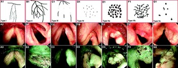
In the narrow band imaging mode, normal laryngeal mucosa was observed to consist of submucosal vessels (appearing green) connecting with an arborescent vascular network (appearing dark brown) (Figure 1, A3). These arborescent vessels were interconnected and ran parallel to the epithelium, before branching out further into oblique capillaries. These capillaries ran obliquely upward to the level of the squamous epithelium, terminating in the intraepithelial papillary capillary loop located beneath the basement membrane of the epithelium. Abnormalities of these intraepithelial papillary capillary loops were easier to observe and to differentiate using endoscopic narrow band imaging.
Diagrams of microvasculature and endoscopic views of vocal folds, illustrating classification of intraepithelial papillary capillary loop features using narrow band imaging. Type I (A1, A2 and A3): thin, oblique and arborescent vessels are interconnected and intraepithelial papillary capillary loops are almost invisible. Type II (B1, B2 and B3): diameter of oblique and arborescent vessels is enlarged, and intraepithelial papillary capillary loops are almost invisible. Type III (C1, C2 and C3): intraepithelial papillary capillary loops are obscured by white mucosa. Type IV (D1, D2 and D3): intraepithelial papillary capillary loops can be recognised as small dots. Type Va (E1, E2 and E3): intraepithelial papillary capillary loops appear as solid or hollow, with a brownish, speckled pattern and various shapes. Type Vb (F1, F2 and F3): intraepithelial papillary capillary loops appear as irregular, tortuous, line-like shapes. Type Vc (G1, G2 and G3): intraepithelial papillary capillary loops appear as brownish speckles or tortuous, line-like shapes with irregular distribution, scattered on the tumour surface.

Previous findings indicate that pathology may be diagnosed by evaluating the morphological changes of the intraepithelial papillary capillary loop; such changes have been found to predict the depth of superficial oesophageal cancer invasion.Reference Inoue, Kumagai, Yoshida, Kawano, Endo and Iwai7–Reference Yoshida, Inoue, Usui, Satodate, Fukami and Kudo9
Intraepithelial papillary capillary loop changes, viewed using narrow band imaging, may be classified into five types (I to V) (Figure 1), as follows. In type I, the intraepithelial papillary capillary loops are almost invisible, and oblique and arborescent vessels of small diameter can be clearly seen. In type II, the intraepithelial papillary capillary loops are also almost invisible, but the diameter of the clearly observed oblique and arborescent vessels is enlarged. In type III, the mucosa is white and the intraepithelial papillary capillary loops cannot be seen; if the white patch is thin, the oblique and arborescent vessels may be seen indistinctly, but if the white patch is thick the vessels will be obscured. In type IV, the mucosal intraepithelial papillary capillary loops are visible with a relatively regular arrangement and low density, the capillary terminals are bifurcated or slightly dilated, and the intraepithelial papillary capillary loops appear as scattered, small, dark brown spots; the oblique and arborescent vessels are usually not visible.
Type V changes are subdivided into types Va, Vb and Vc according to the shape, regularity and distribution of vessels. In type Va, intraepithelial papillary capillary loops are significantly dilated and of relatively high density, and appear to be solid or to have hollow, brownish, speckled features and various shapes. In type Vb, the intraepithelial papillary capillary loop itself is destroyed, with its remnants presenting in a snake-, earthworm-, tadpole- or branch-like shape, and the microvessels are dilated, elongated and ‘woven’ in appearance. In type Vc, the lesion surface is covered with necrotic tissue, and the intraepithelial papillary capillary loops present as brownish speckles or tortuous shapes of uneven density which are irregularly scattered on the tumour surface.
Lesions viewed under narrow band imaging were recorded as: (1) malignant (i.e. type V); (2) suspected malignant (i.e. protuberant or ulcerative lesions covered with necrotic tissue, or leukoplakia of unknown type); or (3) benign (types I to IV).
Histological assessment
Tissue was fixed in 10 per cent formalin, embedded in paraffin, and stained with haematoxylin and eosin. Sections were independently evaluated by two pathologists who were blinded to the mucosal and vascular patterns found on endoscopy. The histological diagnosis served as the ‘gold standard’. Histological analysis was based on the World Health Organization 2005 classification.Reference Gale, Pilch, Sidransky, El Naggar, Westra, Califano, Barnes, Eveson, Reichart and Sidransky10 In the present study, laryngeal lesions were divided into the following types according to their histological features: (1) benign (laryngeal polyps and laryngitis); (2) precancerous (squamous cell hyperplasia and mild, moderate or severe dysplasia); or (3) malignant (carcinoma in situ and invasive cancer).
The accuracy of the white light and narrow band imaging diagnostic results was analysed by comparison with the histological results. In addition, we also evaluated the relationship between the intraepithelial papillary capillary loop characteristics, viewed under narrow band imaging, and the histological features.
Statistical analysis
Statistical analysis was performed using the Statistical Package for the Social Sciences version 11.5 software program (SPSS Inc, Chicago, Illinois, USA). The modality, sensitivity, specificity, positive predictive value, negative predictive value and diagnostic accuracy were independently determined. Chi-square testing was used to evaluate the difference between the white light results and the narrow band imaging results. A value of p < 0.05 was considered statistically significant.
Results
Patients and lesion types
Eighty-five patients (80 men and five women), with a mean age of 55 years (range, 36–83 years), were prospectively enrolled and evaluated.
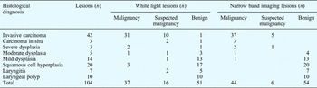
Of these patients, 43 had malignant lesions and 42 nonmalignant lesions. In the patients with malignancies, 45 malignant lesions and six nonmalignant lesions were detected by white light and narrow band imaging. In the patients with nonmalignant lesions, a total of 53 nonmalignant lesions was observed in the same way. Therefore, a total of 104 lesions (45 malignancies and 59 nonmalignancies) was suggested by white light visualisation and narrow band imaging.
The histological diagnoses of these patients were as follows: laryngeal polyp (n = 10; 9.6 per cent); laryngitis (n = 7; 6.7 per cent); squamous cell hyperplasia (n = 20; 19.2 per cent); mild dysplasia (n = 14; 13.5 per cent); moderate dysplasia (n = 5; 4.8 per cent); severe dysplasia (n = 3; 2.9 per cent); carcinoma in situ (n = 3; 2.9 per cent); and invasive cancer (n = 42; 40.3 per cent) (see Table I).
Relationship between endoscopic findings (white light and NBI) and histological diagnosis

NBI = narrow band imaging; pts = patients
Narrow band imaging and histology results
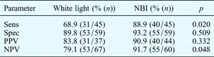
For the total 104 lesions, the diagnostic accuracy of narrow band imaging and white light visualisation was 90.4 per cent (94/104) and 76.9 per cent (80/104), respectively, suggesting significantly improved diagnostic accuracy for narrow band imaging compared with white light visualisation (p = 0.028). The sensitivity of narrow band imaging in detecting malignant lesions (carcinoma in situ or invasive carcinoma) was 88.9 per cent, significantly greater than that of white light visualisation (68.9 per cent) (p = 0.020). The negative predictive value of narrow band imaging (91.7 per cent) was also markedly greater than that of white light visualisation (79.1 per cent) (p = 0.048), as shown in Table II. The specificity and positive predictive value of narrow band imaging were 93.2 and 90.9 per cent, respectively; results for white light visualisation did not differ significantly (p > 0.05). These findings suggest that narrow band imaging has greater accuracy in the diagnosis of malignant laryngeal lesions, compared with white light visualisation.
Diagnostic effectiveness of white light vs NBI for carcinoma in situ and invasive carcinoma

NBI = narrow band imaging; sens = sensitivity; spec = specificity; PPV = positive predictive value; NPV = negative predictive value
The intraepithelial papillary capillary loop features of laryngeal lesions, viewed by narrow band imaging, could be evaluated in most cases. Only six lesions (5.8 per cent) could not be evaluated and classified by narrow band imaging; of these, five lesions comprised invasive carcinoma and one severe dysplasia. This may be related to necrotic tissue or a thick white patch on the lesions, masking the mucosal microvasculature.
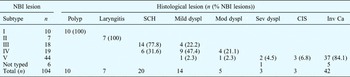
The relationship between the narrow band imaging classification of intraepithelial papillary capillary loop features and the histological classification of laryngeal lesion biopsy tissue is shown in Table III. Laryngeal polyps consisting of normal laryngeal epithelium were observed in 100 per cent of patients with type I lesions. Laryngitis was noted in 100 per cent of patients with type II lesions. In patients with type III lesions, squamous cell hyperplasia was observed in 77.8 per cent and mild dysplasia in 22.2 per cent, a statistically significant difference (p = 0.002). Of patients with type IV lesions, 31.6 per cent had squamous cell hyperplasia, 47.4 per cent mild dysplasia and 21.1 per cent moderate dysplasia. In patients with type IV lesions, low grade dysplasia (mild to moderate) was observed more often than squamous cell hyperplasia (p = 0.023); no type IV patients had severely dysplastic or malignant lesions. Using narrow band imaging, all malignancies were classified as type V lesions, except for five unclassified invasive carcinomas. Of the type V lesions, 2.3 per cent comprised mild dysplasia, 2.3 per cent moderate dysplasia, 4.5 per cent severe dysplasia, 6.8 per cent carcinoma in situ and 84.1 per cent invasive carcinoma. Type V lesions comprised malignancies more often than nonmalignancies (p < 0.001).
Relationship between NBI classification and histological diagnosis

NBI = narrow band imaging; SCH = squamous cell hyperplasia; dyspl = dysplasia; mod = moderate; sev = severe; CIS = carcinoma in situ; inv Ca = invasive carcinoma
The sensitivity and specificity of type I narrow band imaging lesions in predicting histologically diagnosed laryngeal polyps were both 100 per cent; likewise, the sensitivity and specificity of type II lesions in predicting histologically diagnosed laryngitis were both 100 per cent. The sensitivity and specificity of type III lesions in predicting a histological diagnosis of squamous cell hyperplasia were 70.0 and 95.2 per cent, respectively; those of type IV lesions in predicting a diagnosis of low grade dysplasia were 68.4 and 92.9 per cent, respectively, while those of type V lesions in predicting a diagnosis of malignancy were 88.9 and 93.2 per cent, respectively. The sensitivity and specificity of type V lesions in predicting a diagnosis of severe dysplasia or carcinoma in situ were 83.3 and 100 per cent, respectively.
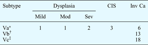
The narrow band imaging type V lesions were further divided into three subtypes. The relationship between these subtypes and the features seen on histological examination are shown in Table IV. In type Va lesions, a histological diagnosis of severe dysplasia, carcinoma in situ or invasive carcinoma was made more frequently than a diagnosis of low grade dysplasia (84.6 vs 15.4 per cent; p = 0.001). The sensitivity and specificity of type Va lesions in predicting a diagnosis of severe dysplasia or carcinoma in situ were 100 and 79.5 per cent, respectively. Type Vb and Vc lesions were noted for 100 per cent of invasive carcinomas. The sensitivity and specificity of type Vb plus type Vc in predicting a diagnosis of invasive carcinoma were 83.8 and 100 per cent, respectively.
Relationship between NBI type V subtype and histological diagnosis
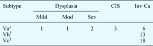
Data represent number of lesions. *n = 13; †n = 13; ‡n = 18. NBI = narrow band imaging; CIS = carcinoma in situ; inv Ca = invasive carcinoma; mod = moderate; sev = severe
Discussion
Narrow band imaging is an innovative optical technology which modifies the central wavelength and bandwidth of an endoscope's light into a narrow band comprising only 415 ± 30 and 540 ± 30 nm. Narrow band imaging markedly improves capillary pattern contrast, and enables in vivo visualisation of changes in microvasculature morphology in superficial neoplastic lesions.
The optical basis for narrow band imaging is that short wavelength light falls within the haemoglobin absorption spectrum, and therefore facilitates clearer visualisation of vascular structures.Reference Song, Adler, Conway, Diehl and Farraye11 The properties of narrow band imaging light produce the greatest contrast between the microvasculature and the surrounding mucosa.
The terminal microvessels located on the epithelium, termed intraepithelial papillary capillary loops, can be clearly visualised using a narrow band imaging system combined with a magnifying endoscope.Reference Yoshida, Inoue, Usui, Satodate, Fukami and Kudo9 The characteristics of these intraepithelial papillary capillary loops, viewed using narrow band imaging, are important determinants of the diagnosis of epithelial lesions. It has been reported that the structure and organisation of blood vessels are dynamic and may undergo considerable change during the transition from a precancerous to a malignant state.Reference Kumagai, Toi and Inoue12 Therefore, the detection of changes in mucosal microvasculature may be beneficial for the early diagnosis of epithelial neoplasia. Currently available evidence indicates that narrow band imaging enables more accurate in vivo diagnosis of neoplasia across a range of organs, using simple, microvasculature-based techniques.Reference East, Tan, Bergman, Saunders and Tekkis13 Narrow band imaging currently shows promise as an effective technique for the early diagnosis of cancer during routine endoscopic examination.Reference Muto, Horimatsu, Ezoe, Hori, Yukawa and Morita3
Approximately 95 per cent of laryngeal cancers comprise typical squamous cell carcinoma. Premalignant epithelial changes in the larynx cover a spectrum of pathological changes, beginning with hyperplasia and progressing through dysplasia and carcinoma in situ to invasive carcinoma.Reference Crissman and Zarbo14
This tumourigenic process is similar to that of oesophageal squamous cell carcinoma. The superficial vascular network of the oesophageal mucosa can be classified into five types, ranging from normal to carcinomatous, according to the intraepithelial papillary capillary loop features seen under high-magnification endoscopy. In addition, the depth of cancer infiltration can be estimated by the extent of destruction of intraepithelial papillary capillary loops and other affected vessels.Reference Inoue, Kumagai, Yoshida, Kawano, Endo and Iwai7 In the diagnosis of superficial malignant lesions, the most notable intraepithelial papillary capillary loop features are well demarcated brownish areas and scattered brown dots. These features have been observed in the early stage of oesophageal cancer, pharyngeal cancer, oral cancer and laryngeal cancer, amongst others.Reference Watanabe, Taniguchi, Tsujie, Hosokawa, Fujita and Sasaki6, Reference Yoshida, Inoue, Usui, Satodate, Fukami and Kudo9, Reference Watanabe, Taniguchi, Tsujie, Hosokawa, Fujita and Sasaki15–Reference Muto, Nakane, Katada, Sano, Ohtsu and Esumi18
Recently, via comparison with histopathological observations, the sensitivity and specificity of narrow band imaging in predicting a diagnosis of laryngeal carcinoma have been found to be 91.3 and 91.6 per cent, respectively.Reference Watanabe, Taniguchi, Tsujie, Hosokawa, Fujita and Sasaki6 This is consistent with our study findings (i.e. 88.9 and 93.2 per cent, respectively). Moreover, the sensitivity and negative predictive value of narrow band imaging were significantly greater than those of white light visualisation.
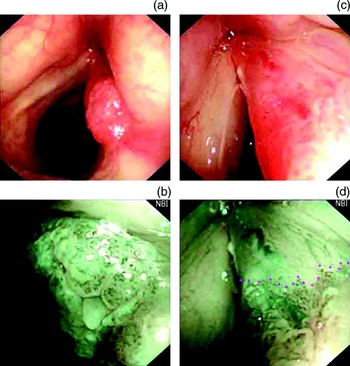
Therefore, we propose that routine laryngoscopic examination incorporating endoscopic narrow band imaging may represent a favourable diagnostic approach in differentiating malignant from nonmalignant laryngeal lesions. Furthermore, the boundaries of malignant lesions can be easily identified using narrow band imaging (Figure 2); this may be beneficial for the pre-operative assessment of the extent of cancer infiltration, and may aid the selection of therapeutic strategies.
Squamous cell carcinoma in the right vocal fold, as seen by white light visualisation (a and c) and narrow band imaging (b and d). Under narrow band imaging, the lesion was classified as type Va based on the intraepithelial papillary capillary loop features (b). The border of the lesion can be clearly identified (indicated by purple dots, d) and the anterior commissure is not involved; this was confirmed post-operatively on histopathological examination.

Previous studies have not described intraepithelial papillary capillary loop features in detail, nor have they used this as a basis for distinguishing different stages of metaplasia, due to poor endoscope resolution. In the present study, however, high resolution bronchoscopy combined with narrow band imaging was employed to investigate the clinical value of endoscopic narrow band imaging in the early diagnosis of laryngeal cancer. The mucosal microvasculature and abnormal intraepithelial papillary capillary loop characteristics could be clearly identified, and were classified into five different types.
Data analysis indicated that intraepithelial papillary capillary loop features were closely related to histopathological findings. The current study considered type I to IV lesions as nonmalignant and type V lesions as malignant. Furthermore, low grade dysplasia was more frequently observed in lesions classified as type IV, severe dysplasia and carcinoma in situ in type Va lesions, and invasive cancer in type Vb or Vc lesions. Thus, the classification of intraepithelial papillary capillary loop features may facilitate the prediction of laryngeal cancer or precancerous lesions.
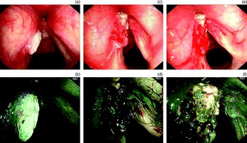
However, the presence of necrotic tissue or a thick white patch on lesions may affect the evaluation of intraepithelial papillary capillary loop features, especially for invasive carcinoma, resulting in false negative findings. In this situation, more attention should be paid to borderline regions and areas with a thinner white patch, in order to delineate any dots or irregular, tortuous, line-like shapes which may aid classification (Figure 3a and 3b).
Endoscopic white light and narrow band imaging views of vocal folds, showing the influence of a laryngeal white patch and bleeding on the observation of intraepithelial papillary capillary loop features (a and b). The tumour in the left vocal fold was white in both white light and narrow band imaging modes. Brown dots (indicated by purple dots) are seen on the edge of the tumour under narrow band imaging. The tumour was classified as a type Va lesion; histopathological analysis indicated a squamous cell carcinoma. Parts (c) to (f) show a tumour in the vocal area before and after bleeding, as seen in white light and under narrow band imaging. After the onset of bleeding, the surface of the vocal folds appears black under narrow band imaging, and the intraepithelial papillary capillary loops are obscured (d). After effective haemostasis, brown dots (indicated by purple dots) and tortuous line shapes (indicate by yellow dots) can be seen (f). Histopathological analysis indicated a squamous cell carcinoma.

In the present study, only six lesions (5.8 per cent) could not be evaluated and classified according to their intraepithelial papillary capillary loop features. Bleeding during endoscope insertion may affect the assessment of intraepithelial papillary capillary loop features (Figure 3c to 3f). Because haemoglobin absorbs blue light, narrow band imaging shows blood on mucosa as black (Figure 3d). Thus, touching surface lesions should be avoided during endoscopic narrow band imaging, particularly when topical anaesthesia is being applied. In addition, the operator should avoid causing severe coughing and directly entering the subglottic area. If bleeding occurs, an adrenaline spray may be used, but the procedure should be delayed for 5–10 minutes (Figure 3f).
• Laryngoscopy plays a key role in the early detection of laryngeal cancer and precancerous lesions
• Narrow band imaging is a new, high-resolution endoscopic technique which has improved the accuracy of in vivo diagnosis of epithelial lesions
• Narrow band imaging enables clear visualisation of the mucosal microvasculature (including the intraepithelial papillary capillary loops), an important determinant in the diagnosis of epithelial lesions
• In this study, intraepithelial papillary capillary loop morphology was classified into five types, which were found to correlate well with the histopathologically confirmed severity of laryngeal lesions
• Narrow band imaging shows promise as an effective technique for the early diagnosis of laryngeal cancer during routine endoscopic examination
Early diagnosis of laryngeal cancer is crucial to maximise the prognosis and to enable the best selection of therapeutic strategies. Laryngoscopy plays a key role in the early detection of laryngeal cancer and precancerous lesions.
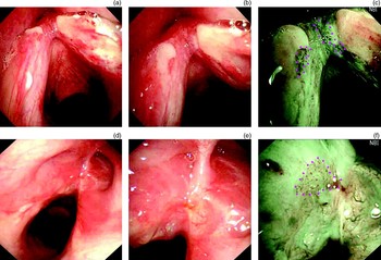
Previous studies have shown that autofluorescent laryngoscopy, used in combination with traditional white light endoscopy, can improve the detection of precancerous and malignant laryngeal lesions;Reference Malzahn, Dreyer, Glanz and Arens19, Reference Baletic, Petrovic, Pendjer and Malicevic20 the sensitivity of this technique in this regard is similar to that of narrow band imaging as assessed in the current study. However, autofluorescent laryngoscopy had a low specificity because some benign lesions (including granulation tissue, papillomas and vocal fold polyps) also display a loss of green fluorescence.Reference Paczona, Temam, Janot, Marandas and Luboinski21 Moreover, mucosal scars and inflammation often cause false positive results,Reference Johnson22 which limits the application of autofluorescent endoscopy in the follow-up examination of laryngeal cancer patients undergoing surgery or radiotherapy. In contrast, our results indicate that scar tissue and inflammation present as type II or III lesions under narrow band imaging, and can easily be differentiated from type V lesions (Figure 4).
Endosopic views of vocal folds under white light and narrow band imaging, illustrating the role of narrow band imaging in post-operative surveillance of laryngeal carcinoma. The vocal area was observed under white light 10 days after endoscopic CO2 laser excision; residual carcinoma was not indicated (a and b). However, under narrow band imaging, brown dots (indicated by purple dots) were observed on the left vocal fold and anterior commissure (c), indicating a type Va lesion. Histopathological analysis indicated carcinoma in situ with mild infiltration. Scar tissue was observed in the vocal area under white light, 3 months after surgery for glottic carcinoma (d and e); no residual or recurrent lesions were identified. Under narrow band imaging, brown dots (indicated by purple dots) were found on the scar of a type Va lesion (f); histopathological analysis indicated carcinoma in situ.

Laryngeal cancer patients are at high risk of developing synchronous and metachronous head and neck carcinomas.Reference Schwartz, Ozsahin, Zhang, Touboul, De Vataire and Andolenko23 The high incidence of secondary head and neck carcinomas may be attributed to a ‘field cancerisation’ effect.Reference Loyo and Pai24 The development of a secondary cancer may markedly decrease the survival rate.Reference Laccourreye, Veivers, Hans, Brasnu, Garcia and Laccourreye25 Furthermore, head and neck cancer is frequently treated with radiotherapy in order to preserve voice function, but high recurrence rates are observed in these patients. To date, CT and conventional MRI still have limitations in diagnosing recurrent disease following chemotherapy or radiotherapy.Reference Zbären, Weidner and Thoeny26 Katada et al. Reference Katada, Nakayama, Tanabe, Koizumi, Masaki and Takeda27 recently reported that narrow band imaging was effective in detecting metachronous superficial squamous cell carcinoma at oropharyngeal and hypopharyngeal mucosal sites following chemoradiotherapy for head and neck cancer. Such surveillance may detect early stage cancers, enabling minimally invasive treatment, which may improve the patient's prognosis and quality of life.
Conclusion
Endoscopic narrow band imaging is a promising approach to in vivo differentiation of nonmalignant and malignant laryngeal lesions, utilising the morphology of mucosal capillaries. In the present study, the vascular features of intraepithelial papillary capillary loops were classified into five types, which were found to correlate well with the severity of laryngeal lesions as demonstrated by histological examination. Furthermore, the intraepithelial papillary capillary loop characteristics of malignant lesions were not influenced by scar tissue or inflammation. Therefore, the evaluation of intraepithelial papillary capillary loop features under narrow band imaging can be recommended during routine pharyngolaryngeal examination in high risk patients, in order to screen for synchronous and metachronous head and neck carcinomas, to monitor the efficacy of radiotherapy, and to check for post-operative recurrence.
Acknowledgement
This study was supported by the Beijing Hope Run Special Fund (LC2008A13). Narrow band imaging endoscopy equipment was provided by Olympus Medical Systems Corp, Tokyo, Japan.