Introduction
Implantation failure after early embryo or blastocyst transfer is a common cause of low pregnancy rate following assisted reproductive technology (ART) techniques. Recurrent implantation failure and repeated abortion can be caused by embryo incompetence, genetic defects, hormonal disorders (negative effect of ovarian stimulation protocols), immunological problems or embryo–endometrium asynchronism during the implantation window.
In the literature, randomized and meta-analysis studies showed that the probability of pregnancy and live birth is significantly higher after blastocyst transfer than early cleavage stage when equal number of embryos are transferred (Levitas et al., Reference Levitas, Lunenfeld, Har-Vardi, Albotiano, Sonnin, Hackman-Ram and Potashnik2004; Papanikolaou et al., Reference Papanikolaou, Camus, Kolibianakis, Van Landuyt, Van Steirtegheim and Devroey2006, Reference Papanikolaou, Kolibianakis, Tournaye, Venetis, Fatemi, Tarlatsi and Devroey2008).
Human embryo co-culture with different feeder systems has been used for more that 20 years to provide embryo selection and increase implantation rate (Menezo et al., 1990; Bongso et al., Reference Bongso, Ng, Fong and Ratnam1991; Quinn et al., Reference Quinn and Margualit1996; Veiga et al., Reference Veiga, Torello, Menezo, Busquets, Sarrias, Coreleu and Barri1999). The use of animal feeder cells was abandoned for health security reasons and different human feeder cells were used in co-culture with human embryos in association with defined culture medium for better embryo growth, quality and implantation (Spandorfer et al., 2004; Ebner et al., Reference Ebner, Moser, Sommergrube, Shebl and Tews2006; Parikh et al., Reference Parikh, Nadkarni, Naik, Naik and Uttamchandani2006; Eyheremendy et al., Reference Eyheremendy, Raffo, Papayannis, Barnes, Granados and Blaquier2010).
Prolonged embryo culture can be performed by sequential, global media or monolayer cell lines. It was reported that feeder cells can be more beneficial to the oocyte and embryo than defined media (Menezo et al., 1998; Kattal et al., Reference Kattal, Cohen and Barmat2008), by detoxifying the culture media and supporting the embryo development via different pathways involving growth factors [such as leukaemia inhibitor factor (LIF), platelet activating factor receptor (PAF-R), epidermal growth factor (EGF) and vascular endothelial growth factor (VEGF)], hormones and cytokines (Parikh et al., Reference Parikh, Nadkarni, Naik, Naik and Uttamchandani2006; Lin et al., Reference Lin, Hwang, Seow, Huang, Chen and Tzeng2009). In sheep, soluble factors secreted by cumulus cells are capable to induce mitotic maturation of early embryo and to reduce necrosis by involving mitogen activated protein kinase activation (Cecconi et al., Reference Cecconi, Mauro, Cappachietti, Berardenelli, Bernabo, Di Vencenzo, Mattioli and Barboni2008) and in the bovine, cumulus cells suppress the apoptosis effects via midkine, a heparin-binding growth differentiation factor (Ikeda et al., Reference Ikeda, Saeki, Imai and Yamada2006). The use of human cumulus cell co-culture has also other advantages, they are autologous and easily obtained via the oocyte retrieval procedure during the in vitro fertilization (IVF) cycle, and do not need very sophisticate culture and maintenance systems (Johnson et al., Reference Johnson, Higdon and Boone2008; Omar et al., Reference Omar and Vlad2008; Goovaerts et al., Reference Goovaerts, Leroy, Van Soon, De Clerq, Andries and Bols2009). In this study we applied autologous cumulus cell embryo co-culture for patients with a minimum of three repeated implantation failures and we compared the blastulation rate and the clinical outcome with classical embryo culture system and day 3 or day 5–6 transfers. We also investigated the expression of LIF and PAF-R on day 3 confluent cumulus cells by fluorescence in situ hybridization (FISH) and RT-PCR techniques to explain the possible contribution of such factors on embryo competency.
Materials and methods
From January 2007 to December 2009, 432 patients with a minimum of three repeated implantation failures and an average age of 33.5 years were accepted in the participating centres for a classical ICSI procedure with a minimum of 1 million spermatozoa per ejaculate (to minimise the paternal effect on embryo development).
Co-culture initiation
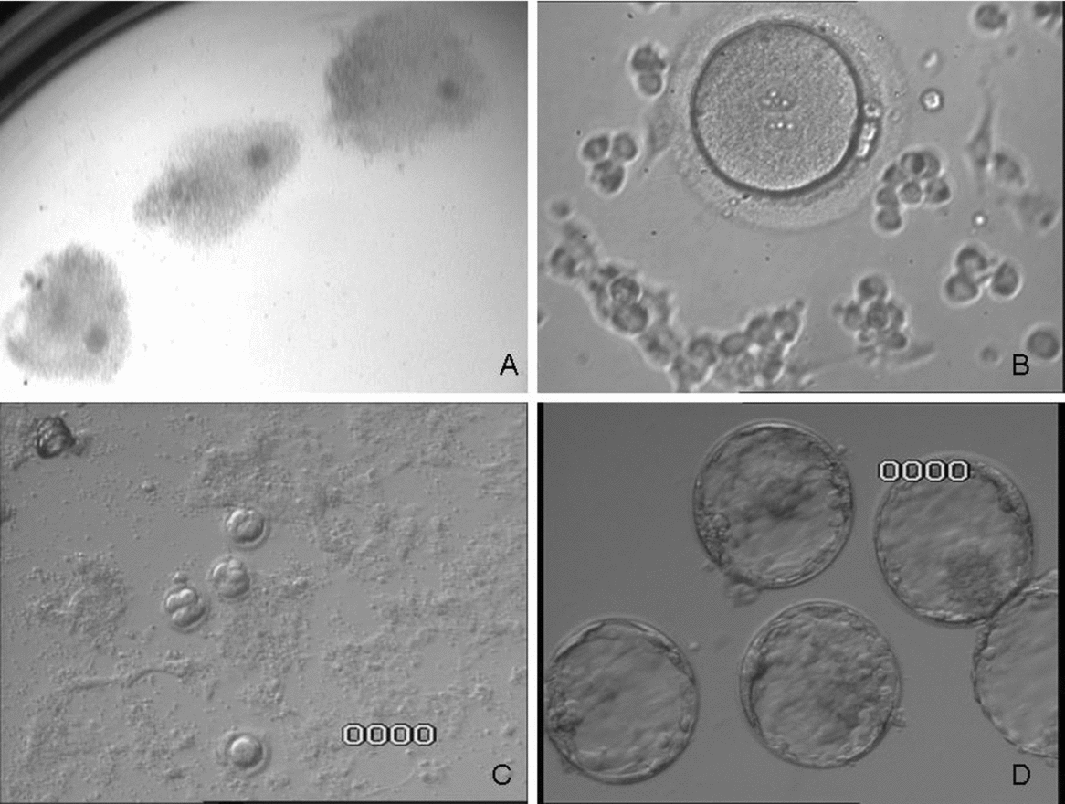
On the day of oocyte pick-up, the cumuli were graded from grade 1 to grade 4 based on the general morphological aspect of the cumulus cell complex. One or two grade 1 cumulus cells (Fig. 1A) were dissected mechanically and then initiated for in situ culture using G2 (VitroLife Sweden) medium supplemented by 10% of dextran synthetic serum (Irvine Scientific) for 18–20 h. After 18–20 h of in situ culture, the cumuli cells were rinsed and supported by fresh G2 medium supplemented and refreshed every 48 h until day 5–6. For co-culture initiation we used G2 supplemented with 10% of dextran synthetic serum than G1 because G2 contain more amino acids and this situation can be more helpful for cumulus cells early attachment, growth and competency to support embryo development.
(A) Grade 1 cumulus selected for dissection and co-culture initiation. (B) 2PN zygote transfer to 24 h cumulus co-culture. (C) Day 3 embryo co-culture. (D) Day 5 blastocysts from co-culture.
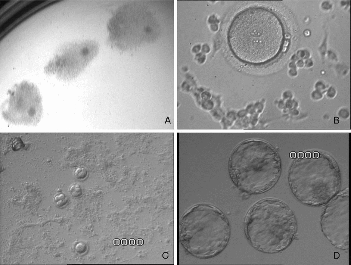
Microinjected metaphase II oocytes were controlled for PN scoring and transferred to the cumulus culture (Fig. 1B) and scored every 24 h (Fig. 1C). Between day 5 and 6 the blastulation rate per patient was calculated (Fig. 1D).
Study groups
The patients were prospectively randomly assigned to four study groups during the starting of ovarian stimulation and based on the quality of ovarian reserve. The randomization was based on groups of four patients including one patient per each culture condition. In the first group, 108 patients underwent intracytoplasmic sperm injection (ICSI), standard culture and day 3 transfer by using G1 media. In the second group, 108 patients underwent ICSI, and from 2PN stage to co-culture for 36 h and day 3 transfer. From this group the non -transferred embryos were co-cultured until day 5–6 prior freezing or vitrification. In the third group, 108 patients underwent ICSI, embryo culture without co-culture and blastocyst transfer on day 5–6 by using sequential culture (G1 and G2).
In this group after fertilization the embryos were cultured with G1 until day 3 and then in G2 until day 5–6 transfer. In the fourth group, 108 patients underwent ICSI, with co-culture and blastocyst transfer on day 5–6.
Molecular and fluorescence in situ hybridization (FISH) analysis
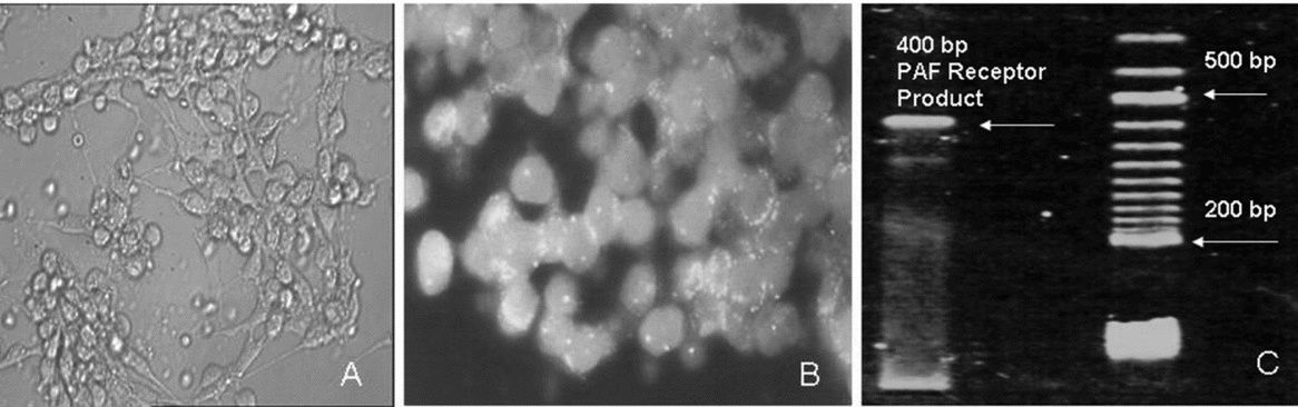
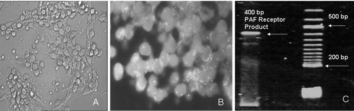
The nearly confluent cumulus cells obtained after 3 days of culture (Fig. 2A) were trypsinized and collected for total RNA extraction and fixation for in situ hybridization. For RT-PCR, total RNA extraction was performed using the RNEasy kit from Qiagen and respecting the manufacturer's recommendations. After total RNA quality control, 5 μg of RNA were reverse transcripted to cDNA (Benkhalifa et al., 2010).
(A) Cumulus cells after 3 days of culture. (B) In situ hybridization of leukaemia inhibitor factor (LIF) gene probe transcript. (C) RT-PCR product of platelet activating factor (PAF) receptor gene (200 bp).
LIF probe production and labelling from cDNA for LIF transcript in situ hybridization
A total of 2 μl of reverse transcripted total RNA product was amplified by PCR for 30 cycles (melting, annealing and extension temperatures: 94, 60 and 72 °C respectively). PCR was carried out in the presence of 5 pm/μl with 5′ and 3′ primers (Bamberger et al., Reference Bamberger, Erdmann, Jenatschke, Bamberger and Schulte1997), 10 mM of dNTPs, and 2.5 units of Taq polymerase in a final volume of 50 μl. The PCR product was loaded onto a 2% of agarose gel to confirm the amplified fragment as 450 bp.
For probe labelling, LIF cDNA fragments were labelled by PCR amplification of 1 μl for 25 cycles in a 50 μl PCR reaction mixture as described above, except that dTTP was replaced by dTTP/fluorescein-16-dUTP (1/1) mixture. Then an aliquot (10 μl for one slide) was ethanol precipitated with 100 μg of yeast RNA and 50 μg of salmon sperm DNA. DNA pellet was dissolved in 10 μl of hybridization solution containing 50% formamide and 10% dextran sulphate in 2× SSC, denaturated for 6 min at 76 °C and preannealed for 90 min at 37 °C.
Slides preparation and hybridization for FISH
Cumulus cell cultures were harvested under laminar flow, DNase and RNase free. Briefly, cells were trypsinized and centrifuged and then treated with trinatrium citrate for hypotonic reactions. After centrifugation, cells were treated with a fixative solution (ethanol/acetic acid) and dropped onto clean slides. After total drying, slides were dehydrated, treated with pepsin, post-fixed with paraformaldehyde and rinsed with phosphate-buffered saline (PBS). The probe was hybridized to the cells for 24–28 h at 37 °C. After hybridization, slides were washed and counterstained with DAPI for fluorescence microscopy analysis.
RT-PCR for PAF receptor analysis
A total of 2 μl of reverse transcripted total RNA products was amplified with 5 units of Taq DNA polymerase and 10 mM each of the human PAF-R primers (Ahmed et al., Reference Ahmed, Dearu, Shams, Li, Sangha, Rola-Pleszcynski and Jiang1998) in 100 μl of reaction mix containing 500 mM KCl, 200 mM Tris–HCl and 1.5 mM of MgCl2 as follow: 95 °C, 30 s; 60 °C, 50 s; 72 °C, 60 s for 40 PCR cycles. The PCR product was analysed by 2% agarose gel electrophoresis to observe the approximately 400-bp product.
Confirmation by in situ hybridization of PAF receptor expression
For probe labelling, PAF-R cDNA fragments were labelled by PCR amplification of 1 μl for 25 cycles in a 50 μl PCR reaction mixture as described above, except that dTTP was replaced by dTTP/fluorescein-16-dUTP (1/1) mixture. Then, an aliquot (10 μl for one slide) was ethanol precipitated with 100 μg of yeast RNA and 50 μg of salmon sperm DNA. DNA pellet was dissolved in 10 μl of hybridization solution containing 50% formamide and 10% dextran sulphate in 2× SSC, denaturated 7 min at 75 °C and preannealed for 80 min at 37 °C
Slide preparation and hybridization for FISH: cumulus cell cultures were harvested under laminar flow, DNase and RNase free. Briefly, cells were trypsinized and centrifuged and then treated with trinatrium citrate for the hypotonic reaction. After centrifugation cells were treated with a fixative solution (ethanol/acetic acid) and dropped onto clean slides. After total drying, slides were dehydrated, treated with pepsin, post-fixed with paraformaldehyde and rinsed with PBS. The probe was hybridized to the cells for 24–28 h at 37 °C. After hybridization, slides were washed and counterstained with 4′,6-diamidino-2-phenylindole (DAPI) for fluorescence microscopy analysis.
Internal controls of RNA transcript of LIF and PAF receptor by in situ hybridization
Cumulus cell slides were prepared for FISH as below but treated for 40 min at 37 °C with RNase solution containing 500 units of RNase per ml to digest the total RNA in the slides, after washing and dehydration. Using the same protocol of fluorescent probes production, the slides were hybridized simultaneously with green LIF fluorescent probe and red PAF receptor fluorescent probe labelled by using the same FISH protocol and analysed by double excitation filter for FITC/Texas Red for microscope analysis.
Results
In this study, 432 patients, considered as repeated implantation failures after a minimum of three transfers with a total of six transferred embryos at day 3, were accepted to undergo IVF–ICSI. After ovarian stimulation we collected an average of 8.5 cumuli per patient. All patients continued to the ICSI procedure and at day 1 we observed a nearly 89% fertilization rate.
Statistical methods
Groups were compared according to the variables, using Student's t-test for quantitative variables and chi-squared test for qualitative variables. For all comparisons, the level of significance was p < 0.05, with bilateral testing.
The power calculation showed that two samples of 108 patients resulted in a power of 80% if the difference in percentages was 15% (30% in a group and 45% in the other). The statistical power decreased to 32% if the anticipated difference was 10% between the two groups.
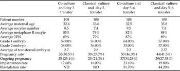
The clinical pregnancy rates per transfer were not statistically different between co-culture and classical culture at day 3 (30.5% vs 27.7%, p = 0.65) or day 5 (46.3% vs 41.5%, p = 0.41). On the opposite There was a difference between transfers at day 5 and day 3 depending on whether co-culture was used (46.3% vs. 30.5%, p = 0.02) or not (41.5% vs. 27.7%, p = 0.04). The largest difference was observed between day 5 transfers with co-culture and day 3 transfer with classical medium (46.3% vs. 27.7%, p < 0.01).
The results followed the same trend for on-going pregnancy rates, statistical significance being only reached between co-culture and day 5 transfer vs. classical medium (G1 use) day 3 transfer. Concerning implantation rate, there was no significant difference between co-culture and classical medium when comparing these parameters at day 3 or at day 5, but comparisons between day 3 and day 5 were highly significant for both co-culture and classical medium. Here also, the largest difference was observed between day 3 transfer with classical medium and day 5 transfers with co-culture (see Table 1).
Clinical data results

Fluorescence in situ hybridization highlighted hybridization of the LIF probe in interphase nucleus (DNA) and the primary transcript of LIF gene in the cytoplasm (Fig. 2B). The PCR using specific primer of PAF receptor gene from total reverse transcripted RNA demonstrates the presence of PAF receptor gene product of 200 bp (Fig. 2C).
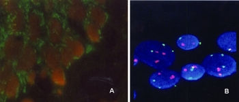
The control of PAF-R expression by in situ hybridization showed clearly the expression of the gene (Fig. 3A). After slide RNase treatment to eliminate the native transcripts in the cytoplasm, double in situ hybridization using two-colour FISH (LIF – green, PAF receptor – red), showed clearly that there was negative hybridization of both probes in the cytoplasm but a positive signal in the nucleus (Fig. 3B).
(A) In situ hybridization of platelet activating factor (PAF) receptor gene probe transcript. (B) Internal controle of leukaemia inhibitor factor (LIF) (green), PAF receptor probe (red) hybridization after RNase treatment. (See online for a colour version of this figure.)

Discussion
The main goal of in-vitro fertilization, embryo culture and selection is to provide high quality embryos with good competency for development and implantation by giving acceptable viable births (Gardner et al., Reference Gardner and Lane1997; Munné et al., Reference Munné, Howles and Wells2009). At early embryo development before genomic activation at day 3 there is no transcriptional activity. For this reason the oocyte should contain all proteins and/or transcripts that code for all enzymes required for the metabolic pathways needed to ensure early embryo cleavage. The gene activation of the first cell cycle of the early embryo is the longest during preimplantation development, any delay in this cycle will result in a decrease of the level of mRNA to one below critical thresholds. Moreover, culture conditions have a direct impact on transcription and translation (Jung et al., Reference Jung1989; Ho et al., Reference Ho, Doherty and Schultz1994), the metabolism of the embryo is depressed in vitro (Leese et al., Reference Leese1995) and protein turnover is accelerated (Goodeaux et al., Reference Goodeaux, Voelkel, Anzalone, Ménézo and Graves1989).
The effect of the co-culture system in human IVF was investigated by a meta-analysis study (Kattal et al., Reference Kattal, Cohen and Barmat2008) and showed that there was a significant improvement in embryo development, implantation, clinical and ongoing pregnancy rates. Co-cultured embryos usually have higher cell numbers than those cultured in simple culture medium (Vlad et al., Reference Vlad, Walker and Kennedy1986; Goodeaux et al., Reference Goodeaux, Voelkel, Anzalone, Ménézo and Graves1989). Randomized experiments (Vlad et al., Reference Vlad, Walker and Kennedy1986; Van Blerkom et al., Reference Van Blerkom1993; Mansoor et al., Reference Mansoor, Aboulghar, Serour and Abbas1994; Carrel et al., Reference Carrel, Peterson, Jones, Katasaka, Udoff, Cornwell, Thorp, Kuneck, Erickson and Campbell1999; Levitas et al., Reference Levitas, Lunenfeld, Har-Vardi, Albotiano, Sonnin, Hackman-Ram and Potashnik2004; Papanikolaou Reference Papanikolaou, Camus, Kolibianakis, Van Landuyt, Van Steirtegheim and Devroey2006, Reference Papanikolaou, Kolibianakis, Tournaye, Venetis, Fatemi, Tarlatsi and Devroey2008) showed the importance of the biological aspects of co-culture on human embryo development. It is more than possible that the metabolism in feeder cells supplies small molecules that allow the cell machinery to work with maximum efficiency, such as lactate/pyruvate ratio optimization, or glucose and glutamine concentration regulation (Ouhibi et al., Reference Ouhibi, Hamidi, Guillaud and Ménézo1990; Edwards et al., Reference Edwards1997; Ebner et al., Reference Ebner, Moser, Sommergrube, Shebl and Tews2006).
There is also a contribution of amino acids by the feeder that leads to a better equilibrium of amino acids, allowing their efficient uptake. Co-culture provides a good redox potential, which is rarely taken into account. Feeder cells synthesize and release reducing substances such as glutathione and, in particular, hypotaurine, that are efficient for improving early preimplantation embryo development (Barnet et al., Reference Barnette and Bavisier1992). These reducing compounds are necessary to avoid undesirable oxidation of lipids and other compounds such as cysteine and methionine.
In repeated implantation failure, co-culture of human embryos with somatic cells has been reported to promote the improvement of embryos quality, implantation and pregnancy rates after transfer (Spandorfer et al., 2004; Kattal et al., Reference Kattal, Cohen and Barmat2008; Eyheremendy et al., Reference Eyheremendy, Raffo, Papayannis, Barnes, Granados and Blaquier2010). The use of cumulus cell co-culture has some advantages, they are easily obtained via the oocyte retrieval procedure during the IVF cycle and does not need very sophisticated maintenance of culture systems. In addition, they are also autologous (Johnson et al., Reference Johnson, Higdon and Boone2008; Omar et al., Reference Omar and Vlad2008; Goovaerts et al., Reference Goovaerts, Leroy, Van Soon, De Clerq, Andries and Bols2009).
Our data showed clearly the benefit of cumulus co-culture on blastulation rate and clinical pregnancy for patients with repeated implantation failure who are younger than 38 years. A previous prospective randomized study (Levitas et al., Reference Levitas, Lunenfeld, Har-Vardi, Albotiano, Sonnin, Hackman-Ram and Potashnik2004) suggested that in patients with adequate ovarian responses who failed to conceive in at least three IVF/ET cycles, transfer at the blastocyst stage gives significantly higher implantation and pregnancy rates per oocyte retrieval. Our data are in concordance with other publications that demonstrate the benefits of cumulus cell co-culture on blastocyst formation (Quinn et al., Reference Quinn and Margualit1996) and on the improvement of implantation by cumulus cells secretion of cytokines and growths factors required to help the process of blastocyst implantation (Parikh et al., Reference Parikh, Nadkarni, Naik, Naik and Uttamchandani2006). In human, the clinical experience of 5 years’ use of endometrial cell co-culture and blastocyst transfer demonstrated a significant increase in blastulation (58%) and implantation (44%) rate (Mercader et al., Reference Mercader, Garcia Valesco, Escudero, Remohi, Pellicie and Simon2003). In the bovine model Moulavi et al. (Reference Moulavi, Hosseini, Ashtiani, Shahverdi and Nasr-Esfahani2006) reported that co-culture of immature oocytes using Vero cells enhanced their ability for cleavage and production of higher quality blastocysts. In sheep and bovine it was reported that soluble factors secreted by cumulus cells are capable of inducing mitotic maturation of early embryos and of reducing necrosis by involving mitogen activated protein kinase activation (Cecconi et al., Reference Cecconi, Mauro, Cappachietti, Berardenelli, Bernabo, Di Vencenzo, Mattioli and Barboni2008) and suppressing the apoptosis effects via midkine, a heparin-binding growth differentiation factor (Ikeda et al., Reference Ikeda, Saeki, Imai and Yamada2006). Parikh et al., (Reference Parikh, Nadkarni, Naik, Naik and Uttamchandani2006) observed the expression of interleukin 6 (IL6), VEGF and insulin–like growth factor (IGF) until day 10 of cumulus cell culture.
Growth factor receptors such as insulin-like growth factors I and II (IGF-I and IGF-II), insulin, EGF and platelet-derived growth factor (PDGF) (Adamson et al., Reference Adamson1993) are also expressed by early embryos. Transforming growth factor β and basic fibroblast growth factor synergistically promote early bovine embryo development during the fourth cell cycle (Larson et al., Reference Larson, Ignotz and Currie1992). Growth factors are potential candidates for embryo autocrine effects, however it is not obvious that growth factors can work through an internal loop, even if the genes for the ligands and the growth factors are expressed (Watson et al., Reference Watson, Hogan, Hahnel, Wiemer and Schultz1992). Chia et al., (Reference Chia, Winston and Handyside1993) demonstrated a reduction of EGF and their receptors in the human blastocyst. This finding implies the need for an exogenous source of growth factors acting in a paracrine way. Co-cultured cells that express LIF, such as Vero cells or uterine epithelial cells, enhance mouse blastocyst development in vitro (Kauna et al., 1993). It was reported that LIF improves in vitro development of ovine (Fry et al., Reference Fry, Batt, Fairclough and Parr1992) and bovine embryos (Fukui et al., Reference Fukui, Saito, Miyamoto, Yamashina and Okamoto1994).
In the literature many authors have reported that many cytokines and paracrine factors are produced by the embryo, the cumulus cells and the endometrium and participate in oocyte competency and embryo quality (Croteau et al., Reference Croteau, Menezo and Benkhalifa1995; Ahmed et al., Reference Ahmed, Dearu, Shams, Li, Sangha, Rola-Pleszcynski and Jiang1998; Parikh et al., Reference Parikh, Nadkarni, Naik, Naik and Uttamchandani2006; Godard et al., Reference Godard, Pukazhenthi, Wildt and Comizzoli2009; Lin et al., Reference Lin, Hwang, Seow, Huang, Chen and Tzeng2009). One of the major contributions of these different factors can be the participation of embryo growth, cumulus cell competency improvement to support embryo blastulation and endometrium preparation for implantation. It was reported that PAF and VEGF both stimulate the tyrosine phosphorylation of a number of proteins, including focal adhesion kinase, which raises the possibility that PAF directly or via VEGF may regulate endometrium cell motility and adhesion (Ahmed et al., Reference Ahmed, Dearu, Shams, Li, Sangha, Rola-Pleszcynski and Jiang1998). It was shown in vitro that during mouse embryo culture, addition of PAF antagonist to the culture system can reduce the implantation rate by inhibiting trophoblastic cell growth (Pink et al., Reference Pink, Ryan and O'Neil1990), suggesting that PAF may be involved not only in the activation of blastocyst attachment but also in the differentiation of the trophectoderm into invasive trophoblast.
In conclusion, in concordance with the literature our data showed that simple autologous embryo co-culture systems can be beneficial in routine IVF for embryo selection and implantation improvement. More molecular investigations need to be done to better elucidate the complex dialogue between the embryo and feeder cells and the different involved biological pathways. Better design of culture media can be considered, taking the lessons from the contribution of growth factors, hormones and cytokines in early embryo development and competency for implantation improvement.
Author statement
The authors declare that they have no competing interests. All authors were involved in this study and in manuscript preparation and have approved the final version of submission.