Although malaria has been eliminated from most developed countries, it remains a major global cause of disease and death, and disproportionately affects developing, resource-poor regions of the globe. Annually, 300-500 million clinical malaria cases result in approximately 1 million deaths with the primary mortality occurring in children under the age of five in sub-Saharan Africa (Ref. Reference Guerra1). Malaria is a mosquito-borne disease and hence it can be controlled at the level of both human and mosquito. Currently, drug treatment of infected individuals, preventive drug treatment of populations at high risk of disease, and insecticide-treated bed nets and indoor-insecticide spraying for mosquito control constitute the main weapons to control malaria (Ref. Reference Greenwood2). However, malaria control is a never-ending battle and requires long-term sustainability and commitment. History shows that if control efforts are terminated before malaria is completely eliminated, it resurges with a vengeance.
Only four of the numerous malaria-causing Plasmodium parasite species are regularly transmitted to humans: Plasmodium falciparum, Plasmodium vivax, Plasmodium malariae and Plasmodium ovale. Plasmodium knowlesi, a nonhuman primate malaria parasite, can also infect humans (Refs Reference Collins and Barnwell3, Reference Cox-Singh4), but is mainly a zoonotic infection. All human malaria parasites are transmitted when infected female Anopheles mosquitoes bite and deposit the parasite's sporozoite (SPZ) stages into the host skin during salivation. Experimental evidence using rodent malaria parasite-infected mosquitoes indicates that a single mosquito can release up to a few hundred SPZs during a blood meal (Ref. Reference Jin, Kebaier and Vanderberg5). SPZs invade blood vessels and are transported within the blood stream to the liver. Here, they exit the bloodstream, infect hepatocytes and form a liver stage (LS) that grows asymptomatically inside hepatocytes for up to 7 days before releasing tens of thousands of infectious merozoites into the bloodstream. SPZs and LSs together constitute the pre-erythrocytic phase of infection. The erythrocytic stage of infection commences when the merozoites released from the liver infect individual erythrocytes. Parasites then replicate as intraerythrocytic stages, and each infected cell releases up to 20-32 new merozoites to invade new red blood cells, destroying the erythrocyte in the process. During blood-stage infection, some intraerythrocytic parasites develop into sexual stages called gametocytes, which are taken up by mosquitoes during a blood feed. Male and female gametes fuse and form a zygote which then initiates infection in the mosquito, a process that progresses through complex developmental changes, taking 2–3 weeks and ultimately resulting in the accumulation of infectious SPZs in the mosquito salivary glands, which, when transmitted, initiate infection of a new host.
The pathogenesis of malaria is multifaceted, and development of severe disease depends on the parasite species that causes infection and the immune status of the infected host. The majority of deaths are caused by P. falciparum infection and have been attributed to the greater multiplication potential of the parasite to infect all stages of red blood cells and adherence of parasite-infected erythrocytes in the microvasculature (Ref. Reference Miller6). P. vivax infections were thought to cause limited morbidity, but the severity of infection might have been underestimated in the past and is now increasingly well documented (Ref. Reference Baird7).
Repeated natural infection with malaria results in acquired immunity that affords protection against severe disease and high parasitaemia, but does not result in sterilising immunity (Ref. Reference Marsh and Kinyanjui8). Many individuals that have an asymptomatic malaria infection carry gametocytes, and in consequence, directly provide a parasite reservoir for continued transmission (Ref. Reference Drakeley9). By contrast, immunisations with whole live SPZ preparations that were conducted in experimental animal models of malaria and in malaria-naive humans, confer sterile protection against challenge with infectious parasites (Ref. Reference Hoffman10). Both naturally acquired semi-immunity and experimentally induced sterile immunity with SPZs have served as paradigms in the quest for malaria vaccines. Here, we review the state of malaria vaccine research and highlight advances as well as challenges in the development of protective vaccines. We will not discuss transmission-blocking vaccines, but refer the reader to reviews on this topic in the Further Reading section.
Whole-cell pre-erythrocytic parasite vaccination approaches
Irradiation-attenuated sporozoites
In 1967, Ruth Nussenzweig and co-workers demonstrated that immunisation of mice with irradiation-attenuated SPZs (irrSPZs) of the rodent malaria parasite P. berghei completely prevented onset of blood-stage parasitaemia after infectious SPZ challenge (Ref. Reference Nussenzweig11). This was a landmark finding that set the standards for immunological protection against malaria infection. Irradiation introduces random mutations and breaks in the SPZ DNA. When dosed adequately, the parasite survives and remains infectious to the hepatocyte. However, LS development terminates during early hepatocyte infection (Ref. Reference Silvie12). The safety and efficacy of irrSPZs is dependent on a precise irradiation dose; too little irradiation allows the parasite to complete LS development and cause blood-stage infection, too much irradiation completely inactivates the SPZs, and inactivated SPZs do not induce significant protection. In mice, irrSPZ-induced protection is mediated mainly by CD8+ T cells, which target the infected hepatocyte, and antibodies against the major SPZ surface protein circumsporozoite protein (CSP), which block SPZ infection (Ref. Reference Doolan and Hoffman13). Humans immunised with P. falciparum irrSPZs (immunised by the bite of irrSPZ-infected mosquitoes) have been effectively protected from subsequent challenge with homologous and heterologous infectious P. falciparum SPZs (Refs Reference Hoffman10, Reference Clyde14, Reference Rieckmann15). Given the high levels of protection achieved by irrSPZ immunisation in many malaria models, the development of a P. falciparum irrSPZ vaccine has been proposed by Stephen Hoffman (Ref. Reference Luke and Hoffman16), whose company Sanaria (http://www.sanaria.com) has developed a manufacturing process for the aseptic production of irrSPZs in mosquitoes. The irradiation dose is tightly controlled and isolation, purification, formulation and cryopreservation of irrSPZs under good manufacturing practices (GMPs) have been established. A first-generation vaccine called the PfSPZ Vaccine has been produced. Currently, the PfSPZ Vaccine administered by intradermal or subcutaneous inoculation by needle and syringe is being tested in a Phase I clinical study with experimental challenge in malaria-naive volunteers to assess its safety, immunogenicity and protective efficacy (Stephen Hoffman [Sanaria Inc., USA], pers. commun.). Calculations based on the previous P. falciparum irrSPZ immunisation trials, where >1000 cumulative infectious mosquito bites confer sterile protection that lasted for at least 10 months, suggest a protective dose of >100,000 irrSPZs (Ref. Reference Luke and Hoffman16). This is in good agreement with irrSPZ studies in rodent malaria models, where the inoculation dose can be precisely determined. However, a recently published paper demonstrated that as few as three intravenous doses of 750 P. yoelii irrSPZs confer complete sterile protection against challenge with infectious SPZs for at least 2 weeks in mice (Ref. Reference Chattopadhyay17). Thus, fewer than 100,000 irrSPZs might be necessary to induce sterile protection if given intravenously in humans. However, other routes of administration might require high numbers of irrSPZs.
Genetically attenuated sporozoites
The availability of genome sequences for a number of Plasmodium species, the generation of stage-specific gene expression data and the ability to genetically manipulate the parasite, have all enabled the search for genes that have essential roles for parasite survival at distinct points during the life cycle. The identification of such genes has allowed the generation of genetically attenuated parasites (GAPs) by precise genetic engineering techniques. Recently, pre-erythrocytic stages of the rodent malaria parasites P. berghei and P. yoelii were attenuated by deletion of genes encoding proteins called UISs (upregulated in infectious sporozoites), which are expressed at the pre-erythrocytic stage. UIS3 and UIS4 (Refs Reference Mueller18, Reference Mueller19, Reference Tarun20) are proteins of the LS parasitophorous vacuole membrane (PVM), the principal host–parasite interface that forms during liver infection (Refs Reference Mueller19, Reference Mikolajczak21). Deletion of UIS3 and UIS4 led to complete arrest of early LS development after hepatocyte infection, but uis4 − parasites showed occasional breakthrough infections when large numbers of SPZs were used for immunisation. This was not observed with uis3 − parasites (Refs Reference Mueller19, Reference Tarun20, Reference Good22). Deletion of another UIS gene, P52, which encodes a putative glycosylphosphatidylinositol (GPI)-anchored protein (Refs Reference van Dijk23, Reference Ishino, Chinzei and Yuda24) and P36, a gene encoding a putative secreted protein (Ref. Reference Ishino, Chinzei and Yuda24), also resulted in developmental arrest at the early stage of hepatocyte infection; but p52 − and p36 − parasites caused breakthrough infections. However, simultaneous deletion of both genes (p52 −p36 −) resulted in complete attenuation, with no breakthrough infections (Ref. Reference Labaied25). Immunisation of mice with uis3 −, uis4 −, p52 − or p52 −p36 − SPZs induced complete long-lasting protection against infectious SPZ challenge (Refs Reference Mueller18, Reference Mueller19, Reference Tarun20, Reference van Dijk23, Reference Labaied25), demonstrating that rodent malaria GAPs are highly efficacious vaccines. In some instances, protection could be achieved with a single dose of 10,000 GAP SPZs (Ref. Reference Tarun20). The GAP-induced protection was mediated mainly by CD8+ T cells (Refs Reference Tarun20, Reference Jobe26, Reference Mueller27), but antibodies also contributed to protection (Ref. Reference Tarun20). More recently, an additional promising GAP strain has been created. Deletion of the SPZ asparagine-rich protein I (SAP1) resulted in early LS developmental arrest. No breakthrough infection occurred when sap1 − SPZs were injected into mice, and mice immunised with sap1 − SPZs were completely protected against infectious SPZ challenge (Ref. Reference Aly28). Interestingly, the sap1 − SPZs showed lack of expression of a number of genes, including UIS3, UIS4 and P52, making the sap1 − GAP a quasi-multiloci attenuated strain (Ref. Reference Aly28). Together, the rodent malaria GAP data demonstrate that safe and protective attenuated malaria parasites can be created by genetic engineering. Is it possible to genetically engineer attenuated human malaria parasites? To that end, the single and simultaneous deletions of the P52 and P36 loci in P. falciparum were recently reported (Refs Reference van Schaijk29, Reference VanBuskirk30). The p52 −p36 − parasites appeared to be normal throughout most of the life cycle, including SPZ production of the attenuated lines. However, p52 −p36 − parasites exhibited complete LS growth arrest in vitro and in a humanised mouse model carrying human hepatocytes (Ref. Reference VanBuskirk30). Dual gene deletions might alleviate safety concerns for the use of GAPs as a vaccine in humans. To assess safety and preliminary efficacy, the P. falciparum p52 −p36 − GAP line has been selected for advancement into a Phase I clinical study, with experimental challenge in malaria-naive volunteers, to assess its safety, immunogenicity and protective efficacy (Ref. Reference VanBuskirk30).
Together, the evidence garnered from vaccination with live-attenuated SPZs using animal models of malaria and P. falciparum demonstrates that this type of whole-cell immunisation elicits immune responses that completely protect against infection for extended periods of time.
Infectious sporozoites
Why does repeated natural exposure to infectious SPZs in malaria-endemic areas not induce protection against infection? One potential explanation is that normal SPZs are somehow qualitatively different from attenuated SPZs and do not induce or even subvert immune responses. However, immunisation of mice with infectious SPZs and treatment with chloroquine (a drug that kills blood stages) or primaquine (a drug that kills LSs) confers complete protection against challenge after drugs are cleared from the immunised animals (Refs Reference Belnoue31, Reference Putrianti32). Furthermore, vaccination of human volunteers that had been given chloroquine along with three doses of 15 bites from P. falciparum-infected mosquitoes conferred protection against challenge with infected mosquitoes after the drug had waned from volunteers (Ref. Reference Roestenberg33). These infection-treatment vaccination experiments indicate that the aforementioned qualitative difference between normal and attenuated SPZs cannot be substantiated. What then is the apparent reason for the lack of protection against infection in endemic areas? A second potential explanation is the suppression of immunity against pre-erythrocytic stages by blood-stage parasites that circulate in many individuals when they become exposed to new infectious mosquito bites. There is evidence from rodent malaria studies that this suppression does indeed occur (Ref. Reference Ocana-Morgner, Mota and Rodriguez34). However, this mechanism has recently been challenged by research showing that mice with blood-stage infections can develop robust, protective T cell responses against pre-erythrocytic stages (Ref. Reference Hafalla35). A third potential explanation might lie in the SPZ dose that is received during a natural infectious bite. We currently do not know how many SPZs are transmitted per bite in endemic areas. The dose might be as low as one, or as high as the few hundred shown for rodent malaria parasites (Ref. Reference Jin, Kebaier and Vanderberg5). Thus, each SPZ inoculation by individual mosquitoes might not be enough to elicit any protective response at all, or might induce immune tolerance.
Subunit vaccine development for pre-erythrocytic stages
Subunit vaccines targeting sporozoites
CSP is an immunodominant antigen of SPZs (Refs Reference Dame36, Reference Enea37). Most early malaria vaccine reasearch focused on this protein, and today, several formulations (peptide, recombinant proteins, DNA, viral-vector) encompassing different segment(s) of CSP alone or in combination with other antigens have been evaluated. The most advanced malaria vaccine candidate RTS,S is based on CSP. RTS,S consists of a fusion protein between the CSP central repeats (R) and C-terminal regions (which include B cell and T cell epitopes) (T) fused to the hepatitis B surface antigen (HBsAg) (S) and prepared in a 1:4 ratio with unfused HBsAg (S) (Ref. Reference Stoute38). RTS,S formulated with adjuvant system AS-0X has demonstrated anti-infection efficacy (Ref. Reference Stoute38). In Phase IIb trials in children living in Mozambique (Refs Reference Alonso39, Reference Alonso40, Reference Alonso41), RTS,S AS02A delayed the time to new infection by 30% and reduced episodes of severe malaria by 58% in the first 6 months (Ref. Reference Alonso41), which persisted for 21 months after immunisation (Ref. Reference Alonso40). Two new adjuvant formulations enhanced RTS,S efficacy from a 30% reduction of all malaria episodes to up to 56% (RTS,S AS01E) in children 1–4 years old (Ref. Reference Alonso41), and to 65% (RTS,S AS02D) in infants (Ref. Reference Aponte42).
RTS,S vaccine efficacy could be attributed partially to the AS-0X adjuvants that contain monophosphoryl lipid A (MPL) and the saponin derivative QS21. MPL is a powerful Toll-like receptor 4 (TLR4) agonist that stimulates many antigen-presenting cells (Ref. Reference De Becker43). All RTS,S AS-0X formulations induced high levels of CSP-specific antibody and CD4+ T cell responses, but failed to induce CD8+ T cells (Refs Reference Stoute38, Reference Lalvani44, Reference Stoute45). Competition with strong anti-HBsAg T cell responses induced in parallel (Ref. Reference Wang46), and the absence of the N-terminal region of P. falciparum CSP in RTS,S might have limited the anti-infection efficacy of the vaccine (Ref. Reference Bongfen47). A second SPZ protein, thrombospondin-related anonymous protein (TRAP), is also considered to be a primary target of anti-infection immunity (Ref. Reference Dunachie48), but was not protective in field trials (Refs Reference Bejon49, Reference Moorthy50).
An alternative delivery system for SPZ antigens is the use of recombinant viral vectors, which induce strong and persistent immune responses with acceptable safety profiles (Refs Reference Brave51, Reference Ophorst52, Reference Vogels53). Both adenovirus (Ad)5-CSP and Ad35-CSP induced similar antibody titres and better interferon-γ (IFN-γ; IFNG) responses in mice when compared with the RTS,S AS01B vaccine (Ref. Reference Shott54). However, in the first efficacy trial with two doses of Ad5-CSP (1 × 10¹⁰ plaque-forming units) failed to protect naive volunteers from P. falciparum challenge using infected mosquito bites, although significant IFN-γ responses were mounted. Unexpectedly, a second dose of Ad5-CSP did not boost the IFN-γ or antibody responses to the levels achieved by the first dose, which might have contributed to the poor efficacy (Tom Richie [US Military Malaria Vaccine Program, Naval Medical Research Center, USA], pers. commun.).
The major obstacle that the adenoviral approach faces is the possible effect of pre-existing adenovirus immunity. Two methods have been used to address this problem, including: (1) the use of adjuvants and (2) the use of nonhuman viral vectors. The Ad35-CSP vaccine formulated with aluminum phosphate adjuvant significantly increased both T and B cell responses against CSP in mice (Ref. Reference Ophorst52). Simian adenoviral vectors have been shown to induce strong protective immunity against sporozoite challenges in mice with pre-existing anti-Ad5 antibodies (Ref. Reference Reyes-Sandoval55). These new approaches appear to be promising for malaria vaccine development, although the failure of an adenovirus HIV vaccine trial is a concern for the use of adenovirus vectors (Ref. Reference Buchbinder56). However, the difficulties associated with this trial may be HIV-specific and thus not relevant to malaria.
Subunit vaccines targeting liver-stage parasites
Although the CSP-based RTS,S vaccine is the most advanced malaria vaccine candidate, it is not 100% efficacious and will need to be combined with other antigens. Indeed, research has shown that CSP transgenic mice that are immunotolerant to CSP are still protected following irrSPZ immunisation (Ref. Reference Kumar57), and in addition, mice immunised with P. berghei irrSPZs expressing only heterologous P. falciparum CSP are completely protected against wild-type P. berghei SPZ challenge (Ref. Reference Gruner58). Taken together, these results demonstrate that CSP is dispensable for induction of sterile immunity. Adoptive transfer of CSP-specific T cell receptor transgenic T cells can prevent SPZ invasion of hepatocytes but cannot eliminate LS parasites in the liver (Ref. Reference Chakravarty59). These results indicate that non-CSP proteins expressed by SPZs or LSs contribute to the induction of protective immunity.
Liver-stage antigen 1 (LSA1) is the only currently known LS-specific antigen, and naturally acquired anti-LSA1 responses are associated with resistance to P. falciparum infection in malaria-endemic regions (Refs Reference Bucci60, Reference Erunkulu61, Reference Kurtis62). An Ad35-LSA1 vaccine (rAd35.LSA1) or recombinant LSA1 protein induced comparable IFN-γ and antibody responses in mice (Ref. Reference Rodriguez63). A recombinant P. falciparum LSA1 protein vaccine that includes the N- and C-terminal regions and two of the 17 amino acid repeats was immunogenic and induced antibody and cytokine responses in mice. However, this vaccine did not protect malaria-naive individuals from P. falciparum-infected mosquito bites in a challenge trial (Ref. Reference Cummings64).
Liver-stage antigen 3 (LSA3) is expressed by SPZs, LS and blood-stage parasites. Although controversy exists regarding whether LSA3 is predominantly a LS antigen or a blood-stage antigen, recombinant P. falciparum LSA3 protein or long synthetic peptides protected chimpanzees and Aotus monkeys against P. falciparum SPZ challenge (Refs Reference Daubersies65, Reference Perlaza66, Reference Perlaza67). Aotus monkeys were protected by immunisation with the N-terminus alone, or with the N-terminus in combination with the repeat region of LSA3, indicating that T cells induced by LSA3 may confer complete protection (Ref. Reference Perlaza66). Encouragingly, a DNA vaccine encoding LSA3 protected chimpanzees from P. falciparum infection even though both antibody- and T-cell-specific counts were low in the protected animals (Ref. Reference Daubersies68).
Multistage and multivalent vaccines
Combinations of vaccine candidates that target different stages of the parasite life cycle are also being developed. PEV3A, which includes peptides from P. falciparum CSP and AMA1 (apical membrane antigen 1), induced functional antiparasite antibodies that slowed parasite growth in vitro and were associated with a delay of onset and low parasitaemia in vivo (Ref. Reference Thompson69). Ad5-vectored P. falciparum CSP and AMA1 combination vaccines are also being developed through a partnership between the US Military Malaria Vaccine Program, GenVec and USAID (United States Agency for International Development). Concurrently, the US Military Malaria Vaccine Program and GenVec working with the Malaria Vaccine Initiative (MVI) are also developing ‘next-generation’ multivalent adenovirus vectors that contain optimised expression cassettes encoding transgenes, one with CSP, LSA1 and the novel pre-erythrocytic antigen Ag2 CelTOS (Ref. Reference Doolan70), and a second with the C-terminal 42 kDa fragment of merozoite surface protein 1 (MSP142) and AMA1. Further development of the pentavalent vaccine hinges on favourable results from the initial trial (Tom Richie, pers. commun.). MSP1 immunisation also induces partial protection against SPZ challenge in mice (Refs Reference Draper71, Reference Renia72). This suggests that antigens consecutively expressed by all parasite stages are capable of inducing effective anti-infection as well as anti-disease immunity in the host.
Immunological mechanisms of protection
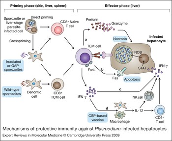
Both live-attenuated irrSPZ (Refs Reference Hoffman10, Reference Nussenzweig11) and GAP vaccines (Refs Reference Mueller18, Reference Mueller19, Reference Tarun20, Reference van Dijk23, Reference Labaied25, Reference Jobe26, Reference Aly28, Reference Kumar73) provide complete, long-lasting protection, and immune mechanisms involved have been extensively analysed (Fig. 1). CD8+ T cells have a critical role in the protection of mice immunised with irrSPZs (Refs Reference Tsuji and Zavala74, Reference Overstreet75), whereas interleukin-4 (IL-4)-secreting CD4+ T cells are essential for the development of CD8+ T cell responses to LS parasites (Ref. Reference Carvalho76). Loss of protective immunity in β2 microglobulin−/− mice (which have no CD8+ T cells or natural killer T cells) and in mice depleted of CD8+ T cells, indicates the indispensable role of CD8+ T-cell-mediated effector mechanisms (Refs Reference Weiss77, Reference White, Snyder and Krzych78). In studies with P. berghei and P. yoelii, adoptive transfer of CD8+ T cell clones specific for CSP epitopes conferred complete protection against SPZ challenge in naive mice (Refs Reference Rodrigues79, Reference Romero80, Reference Weiss81). CD8+ T cell responses can also be detected in humans protected by immunisation with P. falciparum irrSPZs (Ref. Reference Wizel82). Furthermore, CSP-specific CD8+ T cells have been found to efficiently lyse infected hepatocytes in vitro (Refs Reference Rodrigues79, Reference Bongfen83, Reference Weiss84). Since irrSPZs protected perforin (PRF1) and granzyme B (GZMB) double-knockout mice (which lack the ability to kill cells through cytolytic activity) from infectious SPZ challenge, it was speculated that CD8+ T cell killing of infected hepatocytes is primarily through IFN-γ-induced nitric oxide (NO), rather than by direct cytolytic activity (Refs Reference Doolan and Hoffman13, Reference Klotz85). Indeed, it has been shown that IFN-γ responses are correlated with protective immunity against the pre-erythrocytic stage, although the importance of CD8+ T-cell-derived IFN-γ in particular is yet to be fully established (Refs Reference Schofield86, Reference Seguin87). Sterile protection obtained with a P. berghei GAP was found to correlate with IFN-γ-producing CD8+ T cells (Ref. Reference Jobe26). Conversely, IFN-γ-independent CD8+-T-cell-mediated protective immunity has also been demonstrated (Ref. Reference Rodrigues88), and IFN-γ secretion by CD8+ T cells is not essential for protecting mice against P. yoelii SPZ challenge (Ref. Reference Chakravarty89). Recently, Trimnell and colleagues demonstrated that contact-dependent CD8+ T cell elimination of LS-infected hepatocytes is the major effector mechanism in P. yoelii GAP-induced sterile protection (Ref. Reference Trimnell90). Therefore, the relative importance of different CD8+ T cell effector mechanisms might differ between distinct vaccine models.
Mechanisms of protective immunity against Plasmodium-infected hepatocytes. In the protective model of live-attenuated sporozoite vaccines [irradiated sporozoites (irrSPZs) or genetically attenuated parasites (GAPs)], elimination of pre-erythrocytic-stage parasites is primarily dependent on antigen-specific and major histocompatability complex (MHC)-restricted CD8+ T cells, whereas recombinant vaccines, such as the CSP-based RTS,S vaccine, are primarily dependent on CD4+ T cells. Naive CD8+ T cells can be primed by dendritic cells (DCs) that encounter SPZs at skin-draining lymph nodes, by parasite-traversed or -infected hepatocytes in the liver (direct priming), or by other DCs that uptake antigens provided by traversed and infected hepatocytes, or hepatocytes that die in the liver (crosspriming). Primed CD8+ T cells will then proliferate and differentiate to effector (TEM) or central memory (TCM) CD8+ T cells. CD8+ T cells can mediate killing of liver stage (LS)-infected hepatocytes via several mechanisms: (a) release of perforin and granzyme B, which causes necrosis of the infected hepatocyte; (b) apoptosis, a programmed cell death through the Fas–FasL signalling pathway; (c) release of IFN-γ, via signal transducers associated with transcription (STAT) to activate inducible nitric oxide synthase (iNOS), which then induces the NO pathway to destroy the infected hepatocyte; (d) IFN-γ can also promote IL-12 release from DCs, macrophages or other cells to activate the natural killer (NK) and CD4+ T cells that consequently increase the IFN-γ-dependent killing of LS-infected hepatocytes

CD8+ T cell priming might occur in skin-draining lymph nodes after immunisation by the bite of irrSPZ-infected mosquitoes (Ref. Reference Chakravarty59), whereas hepatic CD8+ dendritic cells (DCs) may be responsible for priming CD8+ T cells by intravenous immunisation with irrSPZs (Ref. Reference Jobe91). Both wild-type SPZs and irrSPZs are processed for class I presentation by DCs. However, DCs pulsed with wild-type SPZs appear to stimulate only memory CD8+ T cells, whereas DCs pulsed with irrSPZs are capable of activating both effector and memory CD8+ T cells (Ref. Reference Plebanski92).
Little is known about if and how hepatocytes process and present malaria antigens to CD8+ T cells. Bongfen and co-workers investigated CSP processing in primary mouse hepatocytes exposed to wild-type SPZs or cell-traversal-deficient P. berghei SPZs (Ref. Reference Bongfen83). They demonstrated that parasite-infected hepatocytes process CSP through proteasomes, whereas traversed non-infected hepatocytes utilise aspartic proteases to degrade the CSP left behind by traversing SPZs (Ref. Reference Bongfen93). These observations suggest that the parasite may avoid host immune surveillance by diverting CSP-specific CD8+ T cells to attack traversed but uninfected hepatocytes (Ref. Reference Trimnell90). CSP also suppresses the respiratory burst in Kupffer cells, thus supporting their successful traversal (Ref. Reference Usynin, Klotz and Frevert94). Furthermore, CSP actively promotes LS parasite growth in infected hepatocytes by inhibiting the NF-κB pathway to downregulate pro-inflammatory cytokine release (Ref. Reference Singh95). All these data suggest that CSP is an immunodominant antigen that is the target of protective immune responses but is also responsible for immune evasion to ensure the survival of the parasite within its host cell.
The search for correlates of protection
To date, the most commonly used marker of protection is IFN-γ in conjunction with a few other cytokines and immune cell surface markers. In general, IFN-γ detected by enzyme-linked immunosorbent spot (ELISPOT) assay in 7–14 day peripheral blood mononuclear cell (PBMC) culture (but not in fresh PBMCs) has been associated with naturally acquired or vaccine-induced T cell immunity against infection. Susceptibility to infection, however, has been associated with CD4+ CD25+ regulatory T cells (Ref. Reference Todryk96). In the livers of mice protected by irrSPZs, both effector (TEM) and central (TCM) memory CD8+ T cells were identified (Ref. Reference Krzych and Schwenk97), but long-lasting protection depended on CD8+ TCM cells expressing the IL-15 receptor CD122 (Ref. Reference Berenzon98). However, a single dose of simian adenovirus-induced protection was associated with CD8+ TEM cells, whereas a poxviral vector that did not protect mice from P. berghei infection had predominant CD8+ TCM cells (Ref. Reference Reyes-Sandoval55), indicating that the readiness of TEM cells has a major role in protection against infection. Several cytokine responses (IFN-γ, TNF-α and IL-2) were also greater in mice immunised with adenovirus vector than those in mice immunised with poxvirus vector (Ref. Reference Reyes-Sandoval55). Recently, it was demonstrated that a threshold T cell frequency (≥1% of CSP-specific CD8+ T cells in blood, >106 in spleen and >2 × 10⁵ in liver) is required for long-term protective immunity against P. berghei SPZ challenge. This threshold is 100- to 1000-fold higher than the number of memory CD8+ T cells required for protection against a bacterial or viral pathogen (Ref. Reference Schmidt99). P. berghei is known to be less infectious than P. yoelii in mice, indicating that much higher threshold numbers of CD8+ T cells will be needed for induction of sterile protection against P. yoelii challenge, and potentially against P. falciparum in humans.
Subunit vaccine development for erythrocytic stages
During blood-stage development, malaria parasites have a largely intracellular lifestyle and are only briefly exposed when they are released as merozoites and invade new red blood cells (Ref. Reference Miller6). In the case of P. falciparum and P. vivax, merozoite release occurs once every 48 hours. Erythrocyte invasion occurs very rapidly, within 30–60 seconds (Ref. Reference Dvorak100), and therefore it is assumed that high-titre antibodies will be required for invasion-blocking vaccine approaches to be effective. Once inside the red blood cell, erythrocytes offer a relatively immunoprivileged site for parasite replication because they lack a nucleus and MHC molecules, and therefore are limited in their ability to mount effective antipathogen defences. Nevertheless, humans exposed to malaria parasites gradually acquire considerable anti-blood-stage immunity, which varies with age and transmission intensity (Ref. Reference Marsh and Snow101). Passive immunoglobulin-transfer studies suggest that antibodies are a crucial component of protective immunity (Refs Reference Cohen, Mc and Carrington102, Reference Sabchareon103); however, little is known about the molecular targets and mechanisms of naturally acquired immunity to malaria. Current blood-stage vaccine efforts are focused on a small number of candidate antigens and three types of subunit vaccine approaches: (1) invasion-blocking vaccines, (2) antidisease vaccines for severe and pregnancy-associated malaria, and (3) antitoxin vaccines (e.g. GPI).
Invasion-blocking vaccines
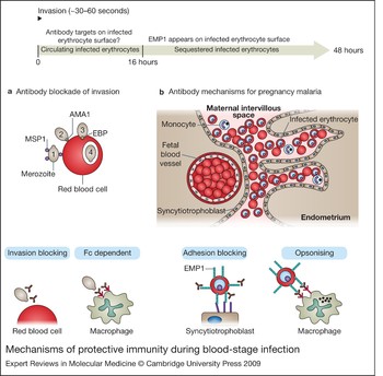
Red blood cell invasion is a multi-step process that involves several proteins at the parasite and erythrocyte surface. Parasite invasion has been divided into four steps: attachment, reorientation, tight junction formation and entry (Fig. 2) (Refs Reference Dvorak100, Reference Gilson and Crabb104). Invasion-blocking vaccine strategies have been highly focused on three proteins, MSP1, AMA1 and the P. vivax Duffy binding protein (PvDBP). MSP1 is believed to function at initial attachment (Ref. Reference Goel105), AMA1 at parasite reorientation and entry (Ref. Reference Mitchell106) and DBP during tight junction formation for P. vivax (Refs Reference Miller107, Reference Singh108) (Fig. 2). Antibodies against all three proteins can inhibit parasite invasion; however, antigen polymorphism, redundancy of invasion pathways and the kinetics of erythrocyte invasion pose significant challenges for vaccine development.
Mechanisms of protective immunity during blood-stage infection. Erythrocyte invasion occurs rapidly, within ~30–60 seconds (Ref. Reference Dvorak100), and once inside the erythrocyte, the parasite appears to be relatively inaccessible to antibodies until approximately 14–16 h after invasion, when new parasite proteins, such as the P. falciparum erythrocyte, membrane protein 1 (EMP1), begins to appear on the infected erythrocyte (IE) surface. (a) Erythrocyte invasion is a complex process that has been divided into four steps: (1) attachment, (2) reorientation, (3) tight junction formation, and (4) entry. MSP1 is the most abundant protein on the merozoite surface and may function at initial attachment (Ref. Reference Goel105), AMA1 at parasite reorientation and entry (Ref. Reference Mitchell106) and P. vivax DBP during tight junction formation for P. vivax (Refs Reference Miller107, Reference Singh108). P. vivax DBP is a member of the erythrocyte-binding protein (EBP) family, which has undergone gene expansion in P. falciparum, leading to functional differences with P. vivax. Antibodies may directly block invasion by inhibiting parasite binding, preventing protein–protein interactions on the merozoite surface, or interfering with proteolytic processing or shedding of invasion ligands. There is also evidence for Fc-dependent mechanisms of protection against MSP1 (Ref. Reference McIntosh121). (b) In pregnancy malaria, P. falciparum-infected erythrocytes sequester in the intervillous spaces of the maternal placenta, but are absent in fetal blood vessels. Infected erythrocytes are found directly adjacent to syncytiotrophoblast cells and also fill the intervillous space. Monocytes are recruited to infected placentas and frequently contain residual hemozoin pigment that may result from ingestion of infected erythrocytes. Potential antibody mechanisms are adhesion blocking or opsonising infected erythrocytes for phagocytosis by monocytes or activated macrophages that were recruited to the infected placenta.

MSP1
MSP1 is synthesised as a GPI-linked 195 kDa precursor protein and undergoes proteolytic processing into four protein fragments (Refs Reference Holder109, Reference Lyon110), which then assemble into a complex with at least two other proteins, MSP6 and MSP7, at the merozoite surface (Refs Reference Kauth111, Reference Kauth112). During merozoite invasion, the C-terminal 42 kDa fragment of MSP1 (MSP142) undergoes secondary processing (Refs Reference Blackman and Holder113, Reference Blackman114). Antibodies that interfere with the secondary processing of MSP142 (Ref. Reference Angov115) or target the C-terminal MSP119 region (Refs Reference Blackman116, Reference John117, Reference Ling, Ogun and Holder118, Reference Majarian119, Reference O'Donnell120) can inhibit erythrocyte invasion or parasite growth, either directly, or possibly through Fc-dependent pathways (Ref. Reference McIntosh121). The most advanced MSP1 vaccine candidates are based on the C-terminal MSP142 fragment. Anti-MSP142 sera can inhibit P. falciparum growth in vitro (Refs Reference Bergmann-Leitner122, Reference Singh123, Reference Woehlbier124) and inhibit the secondary processing of MSP142 (Ref. Reference Angov115), but only conferred strain-specific protection in a P. falciparum-Aotus challenge model (Ref. Reference Lyon125). Although safe and immunogenic in Phase I trials (Refs Reference Ockenhouse126, Reference Stoute127, Reference Thera128), an MSP142-based vaccine conferred no protection in a Phase IIb trial in Kenyan children (Ref. Reference Ogutu129). The reason for vaccine failure is unknown, but could include allelic polymorphism or antibody specificity and/or function.
AMA1
AMA1 is an 83 kDa protein that is targeted to micronemes (Refs Reference Healer130, Reference Narum and Thomas131). Before invasion, it is processed to a 66 kDa form (AMA166), which is transferred to the merozoite surface and subsequently associates with three other proteins, RON4, RON2 and Ts4705 (Refs Reference Cao132, Reference Collins133). Based on studies that were initially performed in the apicomplexan Toxoplasma gondii, the AMA1 complex forms a moving junction that translocates over the zoite surface as the parasite enters the parasitophorous vacuole (Ref. Reference Alexander134). The moving junction is believed to form a barrier that excludes antibodies from entering the parasitophorous vacuole (Ref. Reference Alexander134). Antibodies that inhibit AMA1 complex formation (Ref. Reference Collins133) or disrupt the circumferential redistribution or proteolytic shedding of AMA1 can block parasite invasion (Refs Reference Dutta135, Reference Dutta136), but AMA1 polymorphism severely compromises vaccine efficacy. AMA1 contains at least 64 variable residues, and inhibitory antibodies are highly strain-specific (Refs Reference Hodder, Crewther and Anders137, Reference Kennedy138, Reference Kocken139). Single antigen or bivalent AMA1 vaccines were immunogenic in Phase I trials (Refs Reference Dicko140, Reference Spring141), but invasion-blocking responses were strain-specific and were not protective in human challenge models of naive malaria using a SPZ challenge (Ref. Reference Spring141) or a Phase II trial in African children (Ref. Reference Sagara142). Thus, AMA1 polymorphism poses significant technical and economical challenges to vaccine development, and it is not yet clear whether conserved regions can form the basis for a vaccine. The safety and immunogenicity of a fusion protein between MSP119 and domain III of AMA1 has also been tested (Ref. Reference Hu143), but this approach must also overcome obstacles faced by the individual AMA1 and MSP1 components.
Plasmodium vivax DBP
P. vivax requires the Duffy blood group antigen/receptor for chemokines (DARC) to invade reticulocytes (Ref. Reference Miller144) and has nearly disappeared from West Africa, where most individuals are DARC negative. P. vivax binds to DARC via DBP (Ref. Reference Fang145). P. vivax DBP is a member of a family of erythrocyte-binding proteins characterised by cysteine-rich binding domains, termed Duffy-binding-like (DBL) domains that confer red blood cell binding specificity (Refs Reference Chitnis and Miller146, Reference Sim147). Whereas P. vivax has only a single gene encoding the DBP, P. falciparum has four genes that encode proteins that bind different sialoglycoproteins on the erythrocyte surface (Ref. Reference Cowman and Crabb148). Because P. vivax has only a single DBP gene, the parasite might be more dependent on the DARC interaction for invasion and would be unable to switch to other members to escape antibody pressure.
The structure of the DBL domain has been solved from the P. knowlesi DBP (Refs Reference Adams149, Reference Singh150), and used to model polymorphism and functional residues in P. vivax DBP. Interestingly, critical binding residues in P. vivax DBP mostly map to a solvent-accessible groove, but the majority of polymorphism is concentrated on the opposite surface from the predicted DARC-binding site (Refs Reference Chitnis and Sharma151, Reference Tsuboi152, Reference Xainli, Adams and King153). It is currently disputed whether polymorphism is under significant selection for escape from inhibitory antibodies, although an amino acid change at residue 171 affects invasion-blocking antibodies (Ref. Reference VanBuskirk154) and many children in Papua New Guinea, aged 5–14 years, appear to have strain-specific inhibitory antibodies (Ref. Reference King155). The most advanced P. vivax DBP immunogen is based on a bacterial refolded protein from the DBL-binding domain or region II (P. vivax DBPII) (Ref. Reference Singh156). Antibodies to P. vivax DBPII can inhibit its interaction with DARC in vitro (Refs Reference Devi157, Reference Michon, Fraser and Adams158, Reference Yazdani159), as well as inhibiting parasite invasion in an in vitro invasion assay (Ref. Reference Grimberg160). However, complete Freund's adjuvant was required to protect in a P. vivax and Aotus primate challenge model (Ref. Reference Arevalo-Herrera161).
Antidisease vaccines for severe and pregnancy-associated malaria
Once the parasite has entered the red blood cell, it might be expected that it would not display proteins at the infected erythrocyte (IE) surface, but curiously all Plasmodium species appear to express different clonally variant antigens at the IE surface (Ref. Reference Kyes, Horrocks and Newbold162). The best characterised gene family is called var, or P. falciparum erythrocyte membrane protein 1 (EMP1) (Ref. Reference Kraemer and Smith163) Unlike other Plasmodium species that infect humans, P. falciparum begins to sequester from the circulation approximately 14–16 h after invasion and the EMP1 extracellular domain expressed on the IE surface is responsible for this (Ref. Reference Udomsangpetch164). P. falciparum EMP1 proteins encode multiple receptor-like domains related to the binding region in the P. vivax DBP protein (Refs Reference Baruch165, Reference Su166). Each parasite genome encodes ~60 var genes (Ref. Reference Gardner167), but expresses only one at a time. Switches in var gene expression modify the antigenic and binding properties of IEs (Ref. Reference Smith168). This trait is a major virulence determinant that allows parasites to sustain chronic infection and contributes to severe disease when P. falciparum IEs accumulate in brain or placenta.
The best understood paradigm for IE sequestration is placental malaria. P. falciparum infection during pregnancy is considered the most important nongenetic factor contributing to low birth weight in first pregnancies in Africa, and is believed to cause ~20,000 maternal deaths annually and may contribute to the deaths of 100,000–200,000 infants (Refs Reference Brabin169, Reference Steketee170). Placental malaria has been associated with the expression of a single var gene, termed VAR2CSA, which is believed to mediate IE binding to chondroitin sulphate A (CSA) in the placenta (Refs Reference Duffy171, Reference Magistrado172, Reference Salanti173, Reference Tuikue Ndam174). VAR2CSA is unusual for the var gene family, because a gene copy is found in all P. falciparum strains (Refs Reference Salanti173, Reference Trimnell175), and it is thus being investigated as a syndrome-specific vaccine intervention.
VAR2CSA is a large (~350 kDa) and polymorphic protein. It is technically challenging to express the entire extracellular domain as a recombinant protein and therefore vaccine efforts have focused on expressing individual domains (Refs Reference Avril176, Reference Barfod177, Reference Fernandez178, Reference Nielsen179, Reference Oleinikov180). Sequence and serological comparisons suggest that there is antigenic overlap between geographically diverse placental isolates (Refs Reference Beeson181, Reference Bockhorst182, Reference Fried183), but it is not yet clear how many vaccine components will be required for an effective vaccine. Infected women frequently have five or six different placental genotypes, which could contribute to the breadth of antibody reactivity, even after a single pregnancy, and women may never acquire sterile immunity (Ref. Reference Brabin169). Achieving the same breadth of antibody reactivity as found in protected women by vaccination with VAR2CSA immunogens may require defining highly conserved functional epitopes, should these exist, or multivalent vaccine mixtures. Monoclonal antibodies from pregnant women appear to predominantly target polymorphic epitopes in VAR2CSA (Ref. Reference Barfod184), although some conserved regions in VAR2CSA might be recognised by antibodies (Ref. Reference Andersen185). Antibodies are thought to protect against placental malaria by inhibiting IE binding to CSA (Refs Reference Fried183, Reference Duffy and Fried186, Reference O'Neil-Dunne187, Reference Ricke188) or opsonising IEs for phagocytosis (Refs Reference Jaworowski189, Reference Keen190, Reference Mount191, Reference Staalsoe192) (Fig. 2), but adhesion-blocking epitopes in maternal sera have not been mapped. Recent evidence suggests that antibodies to several different VAR2CSA domains can partially inhibit IE binding to CSA, but the greatest inhibitory activity was against the VAR2CSA DBL4 domain (Ref. Reference Nielsen179). This led to the proposal that it may be possible to base a vaccine on a single VAR2CSA DBL domain. However, the breadth of inhibitory activity and the basis for this effect is not yet understood, and it has been difficult to generate adhesion-blocking responses to VAR2CSA recombinant proteins in many studies (Refs Reference Avril176, Reference Barfod177, Reference Fernandez178, Reference Oleinikov180).
Antitoxin vaccine strategies
Another antidisease intervention is to target the malaria toxin. Despite intensive research efforts to characterise parasite factors responsible for proinflammatory and febrile responses associated with schizont rupture, the actual ‘malaria toxin’ is still a matter of debate. It has been proposed that the P. falciparum GPI is an important parasite factor that activates the host innate immune system, mainly through TLR2 and to a lesser extent TLR4 (Refs Reference Krishnegowda193, Reference Schofield and Hackett194), whereas haemozoin carrying parasite DNA was found to be a TLR9 ligand (Ref. Reference Parroche195). Although the primary function of the innate immune response and fever is to fight off the pathogen, excessive proinflammatory cytokines, such as tumor necrosis factor (TNF), might be involved in the pathogenesis of fatal malaria (Refs Reference Grau196, Reference Kwiatkowski197) and could exacerbate inflammation at adherence sites, such as brain, by upregulating cell-adhesion molecules (Ref. Reference Schofield198). Based on this concept, a synthetic malaria GPI glycan is being explored as an antitoxic therapeutic target in experimental models of malaria in mice (Ref. Reference Schofield199). Immunisation of mice with synthetic GPI glycan prevented pulmonary oedema and cerebral malaria complications, but did not have an antiparasite effect, with the result that mice died of massive parasitaemia with normal kinetics (Ref. Reference Schofield199). The major remaining questions for antitoxin vaccines are whether GPI is the only malaria toxin, and whether this approach could prevent the inflammatory complications of infection without losing the potential beneficial effects of fever or innate responses. If proved to be safe, then antitoxin vaccines would presumably need to be combined with antiparasite vaccine components to be effective.
Clinical implications and outstanding research questions
In summary, all malaria vaccine development approaches discussed in this review face formidable obstacles, but also provide tantalising opportunities. Subunit vaccines do not yet give the desired levels of protection, but would be easiest to manufacture and deliver. Live-attenuated parasites are protective, but we do not know how to best manufacture and deliver them.
Pre-erythrocytic subunit vaccine development faces considerable challenges. Identifying which of the thousands of genes expressed at the SPZ and LSs contribute to protective immunity is one challenge, optimisation of vaccine delivery systems that elicit high-titre functional antibodies to prevent infection and/or induce cellular immunity to infected hepatocytes is another challenge. Recent advances in LS research support for the first time an efficient, pragmatic and systematic approach for the identification and prioritisation of pre-erythrocytic antigens for subunit vaccine development. These include the purification of in vivo LS parasites (Ref. Reference Tarun200), the description of the LS transcriptome and partial proteome of P. yoelii (Ref. Reference Tarun201), the availability of protective attenuated whole-cell vaccines (Ref. Reference Mueller18) and the application of high-throughput immune assays to identify novel pre-erythrocytic antigens targeted by antibodies and T cells in humans (Refs Reference Doolan70, Reference Corradin202, Reference Haddad203, Reference Wang204). These are all potent tools for future investigations which should include: (1) characterisation of the immune mechanisms of protection induced by attenuated parasites; (2) identification of peripheral biomarkers that are associated with acquisition, varying degrees and duration of protective immune responses induced by whole-parasite vaccination using system biology approaches; (3) exploration of the entire Plasmodium SPZ and LS proteome to identify novel pre-erythrocytic antigens that contribute to sterile protection induced by attenuated parasites; (4) high-throughput in vitro assays for determination of antibody- and cell-mediated elimination of LS-infected hepatocytes that correlate with protection; (5) rational selection of vaccine delivery system(s) and adjuvant(s) that induce immune responses of the magnitude necessary to prevent infection.
The lack of immune correlates of protection is a barrier to invasion-blocking vaccine development for blood stages. It is not known whether MSP1 or AMA1 vaccines would be more effective if vaccine antibodies could be focused on functionally critical sites (e.g. complex interaction or proteolytic sites). Whereas most invasion-blocking vaccine effort has focused on P. falciparum, P. vivax might provide a more tractable system to develop ‘proof of concept’ for invasion-blocking approaches because P. vivax has more restricted blood cell tropism (Ref. Reference Miller6). The selective preference for reticulocytes is an important factor limiting P. vivax growth and disease severity, and may provide unique opportunities for vaccine intervention. A challenge for P. vivax DBP vaccine development is that P. vivax cannot be easily grown in vitro; therefore, in vitro growth inhibition assays have used blood collected from naturally infected humans (Ref. Reference Grimberg160). This in vitro invasion assay has relatively low efficiency, and vaccine development has relied on in vitro binding assays with P. vivax DBPII recombinant proteins (Refs Reference Devi157, Reference Michon, Fraser and Adams158, Reference Yazdani159). Major priorities include implementing standardised in vitro binding assays for antibody assessment, developing more robust in vitro invasion assays, optimisation of P. vivax and nonhuman primate challenge models or human challenge models, and a better understanding of how antibodies inhibit P. vivax invasion.
Antidisease vaccines would not prevent infection, but would target specific parasite variants associated with disease. Because severe malaria is a relatively rare complication of malaria infection, estimated to occur in ~2% of infections (Ref. Reference Greenwood, Marsh and Snow205), antidisease vaccine interventions will require large and expensive trials. As many first-time pregnant women acquire severe placental infections, trial design could be smaller and allow the antidisease vaccine concept to be tested. It is thought that a pregnancy malaria vaccine would need to be administered to women in their teenage years, similarly to human papilloma virus vaccines (Ref. Reference Koutsky206). VAR2CSA polymorphism poses challenges, and it is not known whether vaccines can elicit antibodies against conserved regions of the protein that would not vary under vaccine pressure. Current understanding of how antibodies protect against placental malaria is limited and the lack of animal models for P. falciparum sequestration in the placenta are barriers to vaccine development. Whereas vaccine efforts have been highly focused on adhesion-blocking-antibody responses, more consideration may be needed for synergistic vaccines that aim for the broadest achievable adhesion-blocking and opsonising responses.
The current data available for irrSPZ immunisation studies in humans indicate that live-attenuated whole parasites induce sterilising immunity against challenge with homologous P. falciparum via the bite of infected mosquitoes. The protection lasts for up to 10 months and a small number of volunteers, when challenged with heterologous strains, were also protected. Furthermore, as discussed earlier, numerous animal studies support the notion that live-attenuated SPZ vaccinations confer sterile, long-lasting protection. Within the next 2 years, a clinical dose-escalation study with a cryopreserved formulation of irrSPZs that is administered intradermally or subcutaneously and a first-generation genetically attenuated SPZ vaccine that is administered by mosquito bite will probably yield unambiguous data on the safety and protective efficacy of this class of vaccines. In the future, the most formidable obstacle for the live-attenuated vaccine approach lies in the current need for cryopreservation in liquid nitrogen. A cryopreserved malaria vaccine will face challenges when delivered in the context of the ‘Expanded Program on Immunization’ (EPI), which is currently the major platform through which many childhood vaccines are delivered in resource-poor nations. Does this imply that live-attenuated vaccine approaches are a ‘dead end’ that will remain confined to the experimental stage? In this context, it is of interest to consider that delivery of a malaria vaccine for disease elimination will probably require delivery platforms that are distinct from EPI. Furthermore, it should be noted that live-parasite vaccines for veterinary applications are currently delivered in liquid nitrogen in resource-poor countries (Ref. Reference Di Giulio207). Thus, it is conceivable that if the subunit vaccine approaches described in this review continue to fail or confer only limited protection, the delivery of an efficacious whole-cell, live-attenuated malaria vaccine is likely to be a critical tool by which we can hope to eradicate malaria.
Acknowledgements
We thank Dr Ashley Vaughan and our peer reviewers for critically reviewing the manuscript. The authors are funded by grants from the Foundation for the National Institutes of Health through the Grand Challenges in Global Health Initiative, the Bill and Melinda Gates Foundation and the National Institutes of Health (RO1 AI047953-06, RO1 AI053709-07).