Introduction
Mucosa-associated lymphoid tissue lymphomas of the head and neck arise in extranodal organs, such as the salivary and thyroid glands and Waldeyer's ring, and occur with chronic inflammation.Reference Isaacson and Wright1–Reference Yoshino and Akagi4 We assessed the clinical features of patients with newly diagnosed mucosa-associated lymphoid tissue lymphoma in these sites.
Low-grade or indolent malignant lymphoma (e.g. mucosa-associated lymphoid tissue lymphoma) is often observed over a prolonged period. Therefore, it is important to carry out frequent biopsies or resections of the mass for diagnosis.
Materials and methods
We conducted a retrospective analysis of new patients who were diagnosed with mucosa-associated lymphoid tissue lymphoma arising in the head and neck region and who were treated at our institution between January 1990 and December 2009.
At primary presentation, most patients had enlargement of the salivary glands, neck lymph nodes or tonsils, and/or ocular symptoms. Mucosa-associated lymphoid tissue lymphoma was histologically diagnosed according to the criteria of Isaacson and Wright, on the basis of evaluation of haematoxylin and eosin stained specimens obtained following resection or excisional biopsy.Reference Isaacson and Wright1 Furthermore, we performed immunohistochemical analyses of paraffin sections using antibodies against cluster of differentiation 3, 5, 10, 20, 21 and 79a glycoprotein, cyclin D1, kappa and lambda light chains, and Bcl-2 (B-cell lymphoma 2); our patients had results consistent with a diagnosis of extranodal marginal zone B-cell lymphoma of mucosa-associated lymphoid tissue type.
Examination of medical records revealed that 20 patients had been diagnosed during the study period.
Patients' tumours were evaluated on the basis of complete diagnostic investigation, including biopsy, laboratory tests, physical and radiological examinations, and bone marrow biopsy. At the initial presentation, we established the diagnosis and ascertained the lymphoma location, and also obtained relevant patient information, including age, sex, history, complications (if any), clinical staging, treatment and response, recurrence (and time to recurrence), and survival status. Patients were staged according to the Ann Arbor system, and treated with surgery, radiotherapy, chemotherapy or combination therapy.5 Response to therapy was assessed using the standard criteria for complete remission, partial remission, stable disease and progressive disease.
Data were analysed during March 2010.
Results and analysis
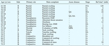
Between January 1990 and December 2009, 20 patients seen at Hamamatsu University School of Medicine were newly diagnosed with primary mucosa-associated lymphoid tissue lymphoma of the head and neck (Table I). There were eight men and 12 women, with ages ranging from 23 to 85 years and a mean age of 59.3 years; the mean ages of the male and female patients were 54 and 62.8 years, respectively. Most patients were in their 60s (seven cases, 35 per cent) or 50s (four cases, 20 per cent); there were two patients each (10 per cent) in their 30s, 40s, 70s and 80s, and one patient in her 20s (5 per cent).
Patient Characteristics: Summary

*Time between first symptoms to referral (ref) to our hospital. Y = years; Assoc = associated; mth = months; M = male; R = right; – = none reported; L = left; F = female; SjS = Sjögren's syndrome; RA = rheumatoid arthritis; OME = otitis media with effusion; ALL = acute lymphocytic leukaemia; MPK = mesopharyngeal carcinoma; SMG = submandibular gland; B = bilateral
The mean time period between patients' first experience of primary symptoms and their referral to our hospital was 7.8 months (range, one to 120 months); however, this time period was 18 months (range, one to 120 months) when patients with ocular adnexa were excluded.
The patients' mucosa-associated lymphoid tissue lymphomas were located in the parotid gland, nasopharynx, ocular adnexa (i.e. conjunctiva), thyroid gland, submandibular gland or tonsils (Table I).
At initial presentation, most patients (80 per cent) were at Ann Arbor stage IA (Table I). Pretherapeutic staging evaluation included radiological studies in all patients, chest radiographs, computed tomography of the whole body, and magnetic resonance imaging of the head and neck. However, the radiological features of the head and neck mucosa-associated lymphoid tissue lymphomas could not be used for diagnosis. Most tumours comprised a well defined, homogeneous mass, and none showed any calcification or multiple small cystic lesions.
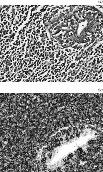
Histological verification was performed in all 20 patients, by means of biopsy and immunohistochemical phenotyping of paraffin sections. Immuno histochemical analysis used antibodies against cluster of differentiation 3, 5, 10, 20, 21 and 79a glycoprotein, cyclin D1, kappa and lambda light chains, and Bcl-2; all patients had results consistent with a diagnosis of extranodal marginal zone B-cell lymphoma of mucosa-associated lymphoid tissue type (Figure 1).
Photomicrographs of mucosa-associated lymphoid tissue lymphoma of the parotid gland, showing (a) H&E-stained specimen and (b) immunohistochemical positivity for cluster of differentation 20 glycoprotein within lymphoma cells around the parotid duct. (×400)

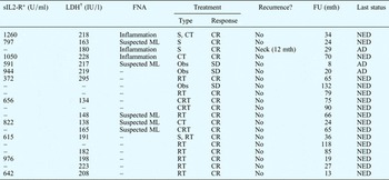
A staging evaluation was carried out in 11 patients, taking into consideration the following laboratory data: full blood count (with differential counts of white and red blood cells), platelet count, biochemical profile (including lactate dehydrogenase) and serum interleukin-2 (IL2) receptor level. The full blood counts and biochemical profiles were within normal limits. However, all of the 11 cases evaluated had extraordinarily high serum IL2 receptor levels (Table II).
Patients' Clinical Features: Summary

*Normal range 115–208 U/ml. †Normal range 220–530 IU/l. sIL2-R = serum interleukin 2 receptor; LDH = lactose dehydrogenase; FNA = fine needle aspiration biopsy; FU = follow-up time; mth = months; S = surgery; CT = chemotherapy; CR = complete remission; NED = no evidence of disease; ML = malignant lymphoma; – = not reported; AD = alive with disease; Obs = observation; SD = stable disease; RT = radiotherapy; CRT = chemoradiotherapy
Despite complications, comprising evidence of autoimmune disease (including Sjögren's syndrome and rheumatoid arthritis) and malignancy (i.e. mesopharyngeal carcinoma and acute lymphocytic leukaemia), all patients made a good recovery from their tumour.
Treatment consisted of: surgical resection alone; surgical resection with consecutive chemotherapy; surgical resection with radiotherapy; radiotherapy alone; and/or therapy with rituximab, cyclophosphamide, hydroxydaunorubicin, vincristine, and prednisolone (Table II).Reference Plosker and Figgitt6 Radiation doses commonly ranged from 20 to 54 Gy, delivered in 10 to 27 fractions over two to six weeks.
Three patients were informed in detail about their diagnosis, and about the risks of treatment and possible complications. Subsequently, these patients underwent observation only, with no active treatment (Table II).
Only one patient experienced disease recurrence, occurring 12 months after treatment and involving a neck lymph node. He was informed of the diagnosis, and no treatment was given. This patient was stable with disease, 17 months after being diagnosed with disease recurrence.
At the time of writing, all the patients were still alive. Information on their survival status was obtained after a median follow-up period of 50.8 months (range, 8–134 months).
Discussion
There is limited information in the literature on the incidence and epidemiology of mucosa-associated lymphoid tissue lymphomas arising in the head and neck. Therefore, much of the following information has been extrapolated from data on mucosa-associated lymphoid tissue lymphomas in other sites.
Mucosa-associated lymphoid tissue lymphoma is more common in women than men, the ratio of incidence being 1.4 to 1, with a mean age at presentation of 64 years.Reference Zukerberg, Medeiros, Ferry and Harris7 The preferred sites in the head and neck appear to be the parotid gland, thyroid gland, Waldeyer's ring, and ocular adnexa, particularly the conjunctiva.Reference Gleeson, Bennett and Cawson2, Reference Watkin, MacLennan and Hobsley3, Reference Ando, Matsuzaki and Murofushi8 Due to the tumour's indolent growth and benign clinical behaviour, it can be difficult to diagnose. The tumour often presents in the head and neck in an indolent fashion, as demonstrated by the prolonged mean referral time for our 20 cases.Reference Isaacson and Wright1–Reference Watkin, MacLennan and Hobsley3, Reference Ando, Matsuzaki and Murofushi8–Reference Wenzel, Fiebiger, Dieckmann, Formanek, Chott and Raderer12 The features of head and neck mucosa-associated lymphoid tissue lymphomas are virtually undetectable.Reference Gleeson, Bennett and Cawson2, Reference Watkin, MacLennan and Hobsley3, Reference Ando, Matsuzaki and Murofushi8–Reference Wenzel, Fiebiger, Dieckmann, Formanek, Chott and Raderer12 Masses or submucosal thickening present for years may be the only clinical sign, further complicating the diagnosis. Therefore, referrals and diagnostic procedures are often heterogeneous and sometimes inappropriate, prior to establishment of the correct diagnosis.
Furthermore, radiological studies are not particularly helpful, other than for planning surgery. However, they are important in the postdiagnostic clinical staging of mucosa-associated lymphoid tissue lymphoma.Reference Ando, Matsuzaki and Murofushi8, Reference Bhattacharyya, Frankenthaler, Gomolin, Kadin and Lauretano9 Recently, mucosa-associated lymphoid tissue lymphoma has been recognised as a subtype of low-grade non-Hodgkin's lymphoma, thus leading to several difficulties with the histopathological analysis of these lesions. Mucosa-associated lymphoid tissue lymphoma is characterised as a type of B-cell marginal zone lymphoma, as noted by Fisher et al. Reference Fisher, Dahlberg, Nathwani, Banks, Miller and Grogan13 Surgical biopsy of a portion of the extranodal tissue is necessary, but this should be coupled with careful pathological evaluation supplemented by immunohistochemical analysis. The resected material must be handled appropriately to enable proper processing for immunohistochemical analysis.
The diagnosis of mucosa-associated lymphoid tissue lymphoma includes analysis of laboratory data, including full blood counts and biochemical profile. In our patients, extraordinarily high serum interleukin 2 (IL2) receptor levels were detected. However, serum IL2 receptor levels have been found to be high not only in mucosa-associated lymphoid tissue lymphoma patients but also in those with malignant lymphoma. Several studies have provided additional immunological, cytogenetic and morphological evidence for the common site and origin of these lymphomas.Reference Zukerberg, Medeiros, Ferry and Harris7, Reference Bhattacharyya, Frankenthaler, Gomolin, Kadin and Lauretano9, Reference Horny, Ferlito and Carbone14
Several biochemical and immunohistochemical markers show promise as possible future aids in the diagnosis of mucosa-associated lymphoid tissue lymphoma, and in its differentiation from other malignant lymphomas. It is important to identify such markers, as it is extremely difficult to differentiate low-grade lymphomas using light microscopy alone.
Mucosa-associated lymphoid tissue lymphomas have a clinical tendency to remain localised for long periods of time. This makes them suitable for localised treatments such as surgery and radiotherapy. Localised treatment was our first choice for stage IA lesions. For advanced-stage disease at multiple sites and disseminated disease, chemotherapy with or without radiotherapy is used. Chemotherapy alone may also be useful, as evidenced by three of our cases. It is noteworthy that some studies have reported complete remission after local excision with clear margins; this was also seen in five of our cases.Reference Bhattacharyya, Frankenthaler, Gomolin, Kadin and Lauretano9, Reference Thieblemont, Berger and Coiffier15 This approach is based on the suggestion that mucosa-associated lymphoid tissue lymphoma which is indolent and low-grade remains localised for prolonged periods of time.Reference Isaacson and Wright1–Reference Watkin, MacLennan and Hobsley3, Reference Ando, Matsuzaki and Murofushi8–Reference Thieblemont, Berger and Coiffier15
Some reports have suggested that patients with mucosa-associated lymphoid tissue lymphoma of the head and neck are at a relatively high risk of early dissemination and subsequent distant recurrence when only local therapies are used.Reference Vega, Lin and Medeiros10, Reference Suh, Huh and Roh11 Despite achieving effective local control via these approaches, subsequent disease recurrence and dissemination may still occur. Nevertheless, localised treatment should be the first choice for these lesions, particularly in the early stages of disease, as evidenced in our series.
• Clinical features and treatment response were assessed for 20 patients with newly diagnosed mucosa-associated lymphoid tissue lymphoma of the head and neck
• Local treatment should be the first choice of therapy in early-stage disease
• Extended follow up is very important to determine the long-term treatment response
There is limited survival data available for mucosa-associated lymphoid tissue lymphomas of the head and neck. Some reports have suggested a five-year survival rate approximating 70–90 per cent for early-stage head and neck disease.Reference Zukerberg, Medeiros, Ferry and Harris7, Reference Bhattacharyya, Frankenthaler, Gomolin, Kadin and Lauretano9, Reference Jacobs, Weiss and Hoppe16 This is similar to survival rates for other low-grade non-Hodgkin's lymphomas in these regions.Reference Zukerberg, Medeiros, Ferry and Harris7, Reference Bhattacharyya, Frankenthaler, Gomolin, Kadin and Lauretano9, Reference Jacobs, Weiss and Hoppe16, Reference Zinzani, Magagnoli, Galieni, Martelli, Poletti and Zaja17 To the best of our knowledge, only a few studies have reported survival data for patients with mucosa-associated lymphoid tissue lymphoma, especially of the head and neck.
We assessed the clinical features and treatment response of 20 patients with newly diagnosed mucosa-associated lymphoid tissue lymphoma of the head and neck. Although 17 patients remained disease-free and three were alive with stable disease following a mean follow-up period of 50.8 months, further follow up is very important to determine the long-term response to treatment.
Conclusion
We reported the clinical study of mucosa-associated lymphoid tissue lymphomas of the head and neck. Localized treatment would be the first choice for these lesions particularly in the early stages of disease.