Cryptogenic cerebral ischemia in young people is often ascribed to paradoxical embolism through atrial septal defects, such as patent foramen ovale, whose prevalence in the general population is approximately 26%.Reference Meier and Lock1 A rare cause of paradoxical embolism is the presence of a pulmonary arteriovenous fistula whose estimated prevalence in the general population is 2–3 per 100,000.Reference Alqoofi, Tyrrell and Feldman2 In young patients with cryptogenic cerebral ischemia, the prevalence of patent foramen ovale rises up to 46%,Reference Meier and Lock1 while a pulmonary arteriovenous fistula, isolated or associated with a patent foramen ovale, may be found in up to 15%.Reference Alqoofi, Tyrrell and Feldman2
Contrast transoesophageal echocardiography may be useful for right-to-left shunt identification in patients with cryptogenic cerebral ischemia. Contrast appearance in the left atrium within three cardiac cycles following complete opacification of the right atrium is supposed to distinguish an intracardiac from an extracardiac shunt.Reference Saric, Armour and Arnaout3 In high-risk patients with patent foramen ovale suffering cryptogenic cerebral ischemia, septal defect closure is a safe and effective alternative to medical treatment.Reference Pristipino, Sievert and D’Ascenzo4 Intraoperative transesophageal echocardiography is usually performed to guide the correct placement of the septal occluder device, while contrast transoesophageal echocardiography at the end of the procedure is not routinary performed.
Case report
A 26-year-old man referred to our Emergency Department complaining motor aphasia in the morning (lasted about 1 hour) followed by migraine with aura. On arrival, he was eupneic, apiretic, with normal pulse oximetry (97%), regular heart rate (65 beats per minute), and bilateral blood pressure of 100/70 mmHg. Physical examination was normal. He was then admitted to the neurology ward for further diagnostic tests. Medical history collection revealed perinatal stroke, previous seizures, recurrent migraine, and a patent foramen ovale, diagnosed the year before by transesophageal echocardiography. Cranial CT scan and MRI were performed and revealed recent ischemic, embolic lesions in the left temporal and frontal cerebral lobe. Additional diagnostic tests were performed to exclude other causes of cerebral ischemia. Carotid Doppler, lower limbs venous ultrasound, electrocardiogram, 24-hour Holter monitoring and laboratory tests were normal.
The evidence of stroke in a patient with previously known patent foramen ovale strongly suggested paradoxical embolism. He was diagnosed with patent foramen ovale-related stroke and he was therefore planned for percutaneous closure of septal defect.
Pre-procedural contrast transoesophageal echocardiography (performed with a Vivid 7 Dimension ultrasound machine, General Electric Healthcare, Norfolk, VA, USA) showed a massive early right-to-left shunt through patent foramen ovale. Percutaneous closure of patent foramen ovale was performed using standardised techniques,Reference Meier5,Reference Diaz, Cubeddu and Rengifo-Moreno6 with transoesophageal echocardiography guidance. Balloon-sizing was avoided in order to prevent septal damage. An 18-mm Amplatzer patent foramen ovale occluder (St Jude Medical, Minneapolis, MN, USA) was implanted.
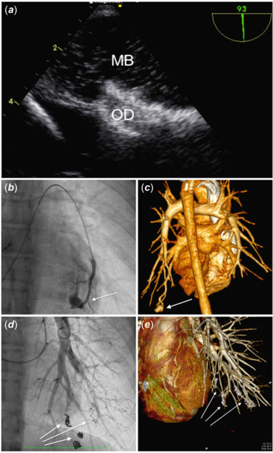
At the end of the procedure, contrast transoesophageal echocardiography (Fig 1A) revealed correct placement of the device with resolution of the early right-to-left shunt, though a persistent and significant late shunt was still evident, with left atrial opacification after five cardiac cycles following right atrial opacification. Since a pulmonary arteriovenous fistula was suspected, an agitated saline injection in the pulmonary trunk was performed, showing left atrial opacification. A CT angiogram was later performed and showed four pulmonary arteriovenous fistulas in the left lung inferior lobe (Fig 1C). The patient underwent percutaneous closure of three pulmonary arteriovenous fistulas (Fig 1B) with embolisation coils. The fourth pulmonary fistula was left untreated because of the inability to access the target site (located in a peripheral position in the superior lobe) and the small size of the lesion (<4 mm). At 6-month follow-up, the patient was asymptomatic, without further episodes of cerebral ischemia. The three pulmonary arteriovenous fistulas were completely closed on both pulmonary and CT angiogram (Fig 1 D/E), while contrast transoesophageal echocardiography showed a trivial residual shunt.
Patent foramen ovale closure with pulmonary arteriovenous fistulas detection and treatment. (a) Intraoperative contrast transoesophageal echocardiography showing correct deployment of the occluder device (OD) but persistence of late shunt (MB, microbubbles) in the left atrium. (b/c) Pulmonary/CT angiogram showing one of the four pulmonary arteriovenous fistulas before its percutaneous closure. (d) Post-procedural pulmonary angiogram showing closed pulmonary arteriovenous fistulas. (e) Six-month CT angiogram showing good results of pulmonary arteriovenous fistulas closure over time.

Discussion
Compared to medical therapy, percutaneous closure of atrial septal defects proved to be effective in the reduction of recurrent neurological events in patients with cryptogenic cerebral ischemia,Reference Pristipino, Sievert and D’Ascenzo4 especially in those with a higher RoPE score.Reference Thaler, Ruthazer and Weimar7 However, despite the significant reduction of risk in patients undergoing patent foramen ovale closure, recurrent ischemic stroke may occur in 5.1% patients, as shown in a recent study by Seeger et al.Reference Seeger, Uber and Wöhrle8 Incident atrial fibrillation is a well-known risk factor for recurrences.Reference Pristipino, Sievert and D’Ascenzo4 Indeed, the presence of undiagnosed pulmonary arteriovenous fistula may explain further ischemic events.Reference Balbi, Canepa, Cheli, Bezante and Brunelli9 Although unusual, contrast transoesophageal echocardiography at the end of patent foramen ovale closure interventions may be useful to detect early and late residual shunts. While early shunts may occur for unsatisfying device deployment, late shunts may be due to the presence of extracardiac shunts such as pulmonary arteriovenous fistula. This simple and safe practice can occasionally identify extracardiac right-to-left shunts that, if left untreated, might explain recurrent thromboembolic events in patients with a technically successful patent foramen ovale closure intervention.Reference Windecker, Wahl and Chatterjee10
Conclusions
In conclusion, the routinary adoption of contrast transoesophageal echocardiography at the end of interventions of patent foramen ovale closure may be useful to detect undiagnosed extracardiac shunt.
Supplementary material
To view supplementary material for this article, please visit https://doi.org/10.1017/S1047951120002152
Authors’ contributions
Alessandro Andreis: data collection, interpretation and analysis, article drafting, critical revision and approval of the final version. Gabriella Agnoletti: interventional cardiologist performing the latter percutaneous procedure described in the case report, interpretation and analysis of data, article drafting, critical revision and approval of the final version. Paolo Scacciatella: interventional cardiologist performing the two percutaneous procedures described in the case report, interpretation and analysis of data, article drafting, critical revision and approval of the final version.
Acknowledgements
None.
Financial Support
This research received no specific grant from any funding agency, commercial or not-for-profit sectors.
Conflicts of Interest
None.
Ethical Standards
The authors assert that all procedures contributing to this work comply with the ethical standards of the Helsinki Declaration of 1975, as revised in 2008, and has been approved by the institutional committee.