Impilcation
The practice of adding inorganic forms of Mn to poultry diets in amounts usually exceeding National Research Council (NRC) (1994) recommendations results in the excretion of large amounts of this element into the environment, causing contamination. Replacing commonly used forms of Mn with nanoparticles will reduce the emissions of this element to the environment because nanoparticles having smaller sizes more easily and faster diffuse into cells and show greater biological reactivity.
Introduction
Manganese is a microelement that participates in many biochemical processes in the cell. It is essential to regulation of the blood sugar level, protection against reactive oxygen species (ROS), blood coagulation, nervous system function, bone growth and immune defences (Horning et al., Reference Horning, Caito, Tipps, Bowman and Aschner2015; Costanzoa et al., Reference Costanzoa, Scolarob, Berlierc, Marengob, Grecchia, Zancanaroa, Malatestaa and Arpiccob2016). Manganese is involved in numerous biochemical reactions, both as an integral part of metalloenzymes and as an activator of enzymes, such as pyruvate carboxylase, glycosyltransferase, glutamine synthetase, alkaline phosphatase, and especially mitochondrial manganese superoxide dismutase (Mn-SOD) (Costanzoa et al., Reference Costanzoa, Scolarob, Berlierc, Marengob, Grecchia, Zancanaroa, Malatestaa and Arpiccob2016). Manganese superoxide dismutase, by catalysing the reduction of superoxide anion to hydrogen peroxide, is involved in protecting cells from damage by ROS. The vast majority of manganese in the blood is bound to erythrocytes (the concentration of manganese in erythrocytes is nearly five times higher than in blood serum), and therefore the highest Mn-SOD activity is also found in these blood cells (Strassburger et al., Reference Strassburger, Bloch, Sulyok, Schuller, Keist, Schmidt, Wenk, Peters, Wlaschek, Lenart, Krieg, Hafner, Kumin, Werner, Muller and Scharffetter-Kochanek2005).
Mn deficiency may lower blood cholesterol, inhibit growth, and cause anaemia, thyroid enlargement and hypogonadism (reproductive system dysfunction) (Costanzoa et al., Reference Costanzoa, Scolarob, Berlierc, Marengob, Grecchia, Zancanaroa, Malatestaa and Arpiccob2016). Genetic studies have shown that the homozygous mutation is lethal, while knockouts of Cu/Zn-SOD and glutathione peroxidase (GPx) are not (Lebovitz et al., Reference Lebovitz, Zhang, Vogel, Cartwright, Dionne, Lu, Huang and Matzuk1996). In a study by Van Remmen et al. (Reference Van Remmen, Ikeno, Hamilton, Pahlavani, Wolf, Thorpe, Alderson, Baynes, Epstein, Huang, Nelson, Strong and Richardson2003), Mn-SOD knockout mice had mitochondrial damage, reduced glutathione (GSH) content and increased content of 8-hydroxydeoxyguanosine (8-OHdG). The authors cited also found that a 50% decrease in Mn-SOD induces an increase in 8-OHdG and DNA damage. The short lifespan of Mn-SOD knockouts is likely linked to the role of this enzyme in maintaining a nanomolar or lower O2 ∙− concentration (Miriyala et al., Reference Miriyala, Spasojevic, Tovmasyan, Salvemini, Vujaskovic, St Clair and Batinic-Haberle2012). Manganese superoxide dismutase activity in the heart has been shown to increase as dietary Mn increases and to be a specific, sensitive criterion for determining the status and requirement of Mn for chickens (Luo et al., Reference Luo, Su, Huang and Liu1992). It is known from the available literature that nanoparticles can induce moderate reactions generating ROS and inflammatory reactions (Li et al., Reference Li, Xia and Nel2008). Moreover, these particles can generate carcinogenic processes, including damage to proteins, lipids and DNA and epigenetic changes (Fu et al., Reference Fu, Xia, Hwang, Ray and Yu2014). There have been reports indicating that an increased level of DNA damage (8-OHdG) is associated with occupational exposure to nanomaterials (Pelclova et al., Reference Pelclova, Barosova, Kukutschova, Zdimal, Navratil, Fenclova, Vlckova, Schwarz, Zikova, Kacer, Komarc, Belacek and Raman2015). Epigenetic changes affecting genetic regulation and cell differentiation can lead to embryonic changes, accelerate the ageing process and induce various diseases (Jaenisch and Bird, Reference Jaenisch and Bird2003). The best-known epigenetic modification is cytosine methylation, whose role is not fully understood. However, it is assumed that cytosine methylation controls gene expression and protects the host organism against the expression of unwanted sequences (De Prins et al., Reference De Prins, Koppen, Jacobs, Dons, Van de Mieroop, Nelen, Fierens, Int Panis, De Boever, Cox, Nawrot and Schoeters2013).
Due to these physiological functions of Mn, poultry diets are enriched with this element, usually by adding various chemical forms of Mn, including MnO and Mn2O3 (Bozkurt et al., Reference Bozkurt, Bulbul, Bozkurt, Bulbul, Maralcan and Qeukeloglu2015). It is accepted that in the case of growing turkeys, the addition of Mn to the diet should amount to 60 mg/kg of feed throughout the rearing period (NRC, 1994). British United Turkeys recommendations are much higher, amounting to 120 mg/kg in the first 4 weeks of rearing and 100 mg/kg after 4 weeks. The practice of adding inorganic forms of Mn to poultry diets in amounts usually exceeding NRC (1994) recommendations results in the excretion of large amounts of this element into the environment, causing contamination (Powers and Angel Reference Powers and Angel2008; Wang et al., Reference Wang, Cerrate, Yan, Sacakli and Waldroup2008).
The hypothesis of the research was the assumption, that Mn nanoparticles can affect the body in the same way as macromolecules. Their smaller size and greater biological reactivity will potentially allow the Mn addition to the diet to be reduced and, consequently, less excretion of this element into the environment. Therefore, the aim of the study was to determine whether the use of Mn nanoparticles would make it possible to reduce the level of this micronutrient added to turkey diets without adversely affecting redox reactions in cells and epigenetic changes.
Material and methods
Nanomaterial
Manganese nanopowder (NP-Mn2O3, purity 98+%, 40~60 nm, spherical, specific surface area 13.5 m2/g, bulk density 1.2 g/cm3) was purchased from SkySpring Nanomaterials (Houston, TX, USA). Phosphate-buffered saline (PBS) and all other common reagents were obtained from Sigma Aldrich (Germany). Rapeseed oil (ZT Kruszwica, Kruszwica, Poland) as a dispersant for nanoparticles suspension was used. Hydrodynamic size distribution and zeta potential (ξ) of nanoparticles were determined using dynamic light scattering (DLS) Zetasizer Nano ZS (Malvern Instruments, Malvern, UK). Nanopowder spectra, zeta potential and DLS were performed during the incubation of NP-Mn2O3 both in acidic conditions (citrate buffer pH 5) and neutral pH (PBS) and in rapeseed oil. In experimental procedure 50 mg of NP-Mn2O3 was suspended in 10 ml of the buffer or oil in a 20-ml scintillation vial (neoLab, Germany). The suspension was transferred into an ultrasonic batch and sonicated for 1 h (three times in 20 min with 5 min pause). Then the sample was diluted 10 times and analysis was performed immediately at room temperature.
Animals
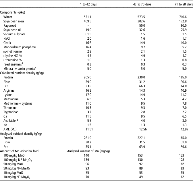
The experimental procedure was approved by the Local Ethics Committee for Experiments with Animals in Olsztyn, Poland (approval no. 30/2015). The subject of the study was 1080-day-old Hybrid Converter turkey hens purchased at the Grelavi hatchery in Kętrzyn. The birds were randomly assigned to 60 pens with an area of 4.0 m2, at 18 individuals/pen. The pens were lined with wood chips. The stocking density of the birds was initially (up to 6 weeks) 4.50 birds/m2 and then 3.75 birds/m2. The birds had unlimited access to feed and water, and environmental conditions were adjusted to the age of the birds and Hybrid recommendations. The experiment was conducted on six groups with 10 replications, in a two-factor design with three dosages of manganese: 100, 50 and 10 mg/kg, and two sources: manganese oxide (MnO) and manganese nanoparticles (NP-Mn2O3). The feed was produced by Agrocentrum sp. z o.o. in two stages: (1) as basal feeds without the addition of a premix, and then (2) with vitamin mineral premixes containing the appropriate amount and form of Mn added to the feed for each experimental group (Table 1).
Composition and nutritional value of compound feed fed to turkeys

AME=apparent metabolizable energy; NP=nanoparticles.
1 Phytase – 750 FTU/kg, xylanase – 300 FXU/kg.
2 Mineral–vitamin premix per kilogram of diet (without Mn supplement): vitamin A – 15 000 IU; vitamin D3 – 5000 IU; vitamin E – 100 IU; vitamin K3 – 4 mg; vitamin B1 – 5 mg; vitamin B2 – 15 mg; vitamin B6 – 6 mg; vitamin B12 – 0.04 mg; nicotinic acid – 100 mg; calcium pantothenicum – 32.7 mg; folic acid – 4 mg; choline – 700 mg; biotin – 0.35 mg; Se – 0.3 mg; Fe – 60 mg; Zn – 100 mg; Cu – 20 mg; J – 1.5 mg; Ca – 1.04 g.
Growth trial and sample collection
During the experiment, BW and feed consumption were recorded and calculated on a pen basis. Daily feed intake per bird was calculated based on total feed consumption for the pen for the entire experimental period divided by the number of days in the period. Feed conversion ratio (FCR; kg of feed/kg of BW) was calculated on a pen basis from BW gain and feed consumption.
At the end of the 98th day of life, 10 birds representing the average BW of each group were selected, tagged, and fasted for 8 h. Blood samples were taken from 10 birds from each group (one bird for each replication) with BW similar to the treatment average. Blood for analysis was collected into test tubes with an anticoagulant (heparin) from the wing vein. Next the blood samples were centrifuged at 3000×g for 10 min and the plasma was collected for further analysis.
Laboratory analysis
Markers of oxidative stress determined in the blood included lipid oxidation indicators, that is, the concentration of lipid hydroperoxides (LOOH) and malondialdehyde (MDA) using kits produced by Cell Biolabs Inc., San Diego, CA, USA. The enzymes Mn-SOD and GPx activity in the blood of the turkeys were determined by spectrometry using Ransel and Ransod diagnostic kits manufactured by Randox (Warsaw, Poland). A diagnostic kit manufactured by Oxis International Inc., Portland, OR, USA, was used to determine activity of catalase (CAT). Activity of ceruloplasmin in the blood plasma was determined using Ceruloplasmin ELISA kit (Biomatik, Wilmington, DE, USA). Also determined in the blood were total glutathione (GSH+GSSG), using Total Glutathione Assay (Cell Biolabs Inc.), total antioxidant status using a diagnostic kit by Randox, albumin content (ALB) using a diagnostic kit by Cormay (Łomianki, Poland), and vitamin C content using ELISA kit (Cell Biolabs Inc.). OxiSelect diagnostic kits (Cell Biolabs Inc.) were used to determine protein carbonyl (PC) derivatives as an indicator of oxidation of amino acid residues, 3-nitrotyrosine (3-NT) as a marker of protein nitration, and 8-OHdG as a marker of oxidation of DNA bases. DNA was isolated from the blood using kits manufactured by QIAGEN (Hilden, Germany). The level of epigenetic changes in the blood of the chickens was determined by analysing global DNA methylation (methylome) using diagnostic kits manufactured by Sigma Aldrich. Manganese content in the feed mixture was determined by inductively coupled plasma optical emission spectrometry.
Statistical analysis
For the statistical analysis of performance parameters, a single pen (n=10) was considered as a replicate experimental unit. For the analysis of blood redox parameters and DNA methylation, individual birds were considered as experimental units. The analysis of blood parameters was performed on 60 birds representing 10 replications from each of six experimental treatments. Two-way ANOVA was performed to determine the effects of Mn additive (10, 50 and 100 mg/kg diet) and source (MnO or NP-Mn2O3) and the interaction between the two factors (additive × source). The significance of differences between means in groups was estimated by Duncan’s multiple range test. Data were processed in the STATISTICA PL 12.0 application.
Results
Nanoparticles characterization
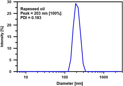
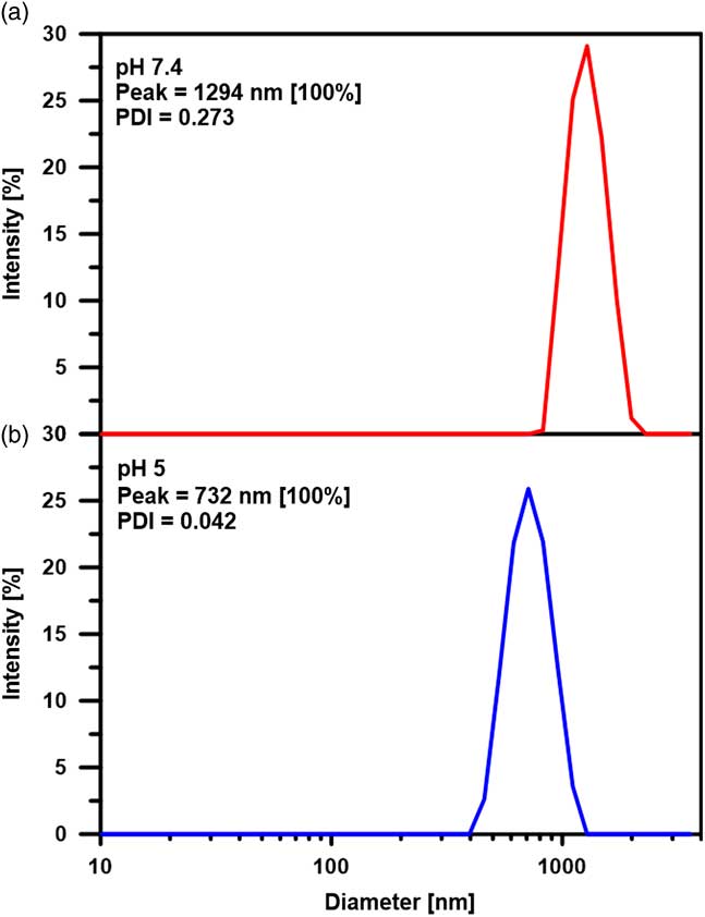
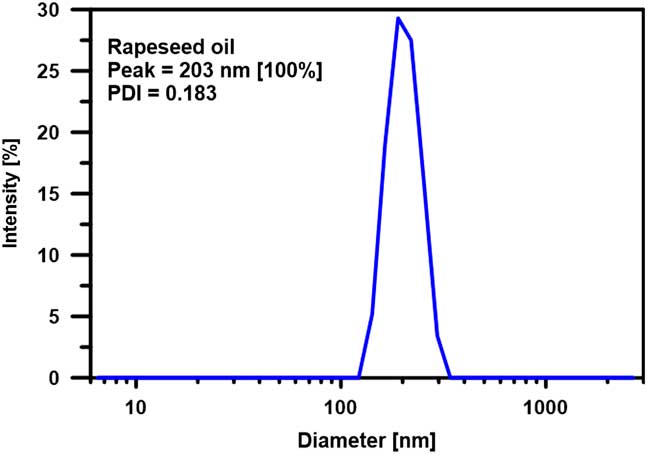
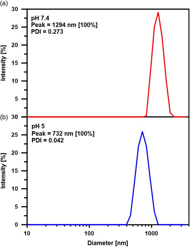
A size of NP-Mn2O3 was characterized by the DLS approach which give insights into the hydrodynamic properties of particles and allows determination of hydrodynamic diameter. Figure 1 presents the DLS characteristics of the nanoparticles in the vehicle (rapeseed oil). As can be seen, the diameter determined by this approach (203 nm) is greater by ~40 to 60 nm than the primary diameter of the metallic core (according to SkySpring Nanomaterials). Dynamic light scattering measurements in water environment shown, that Mn2O3 nanoparticles diameter increased up to 732 nm in pH 5 and to 1294 nm in pH 7.4 (Figure 2). Importantly, the zeta potential of NP-Mn2O3 was determined as −28.6 mV (PBS) and −29,6 mV (pH 5), so they are more anionic than synthetized by Vaidya et al. (Reference Vaidya, Thaplyal and Ganguli2011) or reported by Heinlaan et al. (Reference Heinlaan, Muna, Juganson, Oriekhova, Stoll, Kahru and Slaveykova2017). In rapeseed oil zeta potential was not determined due to low conductivity (~10-4 mS × cm−1) of medium.
Dynamic light scattering (DLS) physiochemical characteristics by intensity size distribution obtained for a manganese nanoparticles (NP-Mn2O3) sample in rapeseed oil. The metal concentration was 0.5 mg×cm−3. PDI=polydispersity index.
Dynamic light scattering (DLS) physiochemical characteristics by intensity size distribution obtained for a manganese nanoparticles (NP-Mn2O3) sample in phosphate-buffered saline (PBS) (a) and citrate buffer (b). The metal concentration was 0.5 mg×cm−3. PDI=polydispersity index.
Content of manganese in diet
In three diets, prepared for periods of 1 to 42, 43 to 70 and 71 to 98 days of turkey rearing, the total content of manganese did not depend on the source of Mn (Table 1). The calculated difference between the total Mn content in diets and the amount of supplement (100, 50 and 10 mg/kg) shows that the basic diet components contained a total of about 45 mg Mn/kg of feed.
Effect of manganese source
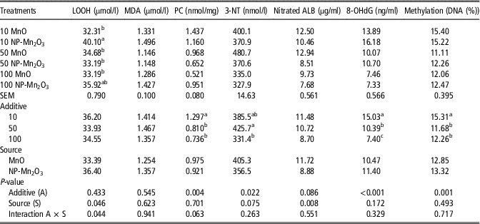
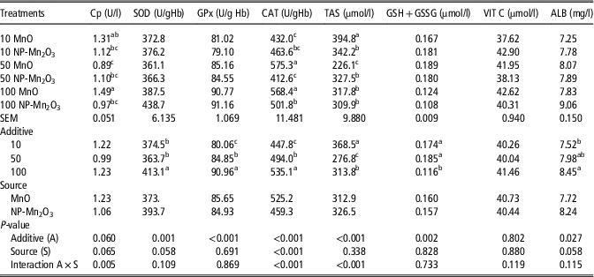
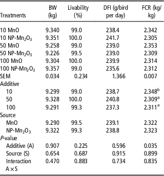
The LOOH content in the blood of the turkeys from the NP-Mn2O3 treatment was higher (P=0.04) than in the turkeys from the MnO treatment. In the case of LOOH content, an interaction of dose × source (P=0.04) was observed, as the plasma concentration of LOOH increased at the lowest dose of Mn administered in the form of NP-Mn2O3 (Table 2). The addition of the NP-Mn2O3 form to the turkey diet decreased (P=0.008) the plasma level of nitrated ALB as compared to the MnO form (Table 2). Catalase activity was lower (P <0.001) in the blood of the turkeys from the NP-Mn2O3 treatment than in the turkeys from the MnO treatment (Table 3). There was no effect of the source of Mn on performance (Table 4).
Indicators of oxidation of lipids, proteins and DNA, and the percentage of DNA methylation in turkeys receiving manganese in the form of oxide (MnO) or nanoparticles (NP-Mn2O3) in their diet
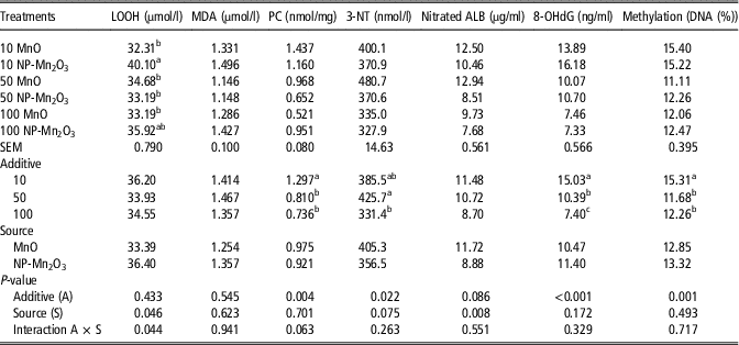
LOOH=lipid hydroperoxides; MDA=malondialdehyde; PC=protein carbonyl; 3-NT=3-nitrotyrosine; Nitrated ALB=nitrated albumin; 8-OHdG=8-hydroxydeoxyguanosine.
a,bMeans within the same column differ significantly (P⩽0.05).
Indicators of antioxidant status in the blood of turkeys receiving manganese in the form of oxide (MnO) or nanoparticles (NP-Mn2O3) in their diet
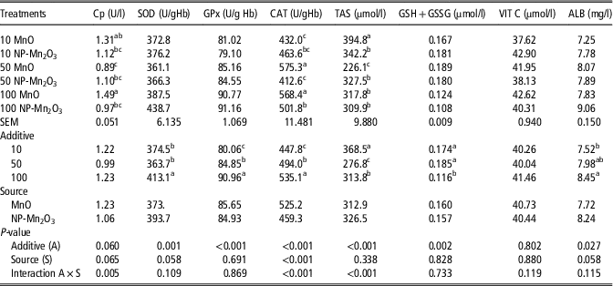
Cp=ceruloplasmin; SOD=superoxide dismutase; GPx=glutathione peroxidise; CAT=catalase; TAS=total antioxidant status; GSH+GSSG=total glutathione; VIT C=vitamin C; ALB=albumin.
a,b,cMeans within the same column differ significantly (P⩽0.05).
Growth performance of turkeys

DFI=daily feed intake; FCR=feed conversion ratio.
a,bMeans within the same column differ significantly (P⩽0.05).
Effect of manganese dosage
The turkeys receiving Mn at a dose of 10 mg/kg had higher (P <0.004) content of PC in the blood than birds receiving diets with the addition of Mn at 50 and 100 mg/kg. As compared to the group receiving the lowest dose of Mn, that is, 10 mg/kg, the administration of Mn at 50 and 100 mg/kg decreased the content of 8-OHdG (P <0.001) and the percentage of cytosine methylation in the DNA (P=0.001) (Table 2). As compared to the group receiving the highest dose of Mn, that is, 100 mg/kg, the addition of Mn to the turkey diet at 10 and 50 mg/kg caused a decrease in the activity of SOD, GPx and CAT (P=0.001, P<0.001 and P<0.001, respectively), while increasing the content of GSH+GSSG (P=0.002) in the blood. A significant interaction of dose × source (P<0.001) was noted in the case of plasma CAT activity, which decreased at Mn dosages of 10 and 50 mg/kg administered in the form of NP-Mn2O3. As compared to the group receiving the highest dose of Mn, that is, 100 mg/kg, the use of Mn at 10 and 50 mg/kg reduced (P=0.027) the content of ALB in the blood (Table 3).
In all treatments, regardless of the size and source of the Mn addition, the liveability of turkeys was very high, falling in the range of 99% to 100% of the number of birds used for the experiment. The final BW of turkey was similar in all groups. It was found that the reduction in the turkey diet additive of manganese from 100 to 50 mg/kg, and then to 10 mg/kg of diet regardless of the used form, increased FCR (P=0.035) (Table 4).
Discussion
In the experiments carried out so far on broiler chickens it was found that the addition of up to 250 mg/kg of various chemical forms to Mn did not improve feed conversion and did not increase chicken growth rate (Bertechini and Hossain, Reference Bertechini and Hossain1992; Olgun, Reference Olgun2017), while Mn was added in high amounts (400 mg/kg and more) an unfavourable effect on chicken production results was observed (Sunder et al., Reference Sunder, Panda, Gopinalth, Mantena, Savaram and Chalasani2006). In the presented experiment, although the decrease in Mn addition increased FCR (with a significant difference between the addition of 100 and 10 mg/kg), however, the final BW of turkeys and the livability of birds did not depend on the Mn level in diets.
Manganese participates in numerous biochemical reactions, both as an integral part of metalloenzymes and as an enzyme activator. As an enzymatic cofactor of Mn-SOD, it protects cells against damage caused by free oxygen radicals by catalysing the one-electron reduction of peroxide anion to hydrogen peroxide. A decrease in Mn content in the cell may result in a reduction in SOD activity, and consequently that of other antioxidant enzymes of the catalytic triad (SOD, GPx and CAT) (Ognik and Krauze, Reference Ognik and Krauze2016). Our study has shown that reducing the dose of Mn in the diet of turkeys from 100 to 50 mg/kg and then to 10 mg/kg reduces the activity of antioxidant enzymes (SOD, GPx and CAT) while increasing the level of GSH+GSSG. Asaikkuttia et al. (Reference Asaikkuttia, Bhavana, Vimalab, Karthika and Cheruparambathc2016) observed that an increase in the dose of manganese oxide nanoparticles in the diet of fish was correlated with an increase in antioxidant enzymes such as SOD, CAT, glutamic oxaloacetate transaminase and glutamic pyruvate transaminase in cells. Campos et al. (Reference Campos, Molognoni, Melo, Galdieri, Carneiro, D’Almeiday, Correa and Jasiulionis2007) report that during induction of oxidative stress in cells as an effect of ROS, an increase in the level of reduced GSH and MDA is also observed, as well as an increase in methylated DNA.
The available literature (Liao et al., Reference Liao, Chung, Lai, Wang, Chiang, Li, Tsou, Li, Lee, Wu, Lin, Hsu, Ho, Chen, Shih, Lin and Liou2014; Pelclova et al., Reference Pelclova, Barosova, Kukutschova, Zdimal, Navratil, Fenclova, Vlckova, Schwarz, Zikova, Kacer, Komarc, Belacek and Raman2015) shows that the increased content of markers indicative of lipid peroxidation is due to the toxic effects of nanoparticles on cells. Our research has shown that the addition of Mn in the form of nanoparticles induces lipid oxidation reactions (increases the LOOH content) in turkeys more than Mn in inorganic form. This effect was particularly evident when the dosage was reduced from 100 to 10 mg/kg. Lu et al. (Reference Lu, Luo, Ji, Liu and Yu2007) found that Mn at a dosage of 100 mg/kg diet for chickens reduces lipid peroxidation reactions (decreases MDA content and increases Mn-SOD activity), while a lack of supplementation with this element intensifies oxidation reactions. Lipid oxidation in cell membranes results in the formation of radicals, that is, peroxyl (LOO●) and alkoxyl (LO●), which can oxidize tyrosine to the ●Tyr radical. The tyrosine radical, which is the substrate for the nitration reaction of this amino acid, is transformed into 3-nitrotyrosine (3-NT) (Bartesaghi et al., Reference Bartesaghi, Wenzel, Trujillo, Lopez, Joseph, Kalyanaraman and Radi2010; Bartesaghi et al., Reference Bartesaghi, Herrera, Martinez, Petruk, Demicheli, Trujillo, Marti, Estrin and Radi2017). There are other mechanisms of oxidation of tyrosine to ●Tyr, induced by ●OH, ●NO2, CO3 ●-, oxo-metal compounds (O=MnIV) and compounds I and II of haem peroxidases, such as myeloperoxidase (Bartesaghi et al., Reference Bartesaghi, Herrera, Martinez, Petruk, Demicheli, Trujillo, Marti, Estrin and Radi2017). Our research has shown that the addition of Mn in the form of nanoparticles reduces the tyrosine nitration process more than Mn in inorganic form. As the dosage of Mn in the turkey diet increased, the total content of ALB increased, while the content of its nitrated form decreased. Nitration of tyrosine may result in changes in the structure of proteins and disrupt their biological functions by reducing the activity of enzymes (e.g. Mn-SOD, prostacyclin synthase and tyrosine hydroxylase, cytochrome c, protein kinase and glutathione S-transferase) (Souza et al., Reference Souza, Peluffo and Radi2008). Our study confirms this relationship, as the lowest SOD activity and the lowest ferric reducing ability of plasma were found in the experimental groups in which the levels of 3-NT and nitrated ALB were highest.
Lipid peroxidation end products and peroxynitrite also react with amino acid residues of proteins to produce PC derivatives (Dotan et al., Reference Dotan, Lichtenberg and Pinchuk2004). Protein carbonylation involves the introduction of carbonyl (aldehyde or ketone) groups to the protein structure. This is most often the result of direct oxidation of lysine, arginine, proline or threonine residues present in the protein chain (Dalle-Donne et al., Reference Dalle-Donne, Scaloni, Giustarini, Cavarra, Tell, Lungarella, Colombo, Rossi and Milzani2005; Purdel et al., Reference Purdel, Margina and Ilie2014). Our study showed that the use of Mn doses lower than 100 mg/kg, irrespective of the form, intensified protein oxidation expressed as increased PC content.
Impairment of the body’s antioxidant defence leads not only to oxidation of lipids and proteins, but also to DNA oxidation. Reactive oxygen species cause the oxidation of DNA guanine to 8-OHdG. Oxidized guanine is one of the most common products of modification of DNA bases by ROS. Guanine modified in this way may form stable base pairs with cytosine, leading to its enzymatic methylation (Liou et al., Reference Liou, Wua, Liaoa, Chenb, Tsaib, Jungb and Leeb2017). When 8-OHdG is adjacent to 5-methylcytosine, methyl-binding protein (MBP) binding is weakened. Moreover, the frequent conversion of 5-methylcytosine to 5-hydroxymethylcytosine alters binding affinity to MBPs, resulting in epigenetic changes (Venza et al., Reference Venza, Visalli, Beninati, De Gaetano, Teti and Venza2015). Global measurements of DNA methylation are a valuable tool for understanding the molecular pathology of cancer (Liou et al., Reference Liou, Wua, Liaoa, Chenb, Tsaib, Jungb and Leeb2017). In our study, the use of Mn doses lower than 100 mg/kg increased DNA oxidation and methylation in turkey cells. Brenneman et al. (Reference Brenneman, Cattley, Ali and Dorman1999) following oral administration of MnCl2 to rats at 0, 25 and 50 mg/kg per day, found that even the highest dose did not increase the level of 8-OHdG.
In summary, as an enzyme cofactor manganese is involved in antioxidant reactions. Decreasing the dosage of Mn in the turkey diet (from 100 to 50 or 10 mg/kg diet) increases oxidation of lipids, proteins and DNA, as well as DNA methylation. In spite of these effects at the cellular level neither replacing inorganic Mn with Mn nanoparticles nor reducing the doses of Mn did not affect the liveability and the final BW of turkeys. It has been observed, that the effects on cellular metabolism are not always reflected in the whole-body responses. Probably during the 15 week rearing period, the antioxidant system and enzymatic mechanisms of DNA repair eliminate the observed effects of reducing Mn or replacing MnO with nanoparticles. It is possible that long-term deficiency of Mn or the use of nanoparticles could disturb the redox status balance (weaken the antioxidant mechanisms) and, consequently, affect the worsening of the growth performance.
Conclusions
In the experiment, in which the diet of turkeys was supplemented with two sources of Mn (MnO or NP-Mn2O3) at three dosages, 100, 50 and 10 mg/kg, the 10 mg/kg dose, especially in the form of NP-Mn2O3, induced lipid oxidation reactions to the greatest extent. Irrespective of the additive of Mn in the turkey diet, Mn in the form of NP-Mn2O3 was found to reduce protein nitration more than Mn in the form of MnO. Reducing the Mn additive in the diet from 100 to 50 mg/kg and then to 10 mg/kg is unfavourable because: proportionally increases protein and DNA oxidation in cells, decreases the activity of antioxidant enzymes, and increases the level of GSH+GSSG. Reducing the dosage from 100 to 10 mg/kg increases global DNA methylation. Replacing MnO with nanoparticles Mn2O3 and reducing the addition of Mn (both forms) to the turkey diet did not worsen growth results of turkeys. The reduction of the Mn level, regardless of the form used, is disadvantageous, because it weakens the defense of the antioxidant system, which consequently can induce oxidative processes in the cells. Although Mn in the form of NP-Mn2O3 reduce protein nitration better than in MnO form, the use of manganese nanoparticles in turkey feeding (even in lower doses) requires further study.
Acknowledgement
The study was conducted under the Biostrateg programme ‘GUTFEED – innovative nutrition in sustainable poultry production’ (No. 267659/7/NCBR/2015).
Declaration of interest
The authors declare that there is no conflicts of interest.
Ethics statement
The experimental procedure was approved by the Local Ethics Committee for Experiments with Animals in Olsztyn, Poland (approval no. 30/2015).
Software and data repository resources
All data are available upon request.