INTRODUCTION
Strongyloidiasis is a soil-transmitted helminthiasis affecting millions of people (probably about 370 million), especially in tropical and subtropical areas, where poor sanitation and warm, humid climate favour the free-living cycle of the nematode in the soil (Bisoffi et al. Reference Bisoffi, Buonfrate, Montresor, Requena-Mendez, Munoz, Krolewiecki, Gotuzzo, Mena, Chiodini, Anselmi, Moreira and Albonico2013). However, the increase in the migration flows to industrialized areas, and in travels to poor-resourced countries, probably makes cases rising in non endemic areas. Previous estimates of prevalence (30–100 million of infected people) (Genta, Reference Genta1989; Jorgensen et al. Reference Jorgensen, Montresor and Savioli1996) have been recently questioned, because they were based on surveys conducted with diagnostic techniques with low sensitivity for the detection of Strongyloides stercoralis (such as stool microscopy, Kato-Katz technique) (Buonfrate et al. Reference Buonfrate, Formenti, Perandin and Bisoffi2015). The accuracy of the laboratory diagnosis of strongyloidiasis is of crucial importance, because the disease can lead to a fatal syndrome in immunocompromised individuals (Mejia and Nutman, Reference Mejia and Nutman2012).
Sensitivity is increased by the Baermann method and agar plate coproculture (APC), which are the fecal-based ‘traditional’ techniques with the highest performance. However, their sensitivity seems not to be sufficiently good (Buonfrate et al. Reference Buonfrate, Formenti, Perandin and Bisoffi2015); moreover, they rely on fresh stools (living larvae are required), and this is often impractical in non-endemic settings.
Serological tests demonstrated a high sensitivity, and they are easy to be used in average-equipped laboratories in non-endemic areas. Recent studies demonstrated the good performance of different serological tests. However, there are still concerns about specificity, particularly about ELISA (enzyme-linked immunosorbent assay) when crude antigenic extracts are used. In fact, cross-reactions can cause false-positive results (Anderson et al. Reference Anderson, Klein, Dornink, Jespersen, Kubofcik, Nutman, Merrigan, Couturier and Theel2014; Bisoffi et al. Reference Bisoffi, Buonfrate, Sequi, Mejia, Cimino, Krolewiecki, Albonico, Gobbo, Bonafini, Angheben, Requena-Mendez, Munoz and Nutman2014). A recombinant antigen (NIE) has been tested in order to increase the specificity of serology, and overcome difficulties due to reliance on crude antigen extracts. In particular, luciferase immunoprecipitation systems (LIPS) based on NIE demonstrated the highest specificity among the serological tests; however, this assay is currently available only in research laboratories (Ramanathan et al. Reference Ramanathan, Burbelo, Groot, Iadarola, Neva and Nutman2008; Bisoffi et al. Reference Bisoffi, Buonfrate, Sequi, Mejia, Cimino, Krolewiecki, Albonico, Gobbo, Bonafini, Angheben, Requena-Mendez, Munoz and Nutman2014).
More recently, molecular techniques have been developed (Verweij et al. Reference Verweij, Canales, Polman, Ziem, Brienen, Polderman and van Lieshout2009; Repetto et al. Reference Repetto, Alba Soto, Cazorla, Tayeldin, Cuello, Lasala, Tekiel and Gonzalez Cappa2013; Sultana et al. Reference Sultana, Jeoffreys, Watts, Gilbert and Lee2013). At the moment there are only in-house methods. The polymerase-chain reaction (PCR) for S. stercoralis is highly specific (Verweij et al. Reference Verweij, Canales, Polman, Ziem, Brienen, Polderman and van Lieshout2009; Schar et al. Reference Schar, Odermatt, Khieu, Panning, Duong, Muth, Marti and Kramme2013; Sultana et al. Reference Sultana, Jeoffreys, Watts, Gilbert and Lee2013; Becker et al. Reference Becker, Piraisoody, Kramme, Marti, Silue, Panning, Nickel, Kern, Herrmann, Hatz, N'Goran, Utzinger and von Muller2015). On the other hand, some authors found a good sensitivity of PCR [particularly the real-time PCR (RT–PCR)] (Becker et al. Reference Becker, Piraisoody, Kramme, Marti, Silue, Panning, Nickel, Kern, Herrmann, Hatz, N'Goran, Utzinger and von Muller2015; Saugar et al. Reference Saugar, Merino, Martin-Rabadan, Fernandez-Soto, Ortega, Garate and Rodriguez2015; Sharifdini et al. Reference Sharifdini, Mirhendi, Ashrafi, Hosseini, Mohebali, Khodadadi and Kia2015; Repetto et al. Reference Repetto, Ruybal, Solana, Lopez, Berini, Alba Soto and Cappa2016), while others reported lower figures, especially in case of a low parasite load (Schar et al. Reference Schar, Odermatt, Khieu, Panning, Duong, Muth, Marti and Kramme2013; Sultana et al. Reference Sultana, Jeoffreys, Watts, Gilbert and Lee2013). It is worth of note that this technique has been compared so far only with other fecal-based methods, while its sensitivity has not been assessed in comparison with a highly sensitive method such as serology.
Aim of this study was to evaluate the sensitivity of RT–PCR in comparison with APC and serology for the diagnosis of strongyloidiasis.
Primary objective
Sensitivity of RT–PCR for the diagnosis of S. stercoralis.
Secondary objective
Concordance among RT–PCR, APC and serology for S. stercoralis.
MATERIALS AND METHODS
Study design
Retrospective analysis of data already available in the electronic database of the Centre for Tropical Diseases of Negrar, Verona (CTD).
Ethical considerations
The study protocol was submitted to the local competent Ethics Committee (Comitato Etico per la Sperimentazione Clinica delle Province di Verona e Rovigo), that gave ethical clearance on 13 July 2016 (protocol number 33910). Each patient who provides biological samples to our hospital is asked to sign an informed consent form for the donation of his/her biological samples for possible future use for study purpose. Biological samples from patients who deny their consent are not stored.
Sample selection
Inclusion criteria
-
• Patients who gave written informed consent to the use of their biological samples for study purpose.
-
• Patients with available results of serology, RT–PCR and APC for S. stercoralis, performed on the basis of a clinical suspicion and/or of exposure history.
-
• For each patient with at least one RT–PCR result, we retrieved the results of S. stercoralis serology and APC, provided that they had been executed no more than two months apart from RT–PCR. For each patient, we included in the analysis only one (the first one, in case more results were available) result from each technique.
Diagnostic methods
DNA extraction and RT–PCR
For DNA extraction, about 200 mg of feces are suspended in 200 µL of phosphate-buffered saline containing 2% polyvinylpolypyrolidone (Sigma-Aldrich) and freezed overnight at −20 °C until the extraction. After thawing and boiling, the samples are run by an automated extractor instrument (Magnapure LC.2, Roche).
The real-time assay is performed as described previously (Verweij et al. Reference Verweij, Canales, Polman, Ziem, Brienen, Polderman and van Lieshout2009). The amplification target is the small-subunit rRNA gene sequence for S. stercoralis. Appropriate positive and negative controls are included in all the experiments. As control for PCR inhibitors and amplification quality, the PhHV-1 control DNA is amplified with the appropriate primers/probe mix in the same reaction as S. stercoralis in multiplex PCR. The reactions, detection and data analysis are performed with the CFX96 detection system (Bio-Rad Laboratories).
Serology
The serological test in use at the CTD is an in-house in-house immunofluorescence test (IFAT), using S. stercoralis filariform larvae. The detailed description of the method has been published previously (Boscolo et al. Reference Boscolo, Gobbo, Mantovani, Degani, Anselmi, Monteiro, Marocco, Angheben, Mistretta, Santacatterina, Tais and Bisoffi2007).
APC
The APC was carried out according to a modified Koga agar plate method (Koga et al. Reference Koga, Kasuya, Khamboonruang, Sukhavat, Ieda, Takatsuka, Kita and Ohtomo1991).
At the CTD, the technique requires collecting a minimum of 50 mL of feces, kept at room temperature and processed within 24 h from collection. Each stool sample is routinely plated onto ten agar plates (about 2 g per plate), in order to increase the sensitivity of the technique. The plates are incubated at 26 °C for 48 h. The plates are first observed macroscopically on both sides. In case of positive culture, steromicroscopy and microscopy observation follow. When the plates macroscopically show bacterial colonies disposed in undulating patterns, but no larvae are observed at microscopy examination, the sample is classified as ‘uncertain’, although the cause is presumably an underlying larval migration (Panosian et al. Reference Panosian, Marone and Edberg1986).
Panel of true positives for the definition of a composite reference standard
APC specificity is recognized to be close to 100% (Salazar et al. Reference Salazar, Gutierrez and Berk1995; Ines Ede et al. Reference Ines Ede, Souza, Santos, Souza, Santos, Silva, Silva, Teixeira and Soares2011; Campo Polanco et al. Reference Campo Polanco, Gutierrez and Cardona Arias2014), therefore we considered positive APC results as true positives. Positive samples at RT–PCR are considered true positives for the purpose of this study. IFAT is positive for values ⩾1:20, but false-positive results may occur. Hence, for the definition of a panel of ‘true positives’ we used titres ⩾1:160 that demonstrated to be virtually 100% specific in a previous study (Bisoffi et al. Reference Bisoffi, Buonfrate, Sequi, Mejia, Cimino, Krolewiecki, Albonico, Gobbo, Bonafini, Angheben, Requena-Mendez, Munoz and Nutman2014).
STATISTICAL ANALYSIS
Sample size
We planned to use a convenience sample, using all available results that met the inclusion criteria.
Analysis of the primary outcome
Sensitivity of the study tests was calculated as the proportion of positive results over all positive samples. Uncertainty was quantified with the 95% confidence intervals. Concordance among tests was calculated with Cohen's κ-test.
RESULTS
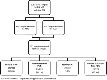
The study sample was retrieved starting from all available RT–PCR results, which were 1293. After evaluation of inclusion criteria, the study sample resulted of 223 patients. Figure 1 resumes the study flow and the results of the three tests, plus a variable IFAT + RT–PCR, which represents the number of samples resulting simultaneously positive to IFAT and RT–PCR.
Study flow chart.

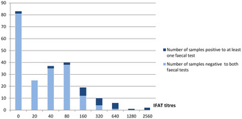
Worth of note, among the 83 cases that resulted negative to IFAT, only two were positive to at least one of the other methods (one positive only to RT–PCR and one to both RT–PCR and APC). Among the 140 samples that resulted positive to IFAT, 115 (82%) were negative to all the other tests. Figure 2 shows the results of RT–PCR and APC (combined) in relation to the IFAT titre: the proportion of APC/RT–PCR negative samples decreases with the increasing IFAT titre.
Results of fecal methods in relation to IFAT serology titres.

Only one of the 25 samples which resulted positive to RT–PCR was negative to both the other two tests.
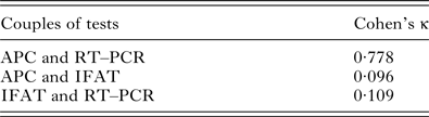
Globally, the concordance (Cohen's κ, Table 1) was found to be poor between IFAT and each one of the other methods, while it was good between APC and RT–PCR.
Concordance between the tests

Interpretation of Cohen's κ results:
Poor agreement = Less than 0·20.
Fair agreement = 0·20–0·40.
Moderate agreement = 0·40–0·60.
Good agreement = 0·60–0·80.
Very good agreement = 0·80–1·00.
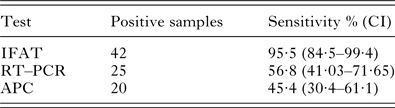
If sensitivity of the tests is calculated in relation to a panel of samples that are considered true positives (composite reference standard, composed by samples positive to RT–PCR and/or to APC and/or with IFAT ⩾160), the analysis is limited to 44 samples (Table 2). IFAT was the test with the highest sensitivity (95·5%), followed by RT–PCR (57%). APC detected less than half of the cases.
Sensitivity of the methods calculated against a composite reference standard for definition of true positive samples

Total number of samples: 44.
DISCUSSION
Serology demonstrated the highest sensitivity, missing only two of the positive results identified by the other methods. RT–PCR performed relatively better than APC (difference not statistically significant), although sensitivity resulted still much lower than that of IFAT. Although there might be concerns about the specificity of serology, the characteristics of strongyloidiasis (potentially fatal disease, highly tolerable therapy) supports the use of a highly sensitive diagnostic tool for screening. According to our preliminary results, neither fecal-based technique can replace serology as a screening method. On the other hand, both APC and RT–PCR have a role as confirmatory methods, in particular in case of a low serological titre.
It must be considered that our modified APC method implies the analysis of a large amount of stool, about 10 times higher than that described by Koga (Koga et al. Reference Koga, Kasuya, Khamboonruang, Sukhavat, Ieda, Takatsuka, Kita and Ohtomo1991), in order to increase the sensitivity of the method. Differences in the execution of routine fecal methods can influence the results of studies evaluating the sensitivity of molecular methods, and this could partly explain the discrepancies among different studies on RT–PCR vs ACP. There is still a need for standardization of the ‘traditional’ methods, too.
The retrospective design of this study entailed some limitations. The main one was the small number of samples that constituted the composite reference standard. However, we believe that the latter is important for a better definition of the denominator for calculation of sensitivity. Although the criteria we used for the definition of true positive samples for the composite reference standard have some limitations, it must be considered that: (1) The results reported in Fig. 2 confirm that the specificity of IFAT increases for high titres, as it was demonstrated previously (Bisoffi et al. Reference Bisoffi, Buonfrate, Sequi, Mejia, Cimino, Krolewiecki, Albonico, Gobbo, Bonafini, Angheben, Requena-Mendez, Munoz and Nutman2014). (2) Specificity of RT–PCR was considered 100% for study purpose, based on the performance of the test declared by Verweji, who implemented the technique (Verweij et al. Reference Verweij, Canales, Polman, Ziem, Brienen, Polderman and van Lieshout2009). However, further studies might be needed for a full evaluation of the accuracy of RT–PCR.
Concluding remarks
In this retrospective study, APC and RT–PCR showed similar sensitivity, while that of serology was found to be higher. Hence, serology might be recommended for screening in non endemic areas, and either APC or RT–PCR can be useful to confirm selected cases. Further studies, prospectively comparing more diagnostics and using a composite reference standard (or other recommended methods when a gold standard is lacking), are required.
ACKNOWLEDGEMENTS
We warmly thank Claudio Duffini for kind assistance for data extraction, and the lab staff (Monica Degani, Maria Gobbo, Manuela Mistretta, Tiziana Serafin and Stefano Tais) for their highly professional work.
FINANCIAL SUPPORT
This research received no specific grant from any funding agency, commercial or not-for-profit sectors.