INTRODUCTION
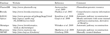
Malaria is a devastating disease claiming more than a million lives each year, mainly in tropical and sub-tropical regions of the world. The most severe form is caused by a unicellular parasite called Plasmodium falciparum. Human malaria research entered the post-genomic era when the sequencing of the P. falciparum genome was completed in 2002 (Gardner et al. Reference Gardner, Hall, Fung, White, Berriman, Hyman, Carlton, Pain, Nelson, Bowman, Paulsen, James, Eisen, Rutherford, Salzberg, Craig, Kyes, Chan, Nene, Shallom, Suh, Peterson, Angiuoli, Pertea, Allen, Selengut, Haft, Mather, Vaidya, Martin, Fairlamb, Fraunholz, Roos, Ralph, Mcfadden, Cummings, Subramanian, Mungall, Venter, Carucci, Hoffman, Newbold, Davis, Fraser and Barrell2002). The availability of the genome sequence (accessible through the PlasmoDB database, Table 1) and the subsequent genomic annotations brought a considerable amount of information for the prediction of biological processes and existing biochemical pathways, offering a global view of the parasite biology. From these initial data, manual and automated metabolic reconstructions have been attempted for predicting the complement of metabolic enzymes of the parasite and the network of reactions that it supports. These are available in several web-accessible databases and provide a global overview of the pathways present in Plasmodium (Table 1). It is particularly useful in predicting potential weak spots of the parasite such as metabolic ‘chokepoints’ or crucial regulatory nodes. Chokepoints are defined as reactions that consume unique substrates or produce unique products, which could be a characteristic of putative drug targets (Yeh et al. Reference Yeh, Hanekamp, Tsoka, Karp and Altman2004; Fatumo et al. Reference Fatumo, Plaimas, Mallm, Schramm, Adebiyi, Oswald, Eils and Konig2009). Indeed, it is expected that the inhibition of an enzyme that consumes a unique substrate will result in the accumulation of the potentially toxic substrate and that the inhibition of an enzyme producing a unique product will result in the starvation of the potentially essential product.
List of databases containing information for Plasmodium metabolic pathways reconstruction

In silico studies using genomic data provide metabolic models mainly based on gene identification by sequence homologies with known enzymes and most are automatically generated. They suffer from several flaws and are rather incomplete (reviewed in Ginsburg, Reference Ginsburg2009). For instance, a number of pathways contain metabolic ‘holes’, for which at least a part of a metabolic pathway is absent. These could be due to a truly non-functional pathway in the parasite, or a cellular function performed by a divergent, yet specific, parasitic enzyme or also to the fact that some metabolites can be directly acquired from the host. Moreover, a database such as KEGG predicts only 685 metabolites for P. falciparum (Pinney et al. Reference Pinney, Papp, Hyland, Wambua, Westhead and Mcconkey2007), yet roughly 50% of Plasmodium genes are still not clearly identified (either because they are homologous to genes of unknown function in other organisms or they show no homology at all with any previously identified genes (Gardner et al. Reference Gardner, Hall, Fung, White, Berriman, Hyman, Carlton, Pain, Nelson, Bowman, Paulsen, James, Eisen, Rutherford, Salzberg, Craig, Kyes, Chan, Nene, Shallom, Suh, Peterson, Angiuoli, Pertea, Allen, Selengut, Haft, Mather, Vaidya, Martin, Fairlamb, Fraunholz, Roos, Ralph, Mcfadden, Cummings, Subramanian, Mungall, Venter, Carucci, Hoffman, Newbold, Davis, Fraser and Barrell2002). Also, an aspect that is overlooked in genome annotation and subsequent pathway reconstruction studies is that the broad substrate-specificity of many of the cellular enzymes tends to underestimate the numbers of predicted metabolites. This calls for a more precise and accurate method of detection of the metabolites themselves that, in conjunction with the biochemical, genomics and transcriptomics data, could elucidate the presence of metabolic pathways and allow for the identification of new ones.
Indeed, when a gene is coding for an enzyme, it is the biologically active metabolite(s) produced by the enzyme that bear the biologically active function. Hence, besides the genomics, transcriptomics and proteomics data, an emergent field termed metabolomics has developed over the recent years to study the cellular complement of low molecular weight metabolites (or metabolome). A true metabolomic approach would theoretically involve the analysis of the whole metabolome under a given set of conditions (Fiehn, Reference Fiehn2002), yet such an exhaustive approach is not a realistic task, owing to the complexity of the metabolome and the diversity of chemistry of the metabolites. The term metabolomics (also sometimes referred to as metabonomics) is thus now rather used to characterize the metabolic phenotypes obtained under specific conditions (i.e. different growth conditions, developmental stages, or after genetic modifications or drug treatment). This usually involves analytical methods that would allow not only the separation and identification, but also the quantification, of the metabolites. A global profile of the metabolites (both identified and unknown) can be analysed and new compounds identified, but most studies are focused on a biochemical pathway or a family of compounds.
These new metabolomic approaches, when applied to a parasite of medical importance such as P. falciparum, could allow the identification of novel parasitic metabolites or fill-in the metabolic gaps of the genomic predictions. Additional to the data provided by the genome projects, metabolomics of Plasmodium could also benefit from a significant knowledge of biochemistry and analytical chemistry of primary metabolites accumulated over the years for this parasite. It certainly offers the potential of discovering new drug targets, or to track the metabolic changes within the parasite caused by treatment with existing drugs, with the aim of optimising the compounds or combating drug resistance. This is why we are currently implementing a metabolomic analysis of water-soluble phospholipids (PL) precursors of intra-erythrocytic stages of P. falciparum.
OVERVIEW OF P. FALCIPARUM PHOSPHOLIPID METABOLISM AND CURRENT QUESTIONS
Malarial lipid biosynthesis is substantially documented for P. falciparum and its blood stages. The Plasmodium parasite's multiplication within red blood cells leads to a production of 16 to 32 merozoites which results in rapid invasion of further erythrocytes. This rate of production demands high metabolic activity particularly through the increased requirement for novel membrane formation. Membranes of this intracellular parasite are essentially composed of PL, with absence of complex lipids or ergosterols/sterols contrary to what may be found in prokaryotic and yeast/mammalian cells for example. Infected erythrocytes show a marked increase in phospholipid by approximately six-fold (Holz, Reference Holz1977; Vial and Ancelin, Reference Vial and Ancelin1992; Vial and Mamoun, Reference Vial, Mamoun and Sherman2005). The predominant lipids are phosphatidylcholine (PC) and phosphatidylethanolamine (PE), representing together 75 to 85% of the parasitic PL composition after infection. Phosphatidylinositol (PI) is also considerably increased, constituting 4–11%, whereas sphingomyeline (SM) and phosphatidylserine (PS) account for much less than 5%. The lack of very sensitive analytic procedures has made it difficult to learn more about the lipids present in trace amounts such as phosphatidic acid (PA), phosphatidylglycerol (PG), cardiolipids and the neutral lipids, even though the latter appear considerably increased (Vial et al. Reference Vial, Ancelin, Philippot and Thuet1990; Vial and Ancelin, Reference Vial and Ancelin1992). Compared to the host erythrocyte, fatty acids grafted on the glycerol backbone are modified in length and degree of unsaturation which are both decreased (Vial and Ancelin, Reference Vial, Ancelin and Sherman1998).
So far, no biochemical experiments have shown that uptake of these lipids from the host serum could be significant. On the other hand, Plasmodium has been biochemically shown to contain all the enzymatic activities for PL biosynthesis, and quantification experiments strongly support that the parasitic machinery provides the bulk of PL composing the P. falciparum membranes (Vial and Ancelin, Reference Vial and Ancelin1992). Genomics studies have confirmed that malaria parasites possess the panoply of corresponding genes (Vial and Mamoun, Reference Vial, Mamoun and Sherman2005). Two points are of interest here: P. falciparum uses a bewildering number of metabolic pathways for PL biosynthesis that, to our knowledge, have never been shown in the same organism. The second point is that rodent and non-rodent malaria parasites differ in their PL metabolic pathways.
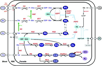
Glycerolipid biosynthesis initiates with 2 molecules of fatty acids being transferred from a donor (usually a fatty acyl-CoA) to a molecule of glycerophosphate forming the most primitive key intermediate PL, phosphatidic acid (PA), which plays critical regulatory roles in lipid biosynthetic pathways. In subsequent steps, the metabolic pathways diverge depending on the organism and the type of esters of cytidine nucleotides used to get an energy-rich bond. Indeed, biochemical pathways involve the activated form of either DAG (i.e. CDP~diacylglycerol), which is the only existing configuration in prokaryotes, or of a polar-head (e.g. CDP~choline), which appears in eukaryotic cells. Thus, after the synthesis of PA, the pathways of glycerolipid biosynthesis separate into two branches in which the next intermediate to be formed is either CDP-diacylglycerol or diacylglycerol (Fig. 1). PA can serve as a substrate for PA phosphatidate cytidylyltransferase (or CDP-DAG synthase, CDS) to yield CDP-DAG, which then serves as a precursor for the synthesis of PS, PI, PG. PA is also a substrate of phosphatidic acid phosphatase (PAP) to produce diacylglycerol, which provides the glycerol backbone and fatty acid moiety for PE, PC and neutral lipids.
. Schematic view of P. falciparum phospholipids biosynthesis pathways. Choline (Cho) entry into the infected erythrocyte (RBC) implies the erythrocytic Cho carrier and the New Permeation Pathway (NPP). Cho is provided to the parasite by a characterized Organic-Cation Transporter (OCT) encoded by yet unknown genes. Ethanolamine (Etn) enters the erythrocyte mainly by passive diffusion. Serine (Ser) is diverted from the host erythrocyte and transported into the parasite, or produced from haemoglobin (Hg) degradation in the food vacuole (FV). The SDPM pathway is differentiated by green arrows. PMT and PEMT (unknown genes) activities were only detected in P. falciparum; SD activity was only detected in P. falciparum and weakly in P. berghei (see (Dechamps et al. Reference Dechamps, Maynadier, Wein, Gannoun-Zaki, Marechal and Vial2009) for details). Lipids are shaded in blue. Enzymes are in red italics.
Metabolites: CL, cardiolipin; DAG, diacylglycerol; CDP~, cytidine-diphospho-; FA, fatty acids; Glu, glucose; G-3-P, glycerol 3-phosphate; Ino, myoinositol; P~, phospho; PA, phosphatidic acid; PC, phosphatidylcholine; PE, phosphatidylethanolamine; PG, phosphatidylglycerol; PGP, phosphatidylglycerolphosphate; PI, phosphatidylinositol; PI-3P, PI 3-phosphate; PI-4P, PI 4-phosphate; PI-4-5P2, PI 4,5-bisphosphate; PS, phosphatidylserine;
Enzymes (with PlasmoDB identifier for putative corresponding gene when identified): CDS, Cytidine diphosphate-DAG synthase (PF14_0097); CK, Cho kinase (PF14_0020); CCT, CTP:P~Cho cytidylyltransferase (PF13_0092); CLS, CL Synthase (MAL6P1.97); EK, Etn kinase (PF11_0257); ECT, CTP:P~Etn cytidylyltransferase (PF13_0253); CEPT, Cho/Etn-phosphotransferase (MAL6P1.145); INS, Ino 1-phosphate synthase (PFE0585c); PAP2, PA phosphatase (PFF1210w); PGPS, PGP synthase; PGPPase, PGP phosphatase; PEMT, PE N-methyltransferase; PIS, PI synthase (MAL13P1.82); PI3-k, PI 3-kinase (PFE0765w); PI4-k, PI 4-kinase (PFE0485w, PFD0965w); PI-4P5-k, PI 4-P 5-kinase (PFA0515w); PLC–ä, phospholipase Cδ (PF10_0132); PLMT, phosphatidyl-N-methylethanolamine N-methyltransferase; PMT, P~Etn N-methyltransferase (MAL13P1.214); PSD, PS decarboxylase (PFI1370c); PSS, PS synthase CDP-DAG dependent (MAL8P1.58); SD, Ser decarboxylase.

The CDP-DAG-dependent ancestral pathways
CDP-DAG is an obligatory intermediate for the CDP-DAG-dependent PL metabolism both in prokaryotes and eukaryotes. In bacteria, CDP-DAG is the precursor of the major anionic PL, PG, cardiolipins, the weakly represented PS and the major zwitterionic PE. In eukaryotic cells, CDP-DAG gives rise to the mitochondrial PL, PG and cardiolipins, but also to PI and, in lower eukaryotes, to PS (Kent, Reference Kent1995; Dowhan, Reference Dowhan1997; Zhang and Rock, Reference Zhang and Rock2008). Plasmodium parasites have only one copy of CDP-DAG synthase, which appears atypical because of a unique and essential N-terminal extension which is cleaved (Martin et al. Reference Martin, Gannoun-Zaki, Bonnefoy, Eldin, Wengelnik and Vial2000; Nagaraku and Vial, unpublished data). In both rodent (Dechamps et al. Reference Dechamps, Maynadier, Wein, Gannoun-Zaki, Marechal and Vial2009) and non rodent Plasmodium species, PS is abundantly synthesised (Vial et al. Reference Vial, Thuet, Broussal and Philippot1982; Elabbadi et al. Reference Elabbadi, Ancelin and Vial1997; Vial and Mamoun, Reference Vial, Mamoun and Sherman2005). CDP-DAG is also used for the abundant PI synthesis (Wengelnik and Vial, Reference Wengelnik and Vial2007) and it is likely that trace levels of PG and cardiolipin are also synthesised.
The CDP-choline and CDP-ethanolamine pathways
As in other eukaryotic cells, Plasmodium is also capable of synthesising PC and PE by the de novo pathways (also known as the Kennedy pathways). Choline is diverted from the host serum into the erythrocyte via the remnant erythrocytic choline carrier while a poly-specific organic cation transporter (OCT) functionally distinct from the known dedicated eukaryotic choline carriers mediates the choline entry into the parasite (Biagini et al. Reference Biagini, Pasini, Hughes, De Koning, Vial, O'neill, Ward and Bray2004). Choline is then phosphorylated into PtdCho, which is subsequently coupled to CTP, thus generating CDP-choline further converted to PC by a parasite CDP-diacylglycerol-cholinephosphotransferase. A similar de novo pathway does exist for PE from ethanolamine. The final stage of both branches of the Kennedy pathway is performed by the same enzyme, CEPT, catalysing the formation of PC and PE from CDP-choline and CDP-ethanolamine, respectively (Vial and Mamoun, Reference Vial, Mamoun and Sherman2005), even though molecular species of DAG recognised by CPET varies for each activities (Vial et al. Reference Vial, Thuet and Philippot1984).
Most eukaryotic cells (particularly Saccharomyces cerevisiae and human liver cells) are able to methylate PtdEtn to PtdCho through the phosphatidylethanolamine N-methyltransferases (PEMT) pathway (Carman and Henry, Reference Carman and Henry1989; Li and Vance, Reference Li and Vance2008). The high capacity of the P. knowlesi-infected erythrocyte to intensively methylate PE into PC unambiguously indicates the presence of PtdEtn N-methyltransferases (PEMT) activity (Moll et al. Reference Moll, Vial, Ancelin, Op Den Kamp, Roelofsen and Van Deenen1988) but the corresponding genes coding for PEMT have not yet been found in any Plasmodium species. Thus, the ‘enzymatic hole’ of the PEMT pathway remains to be clarified.
The SDPM horizontal plant-like pathway
P. falciparum possesses an alternative pathway for the synthesis of PC from serine and ethanolamine, which was termed the serine decarboxylase–phosphoethanolamine methyltransferase (SDPM) pathway. In this pathway, serine, which is diverted from the host or from haemoglobin degradation in the food vacuole, is first decarboxylated to form ethanolamine which is then phosphorylated to produce phosphoethanolamine. The serine decarboxylase (SD) enzymatic activity was first described by our group in P. knowlesi and P. falciparum (Elabbadi et al. Reference Elabbadi, Ancelin and Vial1997). The gene and the related catalytic activity SD activity were subsequently identified in plants (Rontein et al. Reference Rontein, Nishida, Tashiro, Yoshioka, Wu, Voelker, Basset and Hanson2001). The phosphoethanolamine formed is either incorporated into PE via the CDP–ethanolamine pathway and surprisingly, can also be converted into phosphocholine by SAM-dependent triple methylation carried out by a plant-like phosphoethanolamine N-methyltransferase (PfPMT) (EC 2.1.1.103). Phosphocholine is then integrated into the CDP-choline pathway for the synthesis of additional PC. The PfPMT enzyme was characterized by Ben Mamoun and colleagues (Pessi et al. Reference Pessi, Kociubinski and Mamoun2004, Reference Pessi, Choi, Reynolds, Voelker and Mamoun2005). Orthologous PMT genes have been previously detected in plants such as Arabidopsis thaliana and in the nematode Caenorhabditis elegans (see Dechamps et al. Reference Dechamps, Maynadier, Wein, Gannoun-Zaki, Marechal and Vial2009) but not in mammals.
Functional role of PL, regulation and questions for metabolomics
The reason why malaria parasites synthesise only one preponderant class of lipid is unknown. As detailed above, the human malaria parasite is capable of using three different pathways for the biosynthesis of its structural PL: the ancestral CDP-DAG-dependent pathway, the de novo CDP-choline and CDP-ethanolamine (Kennedy) pathways, and the serine decarboxylation-phosphoethanolamine methylation (SDPM) route (Fig. 1). The latter is exciting because it involves SD and a novel methyltransferase class only recently identified and characterized in plants and only sporadically distributed throughout animal genomes, having only being described to date in P. falciparum and C. elegans. In the phylum Apicomplexa, the SDPM-pathway is only conserved in plasmodia and the pathway is deleted in rodent parasites. The lack of a PMT homologous gene at the expected locus in rodent Plasmodium species is likely due to an early deletion, correlated with the separation of rodent from non-rodent Plasmodium branches (Dechamps et al. Reference Dechamps, Maynadier, Wein, Gannoun-Zaki, Marechal and Vial2009).
At present, only a few genetic studies have shown how crucial to the parasite biology are each metabolic pathway. PfPMT could be disrupted, leading to reduced multiplication rate and increased cell death, depending on the presence of external choline (Witola et al. Reference Witola, El Bissati, Pessi, Xie, Roepe and Mamoun2008). The SDPM and de novo pathways use the same initial precursor (phosphocholine) for the synthesis of PC, but their functions are not completely redundant. Recent genetic experiments in the rodent malaria parasite P. berghei have indicated that the genes coding for PbCK, PbCCT, PbECT and PbECPT are essential for the parasite survival (Dechamps et al., unpublished data). Since this rodent model lacks the PMT methyltransferase, extrapolation of a crucial role in the human malaria parasites is thus rather tentative. As for the lipid function and composition in the hepatic, sexual or sporozoite stages, information is rather scanty.
Biochemical studies emphasised the quantitative importance of each metabolic pathway (Vial and Ancelin, Reference Vial and Ancelin1992; Elabbadi et al. Reference Elabbadi, Ancelin and Vial1997; Le Roch et al. Reference Le Roch, Johnson, Ahiboh, Chung, Prudhomme, Plouffe, Henson, Zhou, Witola, Yates, Mamoun, Winzeler and Vial2008). In eukaryotic cells, these pathways involve multiple cellular compartments including the cytosol, the endoplasmic reticulum, the Golgi and the mitochondria, the biosynthetic activity of non-polar metabolites being predominately microsomal, with some activity in the mitochondrial inner membrane (Kent, Reference Kent1995). We showed that there are several distinct pools of PE in P. falciparum-infected erythrocytes, depending on their mode of synthesis (from exogenous ethanolamine, serine, or serine-derived ethanolamine) suggesting a cellular compartmentalisation of these metabolic pathways (Elabbadi et al. Reference Elabbadi, Ancelin and Vial1997). However, the subcellular localisation of most of the malaria enzymes involved in PL synthesis pathways remains to be determined. The cytosolic localisation of CK and EK (Choubey et al. Reference Choubey, Guha, Maity, Kumar, Raghunandan, Maulik, Mitra, Halder and Bandyopadhyay2006; Alberge et al. Reference Alberge, Gannoun-Zaki, Bascunana, Tran Van Ba, Vial and Cerdan2009) is not a surprise. In contrast, P. falciparum PS decarboxylase appears as an ER-resident protein, differing fundamentally from the typical location of type I PSD usually found exclusively in the inner mitochondrial membrane exposed to the intermembrane space (Baunaure et al. Reference Baunaure, Eldin, Cathiard and Vial2004). PfPMT localises to the Golgi apparatus of the parasite, possibly indicating that, at least, the transmethylation reaction would be occurring in the Golgi apparatus (Witola et al. Reference Witola, Pessi, El Bissati, Reynolds and Mamoun2006).
Little is known about the PL synthesis regulation of P. falciparum, but PL components are energetically expensive modules to produce. In all cell types, lipid production is very tightly controlled and regulated, spatially and temporally, in the various cellular compartments to match the growth rate of cells and their differentiation. The different metabolic pathways present in Plasmodium make it difficult to determine the relative amounts of each PL, notably the structural PC and PE synthesised by each pathway and also it raises the question as to whether each pathway would be present in distinct cellular compartments and if Plasmodium possesses regulatory mechanisms to maintain an equilibrium between these pathways.
An example of application: metabolomic studies of PL biosynthetic pathways
We are initiating metabolomic studies that are restricted to the ‘systematic’ study of the unique lipid-related chemical fingerprints i.e. setting up an assay and quantifying all the metabolites that are intermediates within these metabolic pathways. We are focusing on the water-soluble PL precursors and intermediates that can allow understanding the metabolic pathway (limiting steps, regulations, etc …).
Our first approach concerns regulatory steps and internal mechanism of regulation of the various metabolic pathways, deciphered by the relative proportion of metabolites and their repartition when fluxes are changed by modulating precursor concentration or effectors. Characterization of the kinetic properties of the associated enzymes should confirm the limiting and regulatory steps (at the condition that measured metabolites would reflect the levels present in the enzyme compartments). It is possible to study the respective contributions of the different pathways to the production of structural lipids by using deuteriated precursors (i.e. d9-choline and d4-ethanolamine) for metabolic labelling. We have already identified and quantified PC produced by the CDP-choline pathway and by the methylation of PE through isotopic labeling experiments (Enjalbal et al. Reference Enjalbal, Roggero, Cerdan, Martinez, Vial and Aubagnac2004). However, this study, restricted to the lipid components, indicated the relative quantification of PC biosynthesis pathways, but did not reveal the existence of cross-pathway regulation and did not identify the associated mechanism. The profiling of aqueous metabolites of P. falciparum extracts can reveal the origin of each PL and its intermediary metabolites in conditions where an excess of choline or ethanolamine is given and the mechanisms of regulation. Besides, functional disruptions of the enzymes that catalyse key reactions followed by metabolite profiling are powerful new approaches for the elucidations of the relative contribution of metabolic pathways and enzyme isoforms. For instance, we have a few P. falciparum transgenic cell lines modified for the choline biosynthesis pathway (overexpressing cell lines or null mutants) that can be used for assessing the effects of perturbing PC metabolism on the other PL-producing pathways. Finally, metabolite profiling can also be used to analyse qualitatively and quantitatively how the fluxes between the various biosynthesis pathways can be modulated.
A second approach is to utilise metabolomics for drug target identification. There are a number of drugs that interfere with phospholipid metabolism and many steps within PL metabolism are potential targets for malarial chemotherapy. False precursors such as small analogues of polar head groups retaining a free hydroxyl group or unnatural fatty acids can be added to deceive the parasite. After their incorporation into the parasite PL, the false metabolites alter the lipid composition of parasite membranes and their physicochemical properties, leading to parasite death (see Vial and Calas, Reference Vial, Calas and Rosenthal2001 and references therein). Another option is using effectors such as choline analogues that can block the access of the native precursor to the intracellular parasite and prevent synthesis of the essential lipid. These, including a class of potent antimalarial, are now in clinical development for treatment of severe malaria (Wengelnik et al. Reference Wengelnik, Vidal, Ancelin, Cathiard, Morgat, Kocken, Calas, Herrera, Thomas and Vial2002; Vial et al. Reference Vial, Wein, Farenc, Kocken, Nicolas, Ancelin, Bressolle, Thomas and Calas2004). A systematic approach to understand their mechanism of action and characterize the global response of P. falciparum demonstrated a cell cycle arrest and, importantly, revealed that choline analogues might target more than one protein within the PC biosynthetic pathway, and provided evidence for post-transcriptional regulations of parasite metabolism in response to external stimuli (Le Roch et al. Reference Le Roch, Johnson, Ahiboh, Chung, Prudhomme, Plouffe, Henson, Zhou, Witola, Yates, Mamoun, Winzeler and Vial2008). Therefore, it will be interesting to see how metabolic effectors modulate the relative amounts of metabolites and if they trigger an accumulation of intermediary metabolites. All the targets of these drugs are not yet identified (as they could also be multiple); hence, identifying an accumulated metabolite by a metabolomic approach, could help finding the drug target(s) and help optimising the existing compounds to better fit these therapeutic targets.
Metabolomics therefore adds an additional layer of information to data from proteomics and genomics, which will further increase our knowledge of lipid functions and open new opportunities in malaria biology, biomarkers and drug development for various stages of malaria parasites. For data analysis, protocols reminiscent of approaches used in genomics and proteomics are likely to be a major mode of application. This generally includes measurement of paired samples (for example, control and dosed; wild type and transgenic parasite) followed by differential analysis, including correlation and multivariate statistics and metabolic control analysis.
TOOLS FOR PLASMODIUM METABOLOMICS
Several analytical methods can be used for identification and quantification of metabolites. The most commonly used are nuclear magnetic resonance (NMR) spectroscopy and mass spectrometry (MS). These techniques all have their advantages and their disadvantages, so they are complementary and should be chosen depending on what is the purpose of the analysis.
Nuclear magnetic resonance spectroscopy
NMR does not rely on a separation of the compounds prior to analysis and 1H NMR detects a wide spectrum of metabolites (all metabolites containing H atoms), thus permitting a global analysis of metabolites even in complex media. It is also a powerful tool, because isotope-labeled precursors (ie 13C-labelled metabolites) can be used for downstream metabolite identification and fluxomic analysis to decipher the dynamics of the metabolites (Massou et al. Reference Massou, Nicolas, Letisse and Portais2007). NMR is a quantitative and reproducible method and resulting spectra have high information content for multiple compounds. It has been used successfully for the direct identification of P. falciparum metabolites (Teng et al. Reference Teng, Junankar, Bubb, Rae, Mercier and Kirk2009) or for the characterization of changes in the host metabolism induced by a P. berghei infection in the mouse model (Li et al. Reference Li, Wang, Saric, Nicholson, Dirnhofer, Singer, Tanner, Wittlin, Holmes and Utzinger2008). However, its main disadvantage is that it is not very sensitive (typically, micromolar quantities) (Wishart, Reference Wishart2008).
Mass spectrometry
In contrast with the above, MS techniques are highly sensitive (femtomoles can be detected), but the choice of ionisation technique, the mass analyzer, the acquisition mode and instrumental parameters can vary considerably depending on experimental goals (Lokhov and Archakov, Reference Lokhov and Archakov2009). These may range from purely qualitative and exploratory, to quantitative and specifically targeted. Although direct analysis of samples into the mass spectrometer is possible, it may suffer from lack of sensitivity due to matrix effect in complex biological samples such as plasma, urine, or in our case cell extracts. Thus, MS is usually coupled to an upstream chromatographic separation of the compounds from the complex biological extract (see below). The whole range of separation techniques associated with MS comprises capillary electrophoresis (CE)-MS, gas chromatography (GC)-MS and liquid chromatography (LC)-MS.
GC-MS requires the metabolites to be vaporised in order for migration through the capillary to occur. Thus, compounds must be volatile (or amenable to chemical derivatisation to render them volatile) and thermally stable to be analysed by GC-MS. Electron impact ionisation (EI) is the ionisation technique commonly used for GC-MS, and a major advantage is that high inter-instrument reproducibility and the standardised electron energy commonly used for ionisation has rendered possible the creation of spectra libraries for EI-MS data that facilitates identification of compounds.
LC-MS, and related techniques, such as capillary and ultra-performance liquid chromatography (UPLC), can separate a much wider range of chemical species than GC-MS because metabolites do not need to be volatile and they do not need to be derivatised. There are no spectral libraries for LC-MS identification, however the information of the ionic products, which are preserved in LC-MS analyses, can be used to search metabolite databases for identification (especially when coupled to a Fourier transform mass spectrometer, which achieves a high mass accuracy, <2 ppm). The use of LC tandem mass spectrometry (LC/MS/MS) for metabolic studies is relatively recent, but there is a rapid and continuing increase in the number of publications based on this analytical technique (Wilson et al. Reference Wilson, Plumb, Granger, Major, Williams and Lenz2005b; Lenz and Wilson, Reference Lenz and Wilson2007).
Tandem mass spectrometry allows a substantial improvement of the signal to noise ratio in the detection of selected compounds. This method requires the fragmentation of precursor ions selected by a first analyser in order to allow the second analyser to analyse the product ions, and both high selectivity and high sensitivity obtained with LC/MS/MS permit the monitoring and quantification of selected compounds.
Triple quadrupole (TQ) instruments are currently widely used in the field of metabolomics (Idborg-Bjorkman et al. Reference Idborg-Bjorkman, Edlund, Kvalheim, Schuppe-Koistinen and Jacobsson2003; Bajad et al. Reference Bajad, Lu, Kimball, Yuan, Peterson and Rabinowitz2006). Compared to tandem (TOF-TOF) mass spectrometers, TQ-instruments have a limited resolution, which does not allow accurate mass measurements. The main advantage of TQ instruments is the possibility of using different scanning MS/MS modes. In the single reaction monitoring (SRM) mode, both first and second mass analyzers are set to specific masses. By using this mode, it is thus possible to monitor a single transition between a specific parent and a specific daughter ion (the daughter ion is obtained from the parent ion, after collision process taking place in a cell between the two mass analysers). Such a transition, which is characteristic of a certain compound, serves as an evidence for the presence of this compound in the analysed sample. Thus, the SRM acquisition mode allows one to obtain a significant sensitivity and selectivity gain. It implies that the fragmentation properties of the molecules are known and thus, this approach is mainly suitable for identification and quantification of a known molecule in a given sample, such as it is the case with our approach for studying PL precursors.
Recent developments on UPLC, with chromatography columns using 1–2 micron-size particles, have brought a much higher resolution for analyte separation and a lower limit of detection for ions (Wilson et al. Reference Wilson, Nicholson, Castro-Perez, Granger, Johnson, Smith and Plumb2005a). Indeed, the efficiency of the chromatographic process increases proportionally as the matrix's particle size decrease. Consequently, the flow-rate of the mobile phase can be increased without affecting the efficiency of the separation (which is not the case with bigger, 5 or 10 μm, particles). The use of small particles generates high back-pressure (up to 100 MPa), which the UPLC columns are able to cope with. The first applications of UPLC were carried out using TOF mass spectrometry detection for metabolomic and functional genomic studies (Plumb et al. Reference Plumb, Castro-Perez, Granger, Beattie, Joncour and Wright2004; Castro-Perez et al. Reference Castro-Perez, Plumb, Granger, Beattie, Joncour and Wright2005; Wilson et al. Reference Wilson, Nicholson, Castro-Perez, Granger, Johnson, Smith and Plumb2005a). Other recently published works have shown the advantages UPLC over HPLC according to peak resolution, gain of analysis time and sensitivity (Churchwell et al. Reference Churchwell, Twaddle, Meeker and Doerge2005; Jin et al. Reference Jin, Xue, Shi, Xiao, Zhang and Liang2008; Van De Steene and Lambert, Reference Van De Steene and Lambert2008). UPLC seems promising for the future of liquid chromatography, but not all column types are yet available with this particular technology.
In our approach, we chose a HPLC or UPLC separation and detection by a TQ mass spectrometer operated in selected reaction monitoring (SRM) mode for identification and quantification of soluble phospholipids precursors of Plasmodium.
FRACTION USED FOR METABOLOMICS AND METABOLITE EXTRACTION
Isolation of the parasites and quenching of the metabolism
Plasmodium has a complex life cycle. Besides the intra-erythrocytic stages, which are responsible for the potentially deadly symptoms in human malaria, the hepatocyte and the sexual (gametocyte and gamete) stages, as well as the infective sporozoites, are of particular interest for the future of malarial research, because we lack biological data on these stages that are nevertheless of crucial importance in spreading malaria. Only a few laboratories are able to produce liver-stage forms, because of the technical restrictions required to produce infectious sporozoites in Anopheles mosquitoes and the difficulties in generating large numbers of sporozoite-infected hepatocytes. On the other hand, P. falciparum can be cultured in human erythrocytes and parasites can be recovered inside the host erythrocyte from cultures that contain as much as 20% infected erythrocytes. Erythrocytic stages of P. falciparum are auxotrophic for multiple classes of compounds and are unable to survive and replicate outside host cells. This complex interaction with its host cell renders the biochemical studies of the parasites rather difficult, as they would usually necessitate the separation of Plasmodium from the erythrocyte. Thus, some important issues and choices need to be considered when undertaking metabolomic experiments. First, the growth conditions and status of the parasites are crucial determinants, due to their importance for the biological parameters. For instance, it is crucial to ensure that cultures are maintained in similar growth conditions and collected at the same stage of growth if experiments need to be compared. To this end, parasites need to be grown under standard conditions in vitro, with similar batches of culture medium (blood type, albumax or serum), and efficiently synchronised within their intraerythrocytic cycle (Trager and Jensen, Reference Trager and Jensen1976; Lambros and Vanderberg, Reference Lambros and Vanderberg1979).
Sampling is also a crucial step in metabolomics. As the aim of metabolomics is to relate metabolite levels to environmental or genetic changes, one must ensure that the metabolism of the cells is stopped as rapidly as possible to get a true snapshot of the metabolome. Indeed, changes in the concentrations of the metabolites can be induced by unseen variations during the preparation of the samples and most have a rapid turnover: for instance, in yeast cytosolic glucose is converted at a rate of approximately 1 mM/s and ATP at approximatively 1·5 mM/s (de Koning and van Dam, Reference De Koning and Van Dam1992). The ideal quenching step should take place directly when the cells are in the culture medium. Also, if the metabolic analysis involves intracellular compounds, the quenching step should ideally preserve cellular integrity to avoid any leaking of metabolites. Inactivation of metabolism is often achieved by rapidly decreasing the cell temperature to ~−40°C, this is usually done by using an aqueous methanol solution or liquid nitrogen. Hot (boiling ethanol) or acidic (perchloric acid or trichloroacetic acid) solutions are also sometimes used. There is unfortunately no perfect method for quenching cellular metabolism, as all methods lead to the loss of metabolites to a considerable degree (Bolten et al. Reference Bolten, Kiefer, Letisse, Portais and Wittmann2007). As it is already the case for cell wall-containing microbes such as yeast and several bacteria, it is probably even more of a problem for P. falciparum, but more gentle methods such as cold glycerol/saline solutions might be promising (Villas-Boas and Bruheim, Reference Villas-Boas and Bruheim2007).
To date, two main approaches have been used for the metabolomic analysis of Plasmodium. The first one is to compare metabolites from infected erythrocytes versus uninfected erythrocytes, as illustrated by recent mass spectrometry analyses of P. falciparum metabolome (Olszewski et al. Reference Olszewski, Morrisey, Wilinski, Burns, Vaidya, Rabinowitz and Llinas2009; van Brummelen et al. Reference Van Brummelen, Olszewski, Wilinski, Llinas, Louw and Birkholtz2009). It allows for the direct and rapid quenching of the still intracellular parasites with cold methanol. In this case, quenching and metabolite extraction are performed simultaneously. This approach is useful to identify metabolites that are depleted from the host cell because they are being consumed by the parasite, and it would theoretically still permit the identification of parasite-specific metabolites. One main drawback from this approach is that both parasite and host metabolomes are analysed together, thus the high background constituted by the erythrocyte's metabolites would mask any subtle variation of a metabolite also present in the parasite, or prevent the detection of low abundance parasite-specific compounds. If infected erythrocytes are not separated from uninfected cells before the metabolite extraction, values observed for infected suspensions must be corrected for the activity of unparasitised cells present in each preparation. This is crucial, as major metabolic pathways are still present in the erythrocyte, such as glycolysis, pentose phosphate and nucleotide metabolism, or redox components. Also, methods to isolate infected erythrocytes from the uninfected ones, using lysis of mature stages, purification on density gradient or by magnetic affinity (Paul et al. Reference Paul, Roath, Melville, Warhurst and Osisanya1981) have a major disadvantage of being time-consuming and inducing additional stresses on the parasite.
The second approach, which is illustrated by recent papers on intracellular metabolite profiling of P. falciparum erythrocytic stages by NMR (Lian et al. Reference Lian, Al-Helal, Roslaini, Fisher, Bray, Ward and Biagini2009; Teng et al. Reference Teng, Junankar, Bubb, Rae, Mercier and Kirk2009), is to isolate the parasite from its host cell before analysis. Ideally, metabolomics would be used to analyse even the different subcellular fractions of the parasite. Unfortunately, this is unrealistic as the subcellular fractionation of the malaria parasite and isolation of its organites is still technically challenging, time-consuming and inefficient. Separation of the intracellular parasite from the host cell by selective lysis of the different membranes surrounding the parasite using streptolysin O or saponin is also not perfect, potentially leading to contaminations of the fractions. Streptolysin O permeabilises the erythrocyte membrane without perturbing the parasitophorous vacuole and the parasite plasma membranes (Baumeister et al. Reference Baumeister, Paprotka, Bhakdi and Lingelbach2001; Jackson et al. Reference Jackson, Spielmann, Hanssen, Adisa, Separovic, Dixon, Trenholme, Hawthorne, Gardiner, Gilberger and Tilley2007), allowing the recovery of the soluble contents and ghosts of iRBC and the parasite surrounded by its parasitophorous vacuole. This can also be achieved by saponin lysis of the erythrocyte membrane and separation of the parasites by centrifugation. It allows the recovery of a parasitic fraction, which can be used for subsequent analyses. Samples can then be checked microscopically for lysis efficiency. One of the problems with this approach is that, although the saponin lysis is rather quick and can be performed at 4°C to slow down (if not quench) the metabolic reactions, this process takes longer than a direct quenching/extraction and is likely to induce some stresses on the parasites that could lead to metabolic changes. Furthermore, this method can be used for studying water-soluble metabolites, but seems inappropriate for studying membrane-associated metabolites such as lipids, because there can still be some associated erythrocyte membranes with the parasitic fraction. Thus, care must be taken on choosing the appropriate method depending on the type of metabolites to be studied. For studying soluble PL precursors, we are currently using a protocol involving separation of the parasite from the erythrocyte prior to analysis, but we are performing all manipulations at low temperature and limiting the centrifugation steps.
Due to the intracellular nature of the parasite, neither of the available approaches is devoid of pitfalls and developing a cell-free system could provide some solutions. Trager and colleagues tried to develop axenic cultures of P. falciparum in the early 1990s (Trager et al. Reference Trager, Zung and Tershakovec1990; Trager and Williams, Reference Trager and Williams1992), and managed to get free merozoites to go through a single 2 days cycle in RPMI medium supplemented up to 50% with sonicated erythrocyte extracts. However, only a part of the merozoites managed to fully develop, which makes this model unsuitable for quantitative analyses such as metabolomics. Also, given the close interaction between the malaria parasite and its host cell, as illustrated by the extensive host cell remodelling by the parasite (Maier et al. Reference Maier, Cooke, Cowman and Tilley2009), it is possible that this medium would not perfectly mimic the environment of the parasite. This could improve existing methods for parasite isolation and better preserve the metabolic state of the cells. For instance, after saponin lysis and centrifugation, the isolated parasites could be put in a ‘recovery medium’ that would mimic the erythrocyte cytosol (with high potassium content, or with sonicated erythrocyte extracts), to recover from the stresses due to experimental manipulations before final quenching of the metabolism and extraction of the metabolites.
Extraction of the metabolites
Cells contain polar (water-soluble) and non-polar (water-insoluble) metabolites as well as volatile ones. Some classes of metabolites are more efficiently extracted in some solvents than others, thus the extraction procedure is also the source of possible variations. We are mainly focused on the intracellular water-soluble precursors of Plasmodium PL. An efficient way to solubilise these types of compounds is to use organic polar solvents such as methanol that, when used at sufficient concentration (and for long enough), are also efficient at permeabilising the cell membrane. Thus the use of cold methanol allows the development of a rapid protocol, as the solvent serves as a quenching agent as well as a cell-disrupting and extraction solution. One of the advantage is that methanol can also be easily removed by evaporation prior to loading of a chromatography column before LC-MS analysis. It is to note that chloroform can be used in conjunction with methanol to also recover water-insoluble compounds; moreover, chloroform has the advantage of inactivating metabolic enzymes. Although other organic solvents, such as ethanol, can be used at high temperatures (i.e. boiling ethanol) to quench the metabolism rapidly simultaneously to extraction, this can prove detrimental to temperature-sensitive metabolites.
Alternatively, alkaline or acidic extractions can be performed. For instance, agents such as perchloric acid are very efficient at disrupting cells and they allow for the isolation of polar compounds in an aqueous solution. The use of extreme pH can be detrimental to several metabolites which are not stable in these conditions. Teng et al., in their NMR metabolomic study of Plasmodium metabolites, assessed the efficiency of compounds recovery with perchloric acid, methanol/water, methanol/chloroform/water and methanol alone (Teng et al. Reference Teng, Junankar, Bubb, Rae, Mercier and Kirk2009). Extraction methods were more or less efficient, depending on the class of metabolite considered. Generally, perchloric acid was deemed by the authors to be particularly suitable, as it was found to be efficient at quenching cellular metabolism and extracting classes of compounds such as polyamines and some adenine nucleotides, whereas methanol/water was more efficient at recovering amino acids (although they were possibly coming from protein contamination). We preferred a simple protocol implying one cold methanol extraction, followed by two cold methanol/water (80:20) extractions with sonication in a water bath, as used by Olszewski et al. (Olszewski et al. Reference Olszewski, Morrisey, Wilinski, Burns, Vaidya, Rabinowitz and Llinas2009). Indeed, perchloric acid extracts are not easily analysed by LC-MS or HPLC because, after neutralisation they form a potassium perchlorate precipitate which can be difficult to remove. This illustrates the need for adjusting the method of extraction to the class of compound to be studied and the methodology to be used.
FRACTIONATION OF METABOLITES
Metabolomics studies require liquid chromatographic methods that are able to retain and separate polar solutes efficiently, while using mobile phases that are compatible with mass spectrometry conditions. Hydrophilic interaction liquid chromatography (HILIC) is a technique suitable for separation of very polar, basic and hydrophilic compounds. As the elution order is inverted compared to the elution order of the reversed phase liquid chromatography (RPLC), HILIC can be an alternative and complementary to this technique. In fact, compounds that have little or no retention on RPLC columns generally show strong retention on HILIC columns. Contrary to normal phase liquid chromatography, the HILIC columns can be used with semi-aqueous mobile phases. The retention mechanism implicates partitioning between a water-enriched layer partially immobilised on the hydrophilic stationary phase, and the less polar mobile phase. Elution is obtained through increasing the hydrophilicity of the mobile phase by increasing the water content. The retention mechanism is probably due to a superposing of partition, ionic interactions and hydrogen bonding with stationary phase. The most common solvent system is a mixture of water and acetonitrile, with high organic contents. A HILIC buffer typically contains more than 70% acetonitrile. It has been noted that buffer conditions used for HILIC are perfectly compatible with mass spectrometry, and that the high organic contents of these buffers can potentially increase sensitivity in ESI-MS (Naidong,Reference Naidong2003; Guo and Gaiki, Reference Guo and Gaiki2005). In our studies, combined chromatographic approaches are necessary to obtain a more exhaustive representation of the cellular complement of the metabolites that have very diverse chemical properties. We are typically using a RP column (C18) (with heptafluorobutyric acid as an ion pairing reagent to improve the separation and retention of compounds containing an amino group), completed by a HILIC column, with an acetonitrile and triethyl ammonium acetate (TEAAc) buffer at pH 10·2 (this ion pairing reagent probably decreases electrostatic interactions) for the separation of nucleotides and some organic acids. Both methods nicely complement each other, in parallel, for the separation of the soluble precursors of the PL.
ANALYSIS AND QUANTIFICATION OF METABOLITES
Once separated, LC/MS/MS analyses of the metabolites can be carried out using a TQ LC/MS/MS, the ionisation being performed using an electrospray ionization source. An initial step required in developing an LC/MS/MS method is the determination of the best daughter ion to monitor each metabolite of interest. This can be done by infusing a solution containing the metabolite (this implies having it available in a chemically pure form, to be used as a standard) into the electrospray ionization source, and by using an automated fragmentation procedure. Concerning all of our current compounds of interest (PL precursors – ethanolamine, serine, choline and their phosphorylated and nucleosidic derivatives - nucleosides, carboxylic acids, etc …), they can be detected in only two LC runs: one using RP column in positive mode and one using HILIC column in negative mode. We can also quantify the metabolite levels by integrating the peaks of each metabolite. SRM is the method used for mass spectrometric quantification, because it delivers a unique fragment ion that can be monitored and quantified in the midst of a very complicated matrix. SRM plots are very simple (usually containing only a single peak); this characteristic makes the SRM plot ideal for sensitive and specific quantification. Indeed, each metabolite signal can be normalised to the signal of internal standards that have been mixed in known quantities with the sample, and that also separate appropriately on the LC column. As various compounds, with different chemical properties, have to be quantified, several internal standards may be used (for example, d9-choline in positive mode and fluorovaline and 13C4-succinic acid in negative mode). These standards should be added as early as possible at the beginning of the sample processing protocol, to control for variations in the extraction process, the HPLC injection and ionisation.
For quantifying non-endogenous compounds, a standard curve is produced with increasing amounts in the biological matrix by including the internal standards. Thus, the ratios of the non-endogenous compound to internal standards are measured, and the absolute amount of compounds can be calculated. Obviously, the metabolites we are studying are produced endogenously and are thus already present in the biological matrix, but a method in which an addition of increasing amounts of an excess of the corresponding, commercially available, metabolite in the biological matrix can also be used similarly to obtain a calibration curve and extrapolate the lower values. The matrix effect is examined by comparing the MS/MS response (peak area ratio of the analyte versus internal standard) of an analyte spiked after the extraction step, to the MS/MS response of the same analyte prepared at the same concentration in the mobile phase. The matrix effect may be assessed using six independent cell extracts. The slopes of the standard curves constructed in these different extracts are then compared. Theses slopes should be practically identical, which would indicate that the matrix effect does not affect the accuracy of the method.
CONCLUSIONS AND FUTURE PROSPECTS
Metabolic profiling is likely to become in the near future a widespread tool for analysis of microbial pathogens whose genomes have been sequenced. In the case of P. falciparum, whether it is for phenotypic analysis of mutants with the aim of annotating gene function, for the identification of metabolites essential for survival and stage differentiation, or analysing the parasite response to drug treatments, these new approaches will certainly be most helpful. Several technical difficulties still remain to be resolved at the sample preparation stage (especially those intrinsic to the study of intracellular stages of the parasite), but separation and analytical technologies now permit the simultaneous identification and/or quantification of a number of chemically-different classes of compounds. Also, identification of novel classes of compounds and original metabolites from P. falciparum is for the moment rather technically challenging, but the increasing use of high accuracy equipment such as Fourier transform mass spectrometers is promising.
Metabolomics complements traditional biochemical approaches used over the years, which have already identified several areas of the metabolism in which malaria parasites differ from their host and will allow us to dissect more precisely particular features of metabolism, such as the metabolism of Plasmodium PL that we are currently undertaking. It has the advantage that metabolomic studies can, with additional and complementary fluxomic approaches, add a dynamic dimension to the study of the pathways and help elucidate their regulatory and rate-limiting steps. By observing the dynamic metabolic response of the parasite to environmental stimuli, genetic alteration or drug treatment will give a functional readout of the malaria parasite state, provide a better understanding of the physiology of Plasmodium and help identify new areas of intervention for combating the disease. Besides the intra-erythrocytic stages of Plasmodium, considerable future effort should be also applied to expand those studies to the various developmental stages of the parasites in their different hosts; these are also of considerable therapeutic interest, although less easy to cultivate and isolate.
Globally, the data collected should complete the information already available from the other ‘omics’ genome-scale datasets (genomic, transcriptomic and proteomic data) and should be made available to the community through publicly accessible metabolomic database, which could be linked to a genomic sequence repository such as PlasmoDb.
ACKNOWLEDGEMENTS
Work by the authors in this area is supported by the European Union FP6 Network of Excellence BioMalPar LSHP-CT-2004-503578 and Evimalar N°242095. SB is the recipient of a University of Montpellier 2 young researcher support grant. SVD is supported by a fellowship from the RTRS Infectiopole Sud. We thank Sandrine Dechamps for the illustration of the malarial phospholipid metabolism and past and present members of our laboratories for their contribution to some of the studies described in this review.