INTRODUCTION
Marine algae and their products are the focus of medical and biochemical research due to the abundance of useful bioactive compounds that can be isolated and unearthed by various purification techniques (Bazes et al., Reference Bazes, Silkina, Douzene, Faÿ, Kervarec, Morin, Berge and Bourgougnon2009; Badrinathan et al., Reference Badrinathan, Shiju, Sharon, Arya and Pragasam2012). The surfaces of algae nurture diverse communities of organisms which also contribute to various bioactive compounds (Soria-Mercado et al., Reference Soria-Mercado, Villarreal-Gómez, Rivas, Sánchez and Sammour2012). The brown algal genus Sargassum, being invasive for their capacity of adaptation and their high growth rate, supports a diverse community of marine organisms including micro- and macro-epiphytes (see Coston-Clements et al., Reference Coston-Clements, Settle, Hoss and Cross1991). The microorganisms associated with the algae represent a source of secondary metabolites with an array of biological activities originally attributed to the host (Armstrong et al., Reference Armstrong, Yan, Boyd, Wright and Burgess2001; Penesyan et al., Reference Penesyan, Marshall-Jones, Holmstrom, Kjelleberg and Egan2009; Hollants et al., Reference Hollants, Leliaert, De Clerck and Willems2013). Drifting algae facilitate redistribution of organisms in pelagic environment (Turner & Rooker, Reference Turner and Rooker2006). When grown in the vicinity of heavy metal-contaminated sites, Sargassum has been found to be effective in binding heavy metals (Krishnakumar et al., Reference Krishnakumar, Pillai and Valsala1990; Yang & Volesky, Reference Yang and Volesky1999).
Thraustochytrid protists are microorganisms commonly found on algal surfaces (Miller & Jones, Reference Miller and Jones1983; Raghukumar, Reference Raghukumar2002) in addition to bacteria (Soria-Mercado et al., Reference Soria-Mercado, Villarreal-Gómez, Rivas, Sánchez and Sammour2012). These belong to phylum Labyrinthulomycota of the kingdom Stramenopila. These unicellular eukaryotes range from 2–20 µm in diameter and are a common component of marine microbial consortia. There are various reports on the association of these organisms with other macroorganisms like the zooplankton. These serve as prey to the zooplankton and hence supply them with essential nutrients such as polyunsaturated fatty acids (Damare et al., Reference Damare, Damare, Ramanujam, Meena and Raghukumar2013). They have been found to be endolithic in mollusc shell fragments (Porter & Lingle, Reference Porter and Lingle1992) and have been isolated from surface mucus of hermatypic corals (Harel et al., Reference Harel, Ben- Dov, Rasoulouniriana, Siboni, Kramarsky-Winter, Loya, Barak, Wiesman and Kushmaro2008). They are also known to be pathogenic to abalone, certain fishes and seagrasses (see Raghukumar, Reference Raghukumar2002). These protists have been rarely found to be associated with living phytoplankton or plants (Raghukumar, Reference Raghukumar2002) but have been found abundantly in the detritus of Sargassum (Sathe-Pathak et al., Reference Sathe-Pathak, Raghukumar, Raghukumar and Sharma1993).
In the present study, thraustochytrid protists were isolated from drifting pieces of Sargassum cinereum and identified based on 18S rDNA sequencing. Though Sargassum has the capacity to accumulate heavy metals from solution, it is not known if the thraustochytrid isolates derived from this brown alga have the ability to tolerate them. Hence a preliminary screening was done to determine the effect of heavy metals on the morphology of these isolates.
MATERIALS AND METHODS
Isolation of thraustochytrids
Leaf blades of the brown alga Sargassum cinereum floating in the surface waters at Dona Paula Bay, Goa, India were collected, washed with sterile seawater and plated on Modified Vishniac (MV) Agar medium supplemented with antibiotics (Ampilox-C 500 mg – trade name; 0.25 mg ml−1 each of ampicillin and cloxacillin final concentration added in MV). The plates were incubated at room temperature and observed for growth every day for 1 week. The colonies of thraustochytrids were confirmed microscopically and sub-cultured on MV agar plates without antibiotics. Additionally, the washed leaf blades were cut into small pieces under sterile conditions and incubated in sterile seawater baited with pine pollen (baiting technique for isolation). After 2–3 days when thraustochytrid cells were seen (microscopically) growing on the pollen, they were transferred to MV agar plates with antibiotics and sub-cultured thereafter.
Molecular identification of thraustochytrid isolates
Biomass of all isolates was obtained by growing each in MV broth for 5 days and centrifuging at 8000 rpm/15 min. DNA was extracted from the cell pellets using ZR Fungal/Bacterial DNA MiniPrep™ kit (Cat. No. D6005, Zymo Research). Further 18S rRNA gene amplification was carried out using the two sets of primers, viz. (i) 18S001 and NS4 and (ii) NS3 and 18S13 (White et al., Reference White, Bruns, Lee, Taylor, Innis, Sninsky and White1990; Honda et al., Reference Honda, Yokochi, Nakahara, Raghukumar, Nakagiri, Schaumann and Higashihara1999) with the same conditions as previously described by Damare & Raghukumar (Reference Damare and Raghukumar2010). The PCR products were purified using AxyPrep™ PCR Cleanup Kit supplied by Axygen Biosciences (Cat. No. AP-PCR-250). The quality of the purified PCR product was also checked by electrophoresis. The purified PCR products were sequenced using a 3130xl Genetic Analyzer (ABI Sequencer 2200), Applied Biosystems by capillary electrophoresis. The sequences were analysed using the Basic Local Alignment Search Tool (BLAST) provided by NCBI. A phylogenetic tree based on the 18S rDNA sequence was generated by the neighbour-joining (NJ) method using MEGA version 4 (Tamura et al., Reference Tamura, Dudley, Nei and Kumar2007). Apart from molecular identification, identity of these isolates was confirmed by microscopic analysis.
Enzymatic profiles
Seven isolates were tested for enzymatic profiles. These included one isolate belonging to Thraustochytrium sp. (DPSgF18), two isolates of T. kinnei: one from the February sample (DPSgF11) and the other from the March sample (DPSgMr6), Aurantiochytrium mangrovei (DPSgF16), A. limacinum (DPSgF17), Sicyoidochytrium minutum (DPSgF25) and Ulkenia visurgensis (DPSgMr10). These seven isolates (five from February sampling and two from March sampling) were grown in MV broth at 28°C for 3 days and harvested under sterile conditions by centrifuging in sterile centrifuge tubes at 8000 rpm for 10 min. The cells were washed with sterile seawater and then resuspended in API Suspension Medium bringing the turbidity of the suspension between 5–6 McFarland Standard. Each of this was tested for the presence/absence of 19 different enzymes using the semi-quantitative API-ZYM system (Biomérieux; Ref. 25200) (Bongiorni et al., Reference Bongiorni, Pusceddu and Danovaro2005). The strips containing wells were inoculated with small sub-aliquots (65 µl) and incubated at 28°C for 6 h after which the enzyme activities were checked as per the manufacturer's instructions and intensity of all reactions were noted. Cluster analysis was carried out in Statistica v 5.0 software.
Morphological response to heavy metals
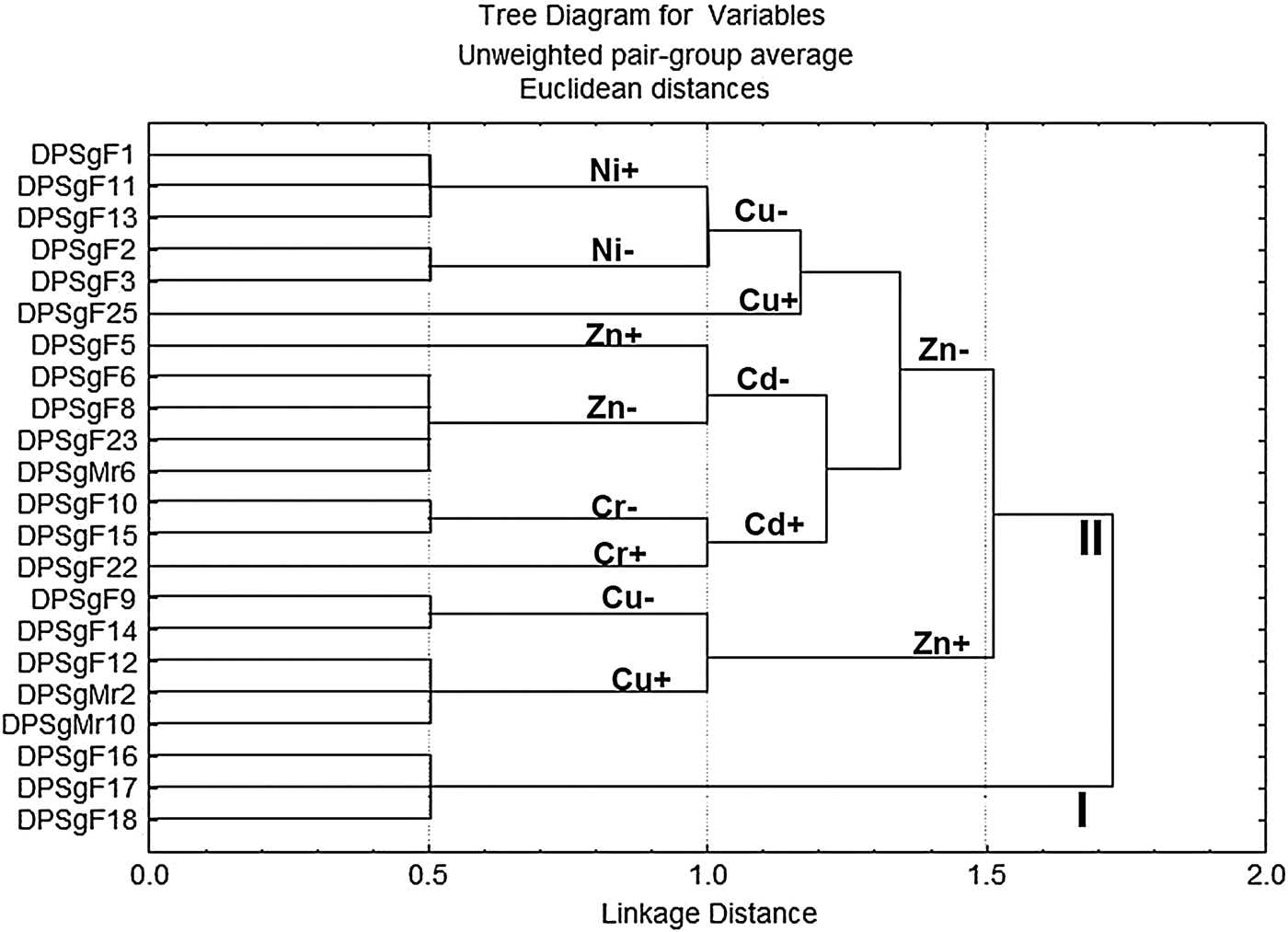
Morphological response of all the isolates to seven different heavy metals was tested: manganese (Mn2+ as MnCl2), zinc (Zn2+ as ZnCl2), nickel (Ni2+ as NiSO4), copper (Cu2+ as CuSO4), cadmium (Cd2+ as CdCl2), chromium (Cr6+ as K2Cr2O7) and lead (Pb2+ as Pb(NO3)2). Stock solutions (1 M or 500 mM) of these metals were prepared. Each metal was incorporated in MV agar medium at a final concentration of 10 mM (Dinu et al., Reference Dinu, Anghel and Jurcoane2011). The isolates were streaked on each of these plates. Growth was monitored on the plates for 15 days by measuring the colony diameter under the microscope (Olympus BX60). Tolerance to Pb2+ was also examined by incorporating Pb(NO3)2 in MV agar at 1, 2, 3 and 5 mM in addition to 10 mM. The growth of the isolates in presence of metals as compared with control (MV without any metal) was monitored for 1 month. The capacity of all the isolates to tolerate 10 mM of each metal was analysed by cluster analysis using the unweighted pair group average method in Statistica v 5.0 software. Plating and microscopy was repeated twice before proceeding for scanning electron microscopic (SEM) analysis.
The isolates with tolerance to metals were grown in MV broth supplemented with respective metal solutions separately and the biomass obtained after 10 days of growth were treated for SEM analysis according to protocol described by Washington State University (http://sbs.wsu.edu/fmic/protocol/fixindex/sem_standard_fix.html). In brief, the cells were washed with sterile seawater, primarily fixed in 1% glutaraldehyde and filtered over pretreated 0.4 µm white polycarbonate filter paper (Millipore). The filter papers were pretreated with 0.1% polylysine hydrobromide (Sigma) and air dried (Kalab et al., Reference Kalab, Yang and Denise2008). The cells were then washed with sterile distilled water and treated with 1% osmium tetroxide for 1 h at RT. The filter papers bearing the cells were passed through an increasing series of dehydration steps (30, 50, 70, 95 and 100% ethanol) of 10 min each. The filter papers were mounted on stubs, freeze-dried (Thermo – Savant, Model Micro Modul YO-230) and sputter-coated with gold. They were then observed with a JEOL, JSM-5800LV scanning electron microscope.
RESULTS
Isolation, identification and enzymatic profiles of thraustochytrid isolates
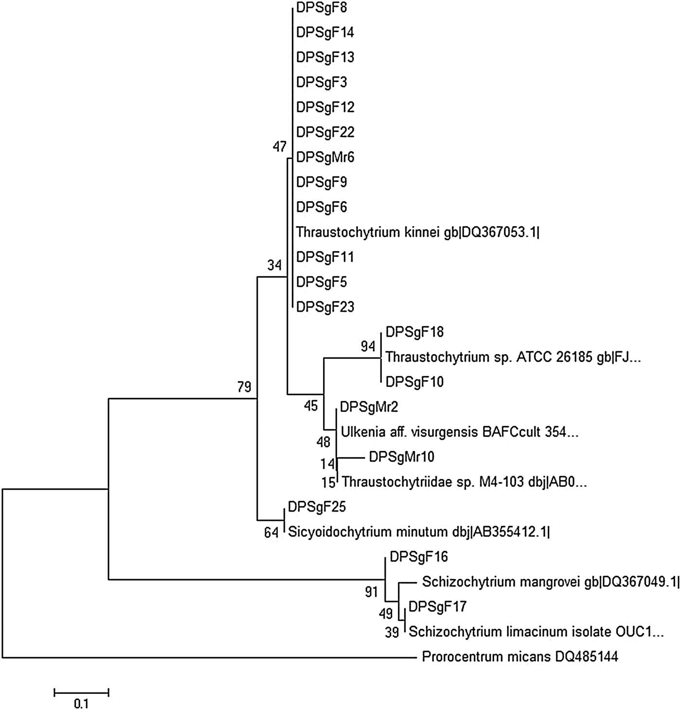
Altogether 35 isolates were obtained in culture from Sargassum samples collected at two different times and only 22 survived repeated sub-culturing. Out of the 22, 19 isolates were attained during February 2012 and three isolates during March 2012. The isolates were designated as DPSgF- for February samples and DPSgMr- for March samples. The topology of the phylogram showed that 22 strains obtained from Sargassum belonged to six species, Thraustochytrium kinnei A. Gaertn., Thraustochytrium sp. ATCC 26185, Aurantiochytrium limacinum (D. Honda & Yokochi) R. Yokoyama & D. Honda, A. mangrovei (Raghuk.) R. Yokoy. & D. Honda, Sicyoidochytrium minutum (Raghuk.) R. Yokoy., Salleh & D. Honda and Ulkenia visurgensis (Ulken) A. Gaertn., among which T. kinnei A. Gaertn. formed the majority of the isolates. Fifteen isolates, namely DPSgF1–3, DPSgF5–6, DPSgF8–9, DPSgF11–15, DPSgF22–23 and DPSgMr6 were T. kinnei. DPSgF10 and DPSgF18 were identified at molecular level (from sequence data) as Thraustochytrium sp. ATCC 26185 and their morphological characteristics resembled the same species as well. Dense lipid bodies in the cytoplasm, cell size between 6–14 µm and lack of clear ectoplasmic net elements were characteristic of Thraustochytrium sp. ATCC 26185 (Weete et al., Reference Weete, Kim, Gandhi, Wang and Dute1997).
Isolates DPSgF16 and DPSgF17 were identified at molecular level to be species of Aurantiochytrium. Microscopic analysis of the two confirmed their identity to be Aurantiochytrium mangrovei (earlier Schizochytrium mangrovei) and Aurantiochytrium limacinum (earlier S. limacinum) respectively. Release of limax-shaped amoebae by DPSgF17 confirmed its identity as A. limacinum. DPSgF16 showed characteristics similar to A. mangrovei (Raghukumar, Reference Raghukumar1988) such as production of amoebae in MV agar. Isolate DPSgF25 was identified as Sicyoidochytrium minutum. Two isolates from March, namely DPSgMr2 and DPSgMr10, were identified as Ulkenia visurgensis.
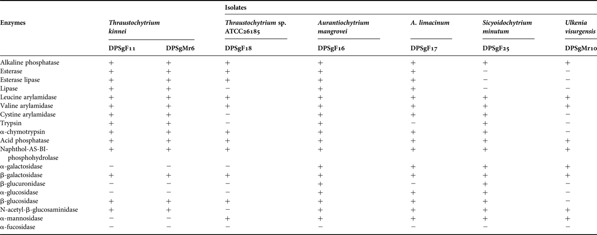
Enzyme profile of the isolates representing each genus/species was distinct from each other (Table 1). Thraustochytrium sp. ATCC 26185 (DPSgF18) was different from that of T. kinnei (DPSgF11 and DPSgMr6). The former produced α-mannosidase and no activity with trypsin, cysteine arylamidase and N-acetyl-β-glucosaminidase as compared with T. kinnei. Profiles of the two species of Aurantiochytrium were similar. However, A. limacinum (DPSgF17) differed from A. mangrovei (DPSgF16) by absence of trypsin and β-glucuronidase. These two differed from S. minutum (DPSgF25) and U. visurgensis (DPSgMr10) by the presence of esterase and lipase. DPSgMr10 differed from the rest by the absence of α-chymotrypsin and β-glucosidase. It produced the least number (9) of enzymes whereas isolate DPSgF17 (A. limacinum) produced the maximum number (18).
Enzymatic profiles of representative thraustochytrid isolates from the present study. (‘ + ’ sign indicates positive activity and ‘ − ’ sign indicates absence of activity).

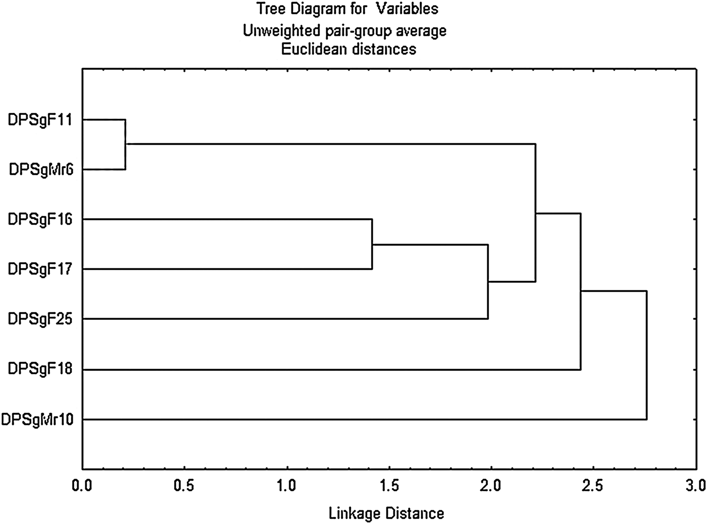
Phylogenetic analysis distributed the isolates into two clusters, one containing isolates belonging to Aurantiochytrium sp. and the other comprising the rest of the genera. The inclusion of several isolates of Thraustochytrium, Sicyoidochytrium and Ulkenia species to clustering algorithm established these thraustochytrids as a highly supported monophyletic group that was distinct from Aurantiochytrium spp. (Figure 1). This major cluster of thraustochytrids consisted of a subcluster of all T. kinnei isolates and of Ulkenia sp. that formed sister lineage to Thraustochytrium sp. ATCC 26185. On the other hand, in the cluster analysis based on enzymatic profiles, isolates of Aurantiochytrium sp. (DPSgF16 and DPSgF17) grouped with isolates of T. kinnei (DPSgF11 and DPSgMr6) and Sicyoidochytrium sp. (DPSgF25) and the isolate belonging to Ulkenia sp. (DPSgMr10) separated out in a distinct clade (Figure 2).
Neighbour-joining (NJ) tree of 18S rDNA of thraustochytrids isolated in the present study along with other thraustochytrid sequences from NCBI database using MEGA 4.0.1. The 18S rDNA sequence of dinoflagellate Prorocentrum micans (Accession no. DQ485144) is used as an outgroup. Some sequences that were too divergent were not considered for construction of the tree. Schizochytrium mangrovei and S. limacinum have been emended as Aurantiochytrium mangrovei and A. limacinum respectively according to Yokoyama & Honda (Reference Yokoyama and Honda2007).
Cluster analysis of representative thraustochytrid isolates based on their enzyme profiles using unweighted pair group average method in Statistica v 5.0 software.
Morphological response to heavy metals
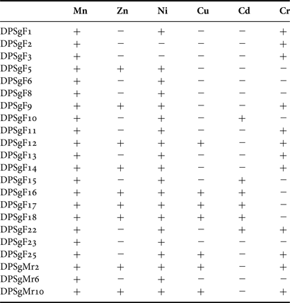
When compared with the control plate (i.e. MV agar without any metals), there was either poor growth or absolutely no growth of thraustochytrid isolates in the agar incorporated with different heavy metals (Table 2). All the isolates grew in the presence of Mn2+ but to varying extent. There was no difference in colony size in the presence of Mn2+ in four out of 22 isolates (DPSgF5, 17, 25 and DPSgMr10) as compared with control, but the cells were loosely packed inside the colony in the case of DPSgF5 and occasionally amoeboid cells were formed in the case of DPSgMr10. In DPSgF17 and DPSgF25, there was no difference in cellular morphology as compared with the control (MV without any metal). Colony size was reduced by 25–70% in the other 18 isolates studied i.e. from an average diameter of 175–200 µm under normal MV, the colony size was reduced to 100–150 µm in the presence of Mn2+. Tolerance of the isolates to Mn2+ was followed by Ni2+, Cr6+ and then Zn2+. In 91% of the isolates (20 out of 22) there was tolerance to 10 mM NiSO4 (Table 2). Twelve out of 22 isolates (55% of the total tested) tolerated Cr6+. Nine isolates (41% of the total tested) showed tolerance to Zn2+. Cu2+ was tolerated by seven isolates (32% of the total) and Cd2+ by six (27%). There was slight tolerance to 1 mM Pb2+ in two isolates DPSgMr2 and DPSgMr10 as marked by very poor growth and shrunken cells. Colony size reduced by 72% (200–225 to 50–60 µm average diameter) in these two isolates. No isolates grew in the presence of 2, 3, 5 and 10 mM Pb2+.
Ability of thraustochytrid isolates to grow on Modified Vishniac (MV) agar incorporated with various metal solutions. (‘ + ’ sign indicates growth or tolerance to that metal and ‘ − ’ sign indicates absence of growth and hence zero tolerance to that metal).
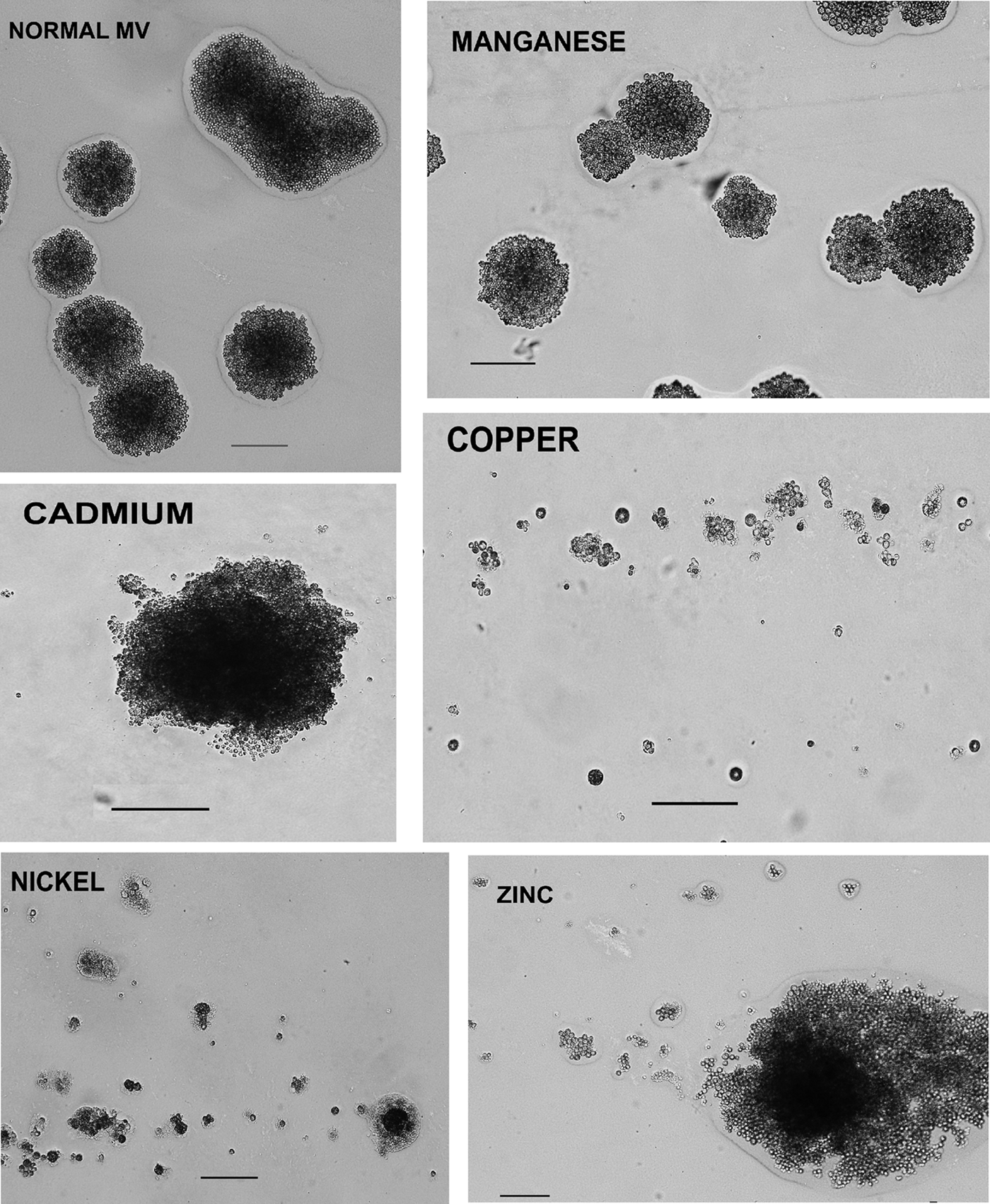
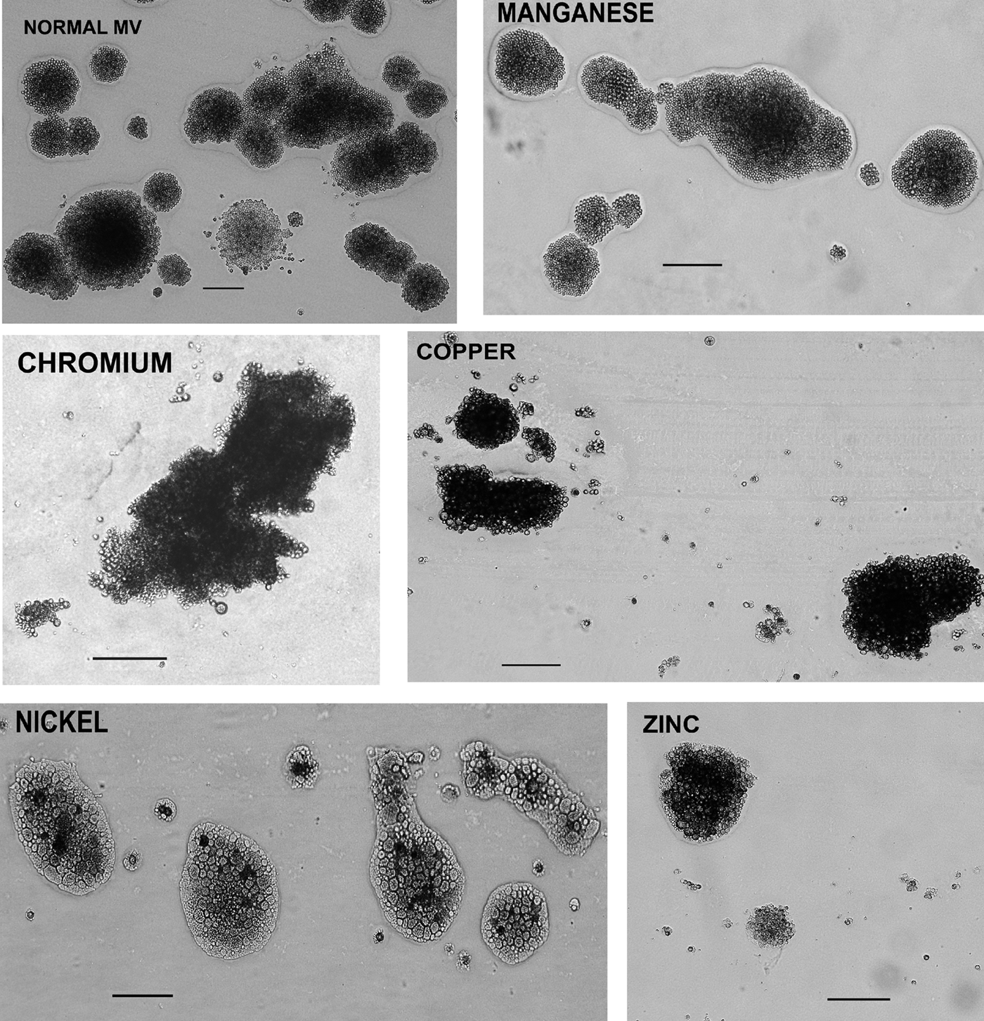
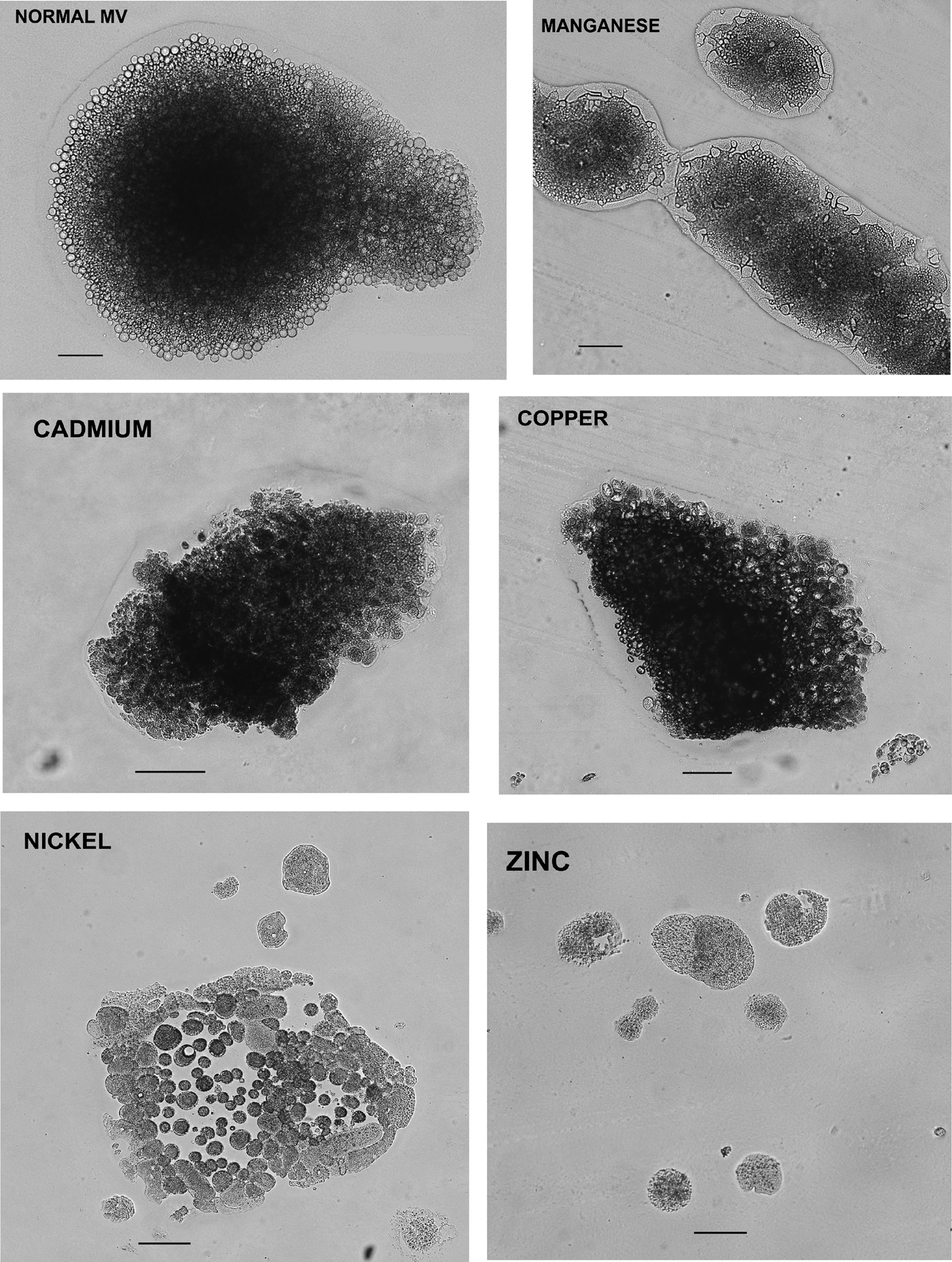
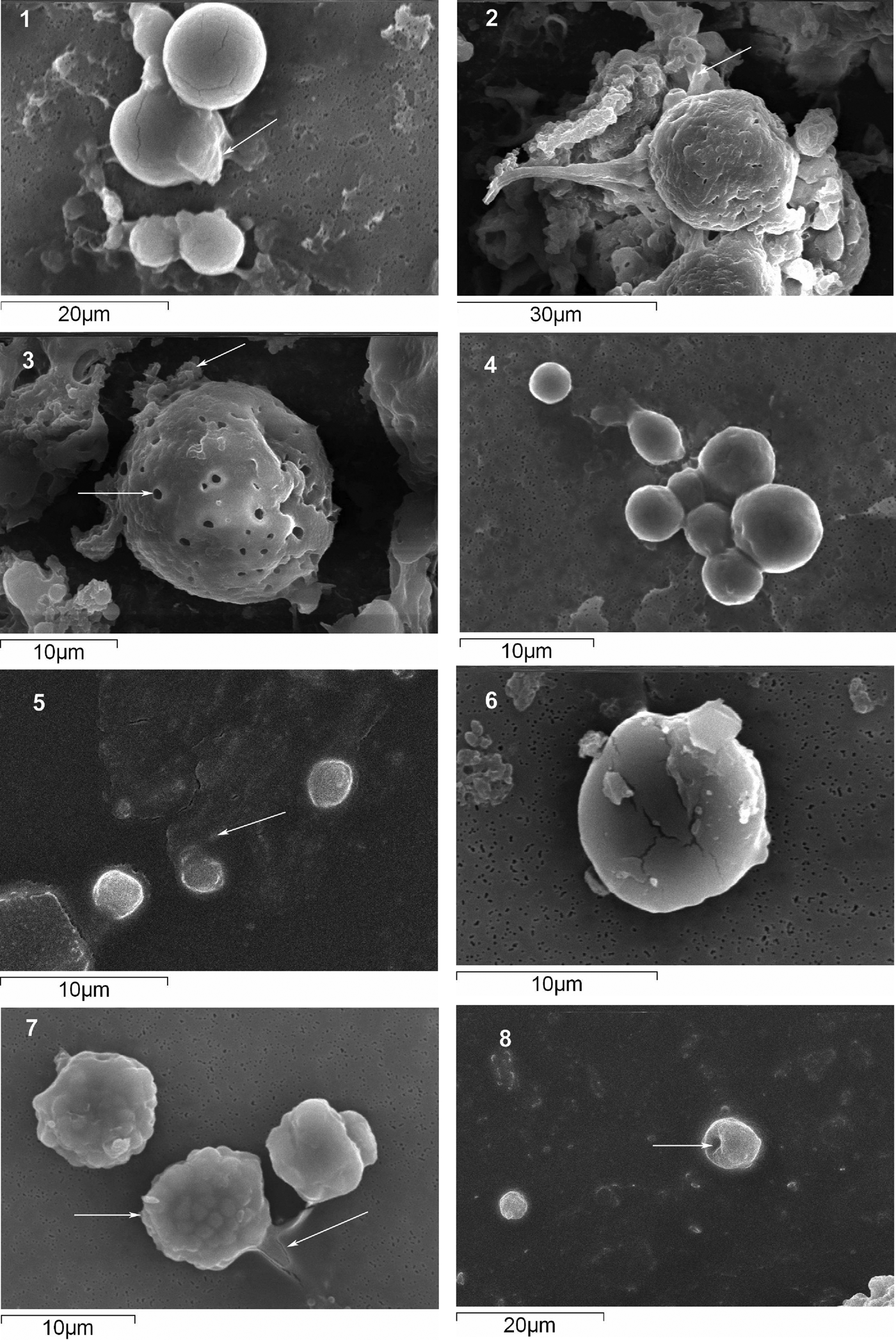
There were differences in morphological characteristics of cells of all isolates in the presence of various metals as compared with normal cells. A few of these isolates are shown in Figures 3–6. No distinct colony formation was observed in some; instead cells were seen spread all over the line of inoculation (as seen in the presence of Cu2+ and Ni2+ in Figure 3, Zn2+ in Figures 4 & 5). Distinct changes in the cell wall in the presence of metals as compared with the normal cells were evident in the electron micrographs. The presence of metals resulted in either depressions in the cells or holes on their cell wall (Figure 6). A significant amount of cell shrinkage was noticed due to the presence of metals as compared with normal healthy cells (e.g. panel 5 and 8 in Figure 6). Average cell size was reduced from 20 to 5 µm in the presence of a few metals like Cu2+, Ni2+.
Microphotographs of the colonies of isolate DPSgF12 growing on Modified Vishniac (MV) agar plates incorporated with different metals. Bar represents 100 µm.
Microphotographs of the colonies of isolate DPSgF18 growing on Modified Vishniac (MV) agar plates incorporated with different metals. Bar represents 100 µm.
Microphotographs of the colonies of isolate DPSgMr2 growing on Modified Vishniac (MV) agar plates incorporated with different metals. Bar represents 100 µm.
Scanning electron microscopy photographs of three isolates grown in the presence of metals; DPSgMr10 isolate in the presence of normal MV (1) and MV supplemented with Ni+2 (2 and 3), DPSgF22 isolate in the presence of normal MV (4) and MV supplemented with Mn+2 (5) and DPSgF16 isolate in the presence of normal MV (6) and MV supplemented with Ni+2 (7) and Cu+2 (8). The arrows pointing leftwards show the EPS surrounding or released by the cells. Cell shrinkage due to metal ions is evident in 5 and 8. The arrow pointing towards the right in 3 shows distinct holes formed in the cell wall. The rightward pointing arrow in 7 shows the distorted cell shape as compared with the spherical cell (normal) in 6. The arrow pointing towards the right in 8 shows depression in the cell wall as an effect of metal.
Cluster analysis based on the metal tolerance capacity of each isolate divided all the isolates into two main clades, one comprising isolates that were sensitive to only one out of six metals (clade I) and the second comprising isolates that were sensitive to one or many metals (clade II). Overall the characters of tolerance to different metals were scattered throughout the clusters (Figure 7). Isolates DPSgF16 and DPSgF17 belonging to Aurantiochytrium sp. and DPSgF18 classified as Thraustochytrium sp. ATCC 26185 clustered in clade I. The rest of the isolates belonging to T. kinnei, Ulkenia etc. grouped together in clade II similar to the NJ tree.
Cluster analysis of all thraustochytrid isolates based on their metal tolerance capacity using unweighted pair group average method in Statistica v 5.0 software. Clade I and II are marked in the figure. Distribution of a few metals in the tree clearly indicates scattering of these characters.
DISCUSSION
The fast-growing brown alga Sargassum is spread by currents or by ships and provides a crucial habitat for a wide variety of marine animals. The polyunsaturated fatty acid (PUFA) signatures of particulate organic matter from the habitats surrounding Sargassum are known to contain high levels of PUFAs like 22:6 n-3 (Turner & Rooker, Reference Turner and Rooker2006). As the macroalgae contain very low quantities of long-chain PUFAs, especially 22:6n-3, the organic matter appears to have originated from other living organisms associated with Sargassum. Thraustochytrid protists are known to produce abundant amounts of this fatty acid intracellularly (Raghukumar, Reference Raghukumar2008). Thraustochytrids could therefore be the source of 22:6n-3 in the organic matter. This suggests the prevalence of these compounds in these habitats and probable association of these organisms with the alga. This was confirmed by the successful isolation of these protists from Sargassum cinereum during both sampling times in the present study.
Thraustochytrids are generally found to be substrate-specific in occurrence rather than free-living in the water column. This substrate-specific nature helps these protists to grow on Sargassum as epibionts (Raghukumar, Reference Raghukumar2002). Though the brown alga produces tannins on distal growing tips that have an inhibitory effect on colonizing epibionts, its effect lasts for a short time after which a succession of other organisms like bacteria, hydroids, bryozoans, etc. rapidly follow (see Coston-Clements et al., Reference Coston-Clements, Settle, Hoss and Cross1991). This explains the prevalence of thraustochytrids with the algae and their subsequent isolation in the present study. Aplanochytrium minuta (S.W. Watson & Raper) C.A. Leander & D. Porter, a member of a sister group of thraustochytrids, called aplanochytrids, was regularly found to be associated as an endobiont with S. cinereum (Sathe-Pathak et al., Reference Sathe-Pathak, Raghukumar, Raghukumar and Sharma1993). But in the present study, this species was not recovered from the samples. Rather, all isolates obtained were found to be thraustochytrids.
Thraustochytrids are also common inhabitants of the detritus originating from Sargassum (Sharma et al., Reference Sharma, Raghukumar, Raghukumar, Sathe-Pathak and Chandramohan1994). Marine detritus appears to be an excellent substrate for growth of these protists (Raghukumar & Damare, Reference Raghukumar and Damare2011). Owing to their absorptive mode of nutrition and production of various degradative enzymes, these organisms are thought to be important remineralizers in the marine ecosystem (Raghukumar & Damare, Reference Raghukumar and Damare2011). In order to explain this role, studies on extracellular enzyme production were carried out. Amongst the 19 enzymes tested, the isolates in the present study showed positive activity with most of them. All the isolates produced alkaline as well as acid phosphatase indicating their ability to dephosphorylate organic molecules for uptake during PO43− replete conditions and also to grow under PO43− starvation as PO43− is an important limiting nutrient for growth of the algae (Kuenzler & Perras, Reference Kuenzler and Perras1965). Except for Sicyoidochytrium minutum and Ulkenia visurgensis, most of the isolates produced esterases and lipases that help to break down short and long chain fatty acids respectively and as a consequence utilize a wide range of substrate molecules possessing amide or ester bonds (Bourne et al., Reference Bourne, Isupov and Littlechild2000). Various proteolytic enzymes such as arylamidases, trypsin and chymotrypsin were produced by most of the test isolates. Amongst the carbohydrate-degrading enzymes, none of the isolates produced fucosidase, an enzyme that breaks down the sugar generally present in the cell wall of the brown algae as well as in the extracellular polysaccharides secreted by the phytoplankton suggesting that thraustochytrids may not be able to degrade the algal cell wall and hence may not initiate an infection. Production of N-acetyl-β-glucosaminidase by all the isolates calls attention towards the ability of thraustochytrids to break down chitin from exoskeletons of marine invertebrates that may inhabit the Sargassum mats (Yu et al., Reference Yu, Lee, Bassler and Roseman1991). This would protect the mats against invaders/consumers (Turner & Rooker, Reference Turner and Rooker2006). Thraustochytrids isolated from seagrasses and green algae that form mats in the water column similar to Sargassum also produce this enzyme (Bongiorni et al., Reference Bongiorni, Pusceddu and Danovaro2005).
The production of various enzymes facilitates absorption of nutrients from the surfaces of Sargassum by breaking down complex organic compounds. β-glucuronidase produced by DPSgF16 and DPSgF25 helps in digesting uronic acid-rich polysaccharides secreted by algae (Dodgson et al., Reference Dodgson, Lewis and Spencer1953). Thus enzyme production by these isolates confirms not only the osmoheterotrophic mode of nutrition of thraustochytrids but also their role as remineralizers in the food chain (Raghukumar & Damare, Reference Raghukumar and Damare2011). Production of a limited number of enzymes by isolate belonging to U. visurgensis indicates a low variety of substrate preference or utilization of more easily available substrates than breaking down of refractory matter.
Thraustochytrids occasionally show ability to withstand and grow in the presence of heavy metals (Raghukumar et al., Reference Raghukumar, Mohandass, Cardigos, D'Costa, Santos and Colaço2008). Generally seaweeds have a high capacity to bind heavy metals. Biosorption of copper, gold, cadmium, zinc, manganese and even radioactive metal such as uranium on Sargassum biomass has been studied and it is therefore recommended as an excellent biomaterial for accumulating and recovering metals from industrial effluents (Krishnakumar et al., Reference Krishnakumar, Pillai and Valsala1990; Volesky & Holan, Reference Volesky and Holan1995; Yang & Volesky, Reference Yang and Volesky1999; Antunes et al., Reference Antunes, Luna, Henriques and da Costa2003). Biosorption on Sargassum biomass may induce metal tolerance in its associated microorganisms (Wagner-Döbler et al., Reference Wagner-Döbler, Beil, Lang, Meiners and Laatsch2002). Hence thraustochytrids from Sargassum might be a good subject to study metal tolerance. There are only two reports that deal with tolerance of thraustochytrids to heavy metals (Raghukumar et al., Reference Raghukumar, Mohandass, Cardigos, D'Costa, Santos and Colaço2008; Lin et al., Reference Lin, Leaño and Pang2010). In their study Lin et al. (Reference Lin, Leaño and Pang2010) observed that Schizochytrium cells were tolerant to Cu2+ and Zn2+ to a certain extent. In the present study the Aurantiochytrium spp. were tolerant to both of these metals along with manganese, nickel and chromium. Deformities in cellular structure were seen as an effect of metals on the cells. Lin et al. (Reference Lin, Leaño and Pang2010) observed leakage of cellular contents due to cell lysis when exposed to high levels of Cu2+ and Zn2+. In the present study the high metal concentration resulted in holes or depressions in the cells as also observed by Singh et al. (Reference Singh, Raghukumar, Parvatkar and Mascarenhas-Pereira2013) in yeasts. Raghukumar et al. (Reference Raghukumar, Mohandass, Cardigos, D'Costa, Santos and Colaço2008) studied the effect of heavy metals including iron, manganese and lead on one thraustochytrid isolate which they identified as Ulkenia sp. They isolated this species from a shallow water hydrothermal vent where heavy metals occur in high concentrations, thus suggesting its adaptation to the environment. The Ulkenia sp. in the present study was tolerant to all the metals studied except cadmium. This was the only species that exhibited tolerance to 1 mM Pb2+. Resistance to heavy metals is usually specific to species (Miersch et al., Reference Miersch, Bärlocher, Bruns and Krauss1997). Generally all the isolates showed wide tolerance to Mn2+ which is a micronutrient and required as a cofactor for several metalloenzymes. In eukaryotes, when manganese is amply available the polypeptides that transport this nutrient in the cell are directed to the lumen of cellular vacuole where they are degraded by vacuolar proteases, thereby minimizing cellular uptake and preventing cell damage (Culotta et al., Reference Culotta, Yang and Hall2005). This would have probably resulted in greater tolerance to manganese than other metals.
The biosorbing capacity of the cells depends not only on the type and concentration of metal ions but also on the type of cell wall that comes in contact with the metals. The functional groups present in the cell wall are mostly responsible for binding heavy metals due to the net charge on them which helps in binding charged metal ions (Das et al., Reference Das, Vimala and Karthika2008). The cell walls of thraustochytrids are made of sulphated polysaccharides containing galactose, glucose, xylose, mannose and also proteins (Chamberlain, Reference Chamberlain1980). In addition, these organisms secrete extracellular polysaccharides (EPS) that contain uronic acids and sulphates, conferring them an overall negative charge (Jain et al., Reference Jain, Raghukumar, Tharanathan and Bhosle2005). The polyanionic nature of the EPS exhibits high binding affinity for cations as well as trace metals and acts as an ion-exchanger (Poli et al., Reference Poli, Anzelmo and Nicolaus2010). Extracellular polysaccharides also help in biofouling and enhance the capacity of source organisms to adhere to marine substrata, for example Sargassum in the present study (Jain et al., Reference Jain, Raghukumar, Tharanathan and Bhosle2005).
Microorganisms living in association with many marine macroorganisms are notable for their capability to produce bioactive compounds earlier ascribed to the hosts (Armstrong et al., Reference Armstrong, Yan, Boyd, Wright and Burgess2001; Penesyan et al., Reference Penesyan, Marshall-Jones, Holmstrom, Kjelleberg and Egan2009; Hollants et al., Reference Hollants, Leliaert, De Clerck and Willems2013). Moreover, these might also cause biosorption of heavy metals from contaminated seawater. Correspondingly the microorganisms living in association with Sargassum, for example thraustochytrids, may be contributing towards its potential to accumulate heavy metals from contaminated seawater. Detailed studies on biosorption of heavy metals on thraustochytrid biomass will shed more light on this aspect.
In conclusion, this study showed that thraustochytrids are commonly associated with Sargassum and could be isolated from this brown alga. By producing various enzymes, these epibiotic organisms survive on the nutrients released from Sargassum or other algal surface-associated organisms and might serve to protect the alga from colonization by parasites. Their tendency to tolerate heavy metals may be used for bioremediation as these organisms contribute to a large extent to microbial biomass carbon owing to their great abundance in the marine environment and hence can be harnessed for this purpose (Damare & Raghukumar, Reference Damare and Raghukumar2008). The heavy metal tolerance capacity of thraustochytrids could be further studied for its bioremediation potential in future.
ACKNOWLEDGEMENTS
The author is grateful to the Director, CSIR-NIO for providing laboratory facilities to carry out this work and to the Department of Science and Technology, India for the financial assistance through SERC Fast Track Proposal for Young Scientists (DST No: SR/FTP/ES-186/2010). Help provided for sequencing of samples by Mr Ram Murti Meena and for SEM analysis by Mr Areef Sardar is highly acknowledged. This is CSIR-NIO's contribution number 5663.