Introduction
Steroid hormones are small hydrophobic signalling molecules derived from cholesterol, more precisely pregnenolone (Ref. Reference Beato and Klug1). Among the steroid hormones, progesterone and oestrogens are considered as the ‘female hormones’ which play crucial roles in the modulation of physiological processes pertinent to development as well as function of the female reproductive system. The uterus holds a unique position in the female reproductive system as it is the site of foetal development. The inner lining of uterus known as endometrium, undergoes cyclic shedding and regeneration during every ovulatory cycle which is tightly regulated by the sequential action of oestrogen and progesterone produced in the ovary (Refs Reference Groothuis2, Reference Hapangama3, Reference Young and Lessey4, Reference Maybin and Critchley5). Endometrium has two major layers, functionalis towards the uterine cavity and basalis, which is adjacent to myometrium. Both these layers comprise of epithelial glands and connective tissue which include stromal fibroblasts and the vasculature (Ref. Reference Figueira6). Oestrogen receptors (ER) – ERα and ERβ and progesterone receptors (PR) – PR-A and PR-B, through which these hormones predominantly act, are expressed in stromal and epithelial cells of both these layers. The expression pattern of these receptors in functionalis varies with the menstrual cycle phases but remains unchanged in the basalis. The hormonal action in basalis and functionalis differs because of the heterogeneity in expression of their receptors (Refs Reference Mote7, Reference Arnett-Mansfield8). The postmenopausal endometrium has lower expression of both ER and PR compared with pre-menopausal endometrium except PR-A, which has a high expression comparable with early secretory phase (Ref. Reference Mylonas9). The status of ER and PR in postmenopausal endometrium indicates that on exposure to hormones, these receptors can transduce the signals and a fully functional endometrium can be revived from post-menopausal endometrium (Refs Reference Gorodeski10, Reference Whitehead11).
The remarkable regenerative capacity of endometrium has led to the identification of stem cells in endometrium and these cells are proposed to regenerate the functionalis layer from the basalis after menstruation (Refs Reference Gargett12, Reference Mutlu13, Reference Masuda14). Exposure of stem cells residing in basalis to oestrogen and progesterone may lead to regeneration of functionalis in post menstrual as well as post-menopausal endometrium (Refs Reference Masuda15, Reference Padykula16, Reference Gargett and Ye17, Reference Ulrich18). Apart from its role in normal endometrial turnover, stem cells of endometrium are also involved in endometrial pathologies like endometriosis – the growth of endometrium outside uterus (Refs Reference Figueira6, Reference Yang and Huang19, Reference Hufnagel20). Endometriosis is a proliferative, endocrine, inflammatory disorder, the aetiology of which is obscure despite various theories being proposed to explain its pathology (Refs Reference Sampson21, Reference Benagiano22, Reference Gruenwald23). The proposals of stem cells as seeds for endometriosis comply with these theories and indirect evidence for involvement of stem cells in endometriosis are available (Refs Reference Gargett24, Reference Pittatore25, Reference Heidari-Keshel26).
The oestrogen–progesterone imbalance associated with endometriosis (Refs Reference Bulun27, Reference Al-Sabbagh28) and the regulatory effect of these hormones on adult stem cells are well known .The association between derailed hormonal signalling and endometrial stem cells (EnSCs) in endometriosis is being explored. In this review, we discuss the impact of oestrogen and progesterone on EnSCs in normal and diseased endometrium. Moreover, it is known that the key developmental signalling pathways – Wnt, Notch and Hedgehog (Hh) – regulate stem cell activities in adult mammalian tissues enabling tissue renewal and regeneration (Refs Reference Clevers29, Reference Artavanis-Tsakonas30, Reference Briscoe and Thérond31). The connection between oestrogen–progesterone signalling and the aforementioned stem cell pathways in normal endometrium is discussed here. The aberrant oestrogen and progesterone signalling in endometriosis could be a trigger for the derailed stem cell signalling or vice versa. Exhaustive studies on this aspect would help to demystify the elusive pathology of endometriosis.
Oestrogen and progesterone influence on stem cells
Stem cells are highly dependent on their niche which is crucial in the determination of their fate. External cues determining the fate of embryonic stem cells (ESCs) mainly consist of growth factor and hormonal signalling. Oestrogen has been shown to induce ESC proliferation probably in an autocrine manner as these cells express ERα and ERβ (Ref. Reference Hong32). A study in zebrafish embryo has shown that the oestrogenic effect on embryo is temporal (Ref. Reference Carroll33). Early exposure to oestrogen, which is prior to the start of circulation and maturation of erythrocytes, regulates the haemoendothelial niche specification in embryo whereas the later exposure enhances haematopoietic stem cell (HSC) number. Oestrogen response also differs between ESC and adult stem cells as oestrogen-mediated induction of stem cell proliferation is seen in embryonic neural stem cells (NSCs) but not in adult NSC of rat, though both express ERα and ERβ (Ref. Reference Brannvall34). The other female hormone, progesterone has been reported to be essential for the differentiation of ESCs to embryoid bodies during human embryonic development (Ref. Reference Gallego35), the action of which is mediated by PR-A expressed in ESCs (Ref. Reference Gallego36). In addition, progesterone induces (Ref. Reference López-González37) or inhibits (Ref. Reference Kang38) differentiation of ESCs into specific lineages.
Studies on ovarian steroid hormonal influence on MaSCs (mammary epithelial stem cells) in mice provided the first in-vivo evidence for the influence of these hormones on adult stem cells. Oestrogen and progesterone treatment in mice cause expansion of mammary epithelial stem cell pool though these cells lack ER and PR (Refs Reference Visvader and Stingl39, Reference Joshi40). Numerous studies have provided direct and indirect evidence for the influence of oestrogen and progesterone on a range of adult stem cells. Oestrogen has been shown to enhance proliferation of HSC (Ref. Reference Illing41) and oligodendrocyte progenitor cells (Ref. Reference Marin-Husstege42) by altering the cell cycle. Oestrogen induces osteogenic differentiation of mesenchymal stem cells (MSCs) (Ref. Reference Gao43), while progesterone promotes differentiation of MSCs to neurons (Ref. Reference Wang44). Progesterone is also shown to influence the immune modulatory effects of MSCs by enhancing the expression and secretion of immunomodulatory proteins such as HLA-G (human leucocyte antigen G), PIBF (progesterone immunomodulatory-binding factor 1), PGE2 (prostaglandin E2) and IL-6 (interleukin 6) in MSC of adipose (Ref. Reference Moslehi45), bone marrow and endometrial origin (Ref. Reference Kyurkchiev46).
Stem cells of endometrium
The presence of cells with stem cell traits in human endometrium is undoubtedly established by various researchers across the globe. Endometrium harbours mesenchymal and epithelial stem cells in the stromal and the epithelial compartments, respectively. The MSCs in the stroma are found to be enriched in CD146+CD140b+ (Ref. Reference Schwab and Gargett47) and Sushi domain-containing protein-2 (SUSD2+) (Ref. Reference Masuda48) population and are well characterised. Epithelial stem/progenitor cells (ESPC) are reported to be enriched in Side Population (SP) (Refs Reference Masuda15, Reference Cervelló49), LGR5+ (Ref. Reference Gil-Sanchis50), SSEA1+ (Ref. Reference Valentijn51) and N-cadherin+ (Ref. Reference Nguyen52) population. However, no definitive markers for ESPCs are presently available. Gene expression profiling and further functional characterisation of cells expressing putative ESPC cell markers would help define the identity of these cells. Present understanding of the identity and role of the EnSCs has been comprehensively reviewed in recent articles (Refs Reference Gargett12, Reference Mutlu13, Reference Masuda14). EnSCs have been proposed to be involved in regeneration of endometrium (Refs Reference Masuda15, Reference Padykula16) from basalis, which is the most accepted harbour for epithelial stem cells (Refs Reference Nguyen53, Reference Gargett54). Study on stem cell traits across menstrual cycle has revealed that the clonogenicity of epithelial and stromal cells does not vary across different phases of menstrual cycle (Ref. Reference Schwab55) which indicates that the number of stem cells may remain the same across the cycle. Whether this is because stem cells undergo asymmetric divisions to maintain endometrium requires further investigation. The EnSCs are also identified in the postmenopausal endometrium (Ref. Reference Ulrich18) which resembles the basalis of pre-menopausal endometrium as both have similar epithelial gene signatures (Ref. Reference Nguyen53). The dormant post-menopausal endometrium retains the capacity to respond to oestrogen and progesterone and a fully functional endometrium can be regenerated from it. This regeneration could possibly be by oestrogen and progesterone mediated induction of a proliferative state in EnSCs, which can survive hormonal deprivation unlike the differentiated epithelial cells (Refs Reference Gargett54, Reference Janzen56).
EnSCs are under the orchestrated influence of oestrogen and progesterone during the reproductive life of females inciting constant proliferation and differentiation, though studies reported the absence/low levels of ERα and PR in these cells of both human (Refs Reference Masuda15, Reference Ulrich18, Reference Cervelló49, Reference Gil-Sanchis50, Reference Valentijn51, Reference Cervelló57) and mouse origin (Ref. Reference Chan58). Studies using mice model have shown that oestrogen alone induces proliferation of stromal stem cells whereas in ESPCs, oestrogen alone (Ref. Reference Hyodo59) or in combination with progesterone can induce proliferation (Ref. Reference Janzen56). The absence of a conclusive marker for the endometrial epithelial cells causes discrepancy in the data regarding hormonal influence on these cells, as one group focused on the SP (Ref. Reference Hyodo59), while the other studied EpCAM(+) CD44(+) ITGA6(hi) Thy1(−) PECAM1(−) PTPRC(−) Ter119(−) cells (Ref. Reference Janzen56). Proliferation of matured epithelial cells may be sufficient for replenishing the epithelial compartment while stromal stem cells are found necessary for replenishing the stromal compartment (Ref. Reference Chan58). It is also shown that the hormone responsiveness of ESPCs located in the basalis and MSCs located in the functionalis of the cycling murine endometrium differ as the oestrogen-mediated induction of proliferation varies in both the cells (Ref. Reference Chan58).
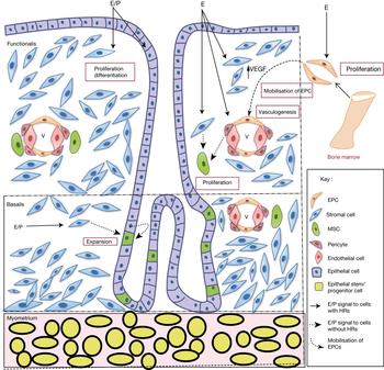
A recent study (Ref. Reference Gunjal60) has shown the presence of pluripotent very small embryonic-like stem cells (VSELs) in mice from which endometrial stem cells are proposed to originate. A common ancestor for both stromal MSC and epithelial progenitor cells has also been proposed which needs extensive research to confirm. The neonatal exposure of VSELs to oestrogen in mice has been shown to cause aberrant proliferation in the endometrium (Ref. Reference Bhartiya61) asserting the influence of oestrogen on these cells. In addition to the stem/progenitor cells discussed above, human endometrium consists of MSCs, hematopoietic stem cells (HSCs) and endothelial progenitor cells (EPCs) of bone marrow origin which are differentiated into stromal fibroblasts, components of blood and endothelial cells respectively (Refs Reference Taylor62, Reference Aghajanova63, Reference Mints64). The EPCs express ERs (Ref. Reference Foresta65) and oestrogen has been shown to induce proliferation of EPCs through ERα activation (Refs Reference Hamada66, Reference Matsubara and Matsubara67). This could be due to up-regulation of matrix metallopeptidase 9 (MMP-9) by oestrogen (Ref. Reference Iwakura68) which leads to the release of soluble Kit ligand (sKitL) from bone marrow stromal cells enabling the transfer of endothelial and HSCs from the quiescent to proliferative niche (Ref. Reference Heissig69). The number of EPCs and sKitL expression has been shown to vary with menstrual cycle in women (Ref. Reference Matsubara and Matsubara67). In addition, mobilisation of bone marrow derived EPCs to the neovascularisation sites involves the vascular endothelial growth factor (VEGF) induction by oestrogen (Ref. Reference Hamada66). Moreover, EPCs from female donors have shown to have more clonogenicity and proangiogenic potential than that of male EPCs (Ref. Reference Fadini70). The available data on correlation between the serum oestrogen level and EPCs is contradictory as one study (Ref. Reference Sugawara71) reports a correlation, while another study (Ref. Reference Robb72) claims that EPC action is independent of circulating oestrogen level. The existing data suggest the influence of oestrogen and progesterone on proliferation and mobilisation of stem/progenitor cells present in endometrium (Figure 1), nonetheless various questions regarding the mode and mechanism of action remain to be answered.
Proposed action of oestrogen and progesterone on stem cells of endometrium. Oestrogen and progesterone (E/P) signal the stromal and epithelial cells of endometrium through their respective hormone receptors (HR) causing proliferation and differentiation of the cells. In the epithelial stem/progenitor cell which lack these hormone receptors, E/P signal is mediated possibly via differentiated stromal and epithelial cells with HRs, to enhance expansion. Mesenchymal stem cells (MSC) located near the vascular region receives oestrogen signal to undergo proliferation, through neighbouring cells expressing hormonal receptors, which includes stromal cells and pericytes. Oestrogen also induces proliferation of hormone receptor positive endothelial progenitor cells (EPC) and enhances mobilisation of these cells to neo-vascularisation sites by inducing VEGF expression in stromal cells.

Hormonal regulation of important stem cell signalling pathways
Stem cell fate is regulated by a number of signalling pathways such as Wnt, Notch and Hh (Refs Reference Clevers29, Reference Artavanis-Tsakonas30, Reference Briscoe and Thérond31, Reference Spitzer73). The influence of oestrogen and progesterone on endometrial stem cell properties by modulating these pathways are discussed below.
Wnt signalling
Significance of Wnt signalling in stem cells has been widely studied and is shown to be essential for maintaining a variety of stem cells, including neural, mammary, embryonic, mesenchymal, haematopoietic, intestinal, gastric and epidermal stem cells, in its undifferentiated state. Nineteen different Wnt proteins have been reported which can bind to the cell surface receptors of the Frizzled family leading to the activation of Wnt/β-catenin signalling pathway (Ref. Reference Clevers29). In post-menopausal endometrium Wnt-β catenin signalling is down-regulated (Ref. Reference Nguyen53) and in cycling endometrium it is established to be essential for endometrial regeneration (Ref. Reference Wang74). Of the different Wnt proteins, WNT7A is found to be crucial for post-menstrual regeneration of endometrium and proliferation of endometrial glands as it is highly expressed in the luminal epithelium and upper glands after menstruation. Therefore, WNT7A inhibition understandably delays the regeneration of the endometrium (Ref. Reference Fan75). Another Wnt protein, WNT5A can induce differentiation of human ESCs to endometrium-like cells further establishing the role of Wnt signalling in endometrium development (Ref. Reference Yu76). Transcriptional profiling of epithelial cells from full-thickness of pre- and post-menopausal endometrium has shown differential expression of Wnt-associated genes in the basalis compared with the functionalis, suggesting a role of Wnt signalling in the maintenance of epithelial stem cells located in the basalis (Ref. Reference Nguyen53) In endometrial stromal stem cells also active canonical Wnt signalling has been reported (Ref. Reference Bukowska77).
The interrelationship between Wnt and sex steroid hormonal signalling is established in various organs under different conditions. This includes direct interaction of ER and β-catenin (Ref. Reference Kouzmenko78) and indirect interaction of ERα and β-catenin through EZH2 (polycomb group protein enhancer of zeste homologue 2) (Ref. Reference Shi79). Moreover, oestrogen up-regulate β-catenin and induce its nuclear translocation (Ref. Reference Chandar80). In addition, Wnt influences oestrogen signalling, as upon stimulation of WNT3A, ERα expression and aromatase enzyme are up-regulated while ERβ is down-regulated, thereby maintaining a fine balance between both the signalling (Ref. Reference Gao43).
In endometrium, oestrogen activates Wnt signalling in proliferative phase while progesterone inhibits this pathway in secretory phase causing suppression of oestrogen-induced proliferation (Ref. Reference Wang81). Oestrogen induces Wnt signalling in ER-dependent and independent manner, by up-regulating WNT4, WNT5A, WNT7A (Ref. Reference Fan75) and FZD2 (Frizzled-2), inducing nuclear localisation of β-catenin in the uterine epithelium as well as by suppressing secreted frizzled related protein (SFRP)-2, an antagonist of Wnt signalling (Ref. Reference Hou82). The regulators of Wnt pathway may also be influenced by oestrogen and progesterone levels as the expression of SFRP4 – the regulator of Wnt-frizzled signalling pathway – varies with menstrual cycle phase (Ref. Reference Abu-Jawdeh83) and Axin2 – the negative regulator of Wnt signalling – is nuclear localised in the less hormonally exposed basalis of cycling endometrium and post-menopausal endometrium (Ref. Reference Nguyen53). The expression of Wnt downstream target, CD44, is up-regulated in women treated with oestrogen alone when compared with the oestrogen- and progesterone-treated women, in an ER-independent manner (Ref. Reference Wang81). Progesterone inhibits Wnt/β-catenin signalling in normal endometrium by decreasing WNT7A expression in secretory phase (Ref. Reference Fan75) and by induction of Dickkopf-1 (DKK1) and Forkhead box protein O (FOXO) (Ref. Reference Wang81). DKK1 can bind to LDL receptor-related protein (LRP) 5 and LRP6 (Ref. Reference Wang84) and FOXO1 to β-catenin (Ref. Reference Essers85) leading to direct inhibition of Wnt signalling.
Hh signalling
Hh signalling is important for the maintenance, expansion and self-renewal of embryonic and adult stem cells (Refs Reference Briscoe and Thérond31, Reference Ruiz i Altaba86). Sonic hedgehog (SHH), DHH (desert hedgehog) and Indian hedgehog (IHH) are the secreted ligands, which bind to Patched (PTCH1), a 12 pass transmembrane spanning receptor, and activates the Hh signalling pathway. Binding to PTCH1 causes de-repression of SMO (smoothened) which in turn activates Gli transcription factors of which GLI1 and 2 are transcriptional activators and GLI3 is transcriptional repressor (Ref. Reference Briscoe and Thérond31). Hh signalling has a critical role in post-natal uterine development as studies with constitutive activation of Hh signalling in mice resulted in abnormal uterine development resulting in an altered endometrium with stratified luminal epithelial cell layer, reduced number of uterine glands and stroma with an altered differentiation profile (Refs Reference Migone87, Reference Franco88). In addition, Hh signalling is found to be integral for the self-renewal and differentiation of MSCs in the endometrium (Ref. Reference Spitzer73). The cycling endometrium shows high expression of SHH and GLI2 in comparison with atrophic endometrium suggesting the need of SHH in maintaining a functional endometrium (Ref. Reference Kim89) which could be through its regulatory effect on critical mediators of cell cycle. Moreover, IHH which is a downstream target of progesterone is reported to correlate with cell division (Ref. Reference Franco90).
The effect of oestrogen and progesterone on Hh signalling is tissue and cell type dependent. For example, oestrogen suppresses Shh–Ptch–Gli expression during prostrate lobe development in a lobe-specific manner in rat (Ref. Reference Pu91), whereas in mice endometrium, oestrogen has no effect on Gli expression (Ref. Reference Matsumoto92). Progesterone is found to up-regulate Ihh and Gli1 in mouse endometrium (Ref. Reference Matsumoto92). IHH, which is a target of PR, acts by increasing expression of PTCH1 and nuclear receptor Subfamily 2 Group F Member 2; NSC: Neural stem cell (NR2F2), the downstream molecules in the Hh pathway (Ref. Reference Simon93) and is obligatory for normal progesterone response in mouse uterus (Ref. Reference Takamoto94). Ihh up-regulation by progesterone in uterine epithelium is mediated by the stromal PRs and IHH induce Ptch–Gli genes in stromal cells by epithelial–stromal cross-talk (Ref. Reference Matsumoto92). In healthy reproductive women, IHH and GLI1 are expressed in both stroma and glands with a high expression in late secretory phase when progesterone is high, low in proliferative phase when oestrogen is high and progesterone is low. Women treated with progesterone modulator CDB-2914 have shown an elevated expression of IHH and GLI1 in endometrium when compared with normal counterparts (Ref. Reference Wei95). The involvement of PR in this IHH induction and the mode of signalling by which IHH induces GLI expression in humans need further investigation. However, there exists a positive correlation between the expression of SHH, PTCH1, GLI-3 and ER/PR status in normal endometrium (Ref. Reference Kim89). Oestrogen activates the Hh pathway in an ERα-dependent manner by induction of SHH (Ref. Reference Koga96) and enhances proliferation as cyclopamine, an Hh pathway inhibitor, inhibits estradiol-induced enhancement in cell proliferation (Ref. Reference Kameda97). Ihh influences oestrogen signalling through NR2F2 by inhibiting the expression and activation of ERα (Ref. Reference Kurihara98) which leads to down-regulation of oestrogen-regulated genes (Ref. Reference Franco90). Constitutive activation of Hh signalling may lead to inappropriate progesterone signalling, which appears to occur independently of any alteration in the PR and ER levels (Ref. Reference Franco88). The interrelationship between progesterone, oestrogen and Hh pathways needs to be explored in detail to better understand the uterine development and the diseases associated with it.
Notch signalling
Notch signalling regulates both embryonic and adult stem cells, in a cell type and context-dependent manner either by enhancing self-renewal ability or by inducing differentiation (Ref. Reference Liu99). The canonical Notch signalling involves interaction of any one Notch receptor [Neurogenic locus notch homolog protein (NOTCH) 1 to 4] with one of the five, membrane-anchored DSL (Delta Serrate LAG2) ligands, Delta-like (DLL) 1, 3, 4, Jagged 1 (JAG1) or 2. When NOTCH binds with its ligand, the intracellular gamma-secretase cleaves NOTCH and the IC domain of NOTCH (NICD) gets translocated to the nucleus to induce transcription (Ref. Reference Artavanis-Tsakonas30). Notch signalling is crucial for uterine development as overexpression of Notch 1 intracellular domain (N1ICD) in mouse uterus leads to the formation of an impaired uterus lacking in uterine glands (Ref. Reference Su100). Canonical Notch signalling is active in human endometrium with both the epithelial and stromal cells expressing Notch receptors 1–4 and ligands JAG1 and DLL4. Notch ligands, JAG1 and DLL4 and Notch target genes, Hairy and enhancer of split-1 (HES1) and Hes-related family BHLH transcription factor with YRPW motif 1 (HEY1) being predominantly expressed in endometrial epithelial cells and scarce in stromal cells suggest that the signalling is more active in epithelial cells (Ref. Reference Mikhailik101).SUSD2, MSC marker in endometrium, is a downstream target of notch signalling thus indicating the importance of Notch in maintenance of EnSCs. Treatment of endometrial cells with DAPT (N-[N-(3,5-difluorophenacetyl)-L-alanyl]-S-phenylglycine t-butyl ester), a gamma secretase inhibitor down-regulates SUSD2 and decreases the percentage of SUSD2+ cells. NOTCH3 and Notch target genes HEY2 and HEYL (Hes-related family BHLH transcription factor with YRPW motif-like) are enriched in SUSD2+ cells in comparison with SUSD2− cells indicating Notch activity in EnSCs (Ref. Reference Murakami102).
Notch signalling is more active in cycling endometrium than post-menopause endometrium as NOTCH1, NOTCH4 and JAG1 are all decreased in post-menopause endometrium (Ref. Reference Cobellis103). NOTCH1and JAG1 has high expression in the secretory phase (Ref. Reference Cobellis103) implying their possible involvement in differentiation, typical of secretory phase while NOTCH4 is high in proliferative phase and plausibly may be associated with cell proliferation (Ref. Reference Suzuki104). The Notch signalling in endometrium is highly dynamic and is spatiotemporal as the expression pattern of Notch receptors and ligands differ according to the cell type and menstrual cycle phase. The expression pattern of NOTCH3 in glandular epithelium, DLL1 plus NUMB (negative regulator of notch) in luminal epithelium, as well as NOTCH2 and JAG2 in epithelial and stromal cells of endometrium, remain constant across the menstrual phases. Conversely, DLL1 plus NUMB in glandular epithelium is elevated at mid-secretory phase and NOTCH3 in luminal epithelium is high during proliferative phase (Ref. Reference Van Sinderen105). Similarly, the epithelial cells of endometrium express DLL4 in the cytoplasm and apical membrane with a high expression in the mid-, late-proliferative phase and secretory phase of menstrual cycle, while the stroma shows a diffused pattern of expression in the cytoplasm irrespective of the cycle phase (Ref. Reference Mazella106).
Influence of oestrogen on Notch signalling is highly cell dependent as seen in ERα positive cells (T47D:A18) where estradiol inhibits Notch activity by inhibiting the gamma secretase-mediated cleavage of NOTCH1 while in ERα negative cells (MDA-MB231) the Notch-induced transcription, which is independent of Notch receptor levels, is high (Ref. Reference Rizzo107). Estradiol treatment up-regulate NOTCH1 and JAG1 in MCF 7 (ERα+ve) cells and HUVEC (ERα-ve) cells in both autocrine and paracrine fashion. This transcriptional up-regulation is via oestrogen-response element (ERE), as demonstrated using oestrogen antagonist ICI 182 780 which can inhibit this effect (Ref. Reference Soares108). Similarly, progesterone via PR is proposed to assert its proliferative effect on PR-negative mammary epithelial cells through notch signalling in a paracrine mode (Ref. Reference Lydon and Edwards109). Evidence for influence of oestrogen and progesterone on notch signalling in endometrium emerged from a study of baboon in which NOTCH1 and N1ICD expression is directly regulated by these hormones. Progesterone along with chorionic gonadotropin up-regulates NOTCH1 and increases the N1ICD level in stromal cells. The study also suggested direct or indirect physical interaction between PR and NOTCH1 (Ref. Reference Afshar110). Oestrogen-mediated up-regulation of NOTCH1 is seen in ER-positive human endometrial cancer cells, Ishikawa, but not in ER-negative KLE cells (Ref. Reference Wei111). Moreover, oestrogen has been shown to reduce DLL4 in stromal cells without any significant effect in glandular cells of endometrium while progesterone reduces DLL4 in all cells (Ref. Reference Mazella106) further indicating the cell-specific response of notch signalling components to hormonal action.
Oestrogen and progesterone signalling are counter-regulated by Notch pathway as NOTCH1 has been shown to increase the transcription of ERα-responsive genes in the presence or absence of oestrogen by a chromatin cross-talk mechanism (Refs Reference Su100, Reference Hao112). In the presence of oestrogen, IKKα (IκB kinase α) and ERα are recruited in a Notch-dependent manner making a supramolecular complex activating ERE transcription, whereas in the absence of oestrogen, p300 is recruited replacing p160 as ERα coactivator in the complex (Ref. Reference Hao112). Overexpression of N1ICD dysregulates progesterone signalling as N1ICD is shown to decrease progesterone biosynthesis in ovary and cause epigenetic modification of the Pgr (PR) by inducing transcription factor PU.1 [SPI1 (Spi-1 (Spleen Focus Forming Virus Proviral Integration) Proto-Oncogene] to recruit DNMT3b (DNA methyl transferase 3 beta) that hypermethylate exon1 of the gene in mouse uterus (Ref. Reference Su100).
Hormone modulators and stem cell signalling
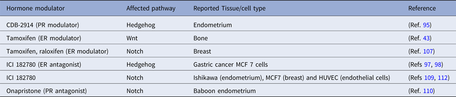
Oestrogen and progesterone modulators have shown to affect stem cell signalling pathways such as Wnt, Hh and notch (Table 1) in a spatiotemporal manner. Hence, an inappropriate clinical usage of these modulators can cause severe adverse effects by mainly perturbing the adult stem cell populations of various organs. The time of exposure and dosage of the modulators should be determined appropriately to curtail the untoward effects. Extensive studies are mandated to understand the modulation of stem cell properties by the hormone modulators.
List of hormone modulators influencing stem cell signalling pathways

ER, oestrogen receptor; PR, progesterone receptor.
Oestrogen, progesterone and stem cells in endometriosis
Hormonally regulated diseases are common in endometrium owing to its dependency on hormonal signal for regeneration and differentiation. Since the EnSCs are indispensable for the normal functioning and architecture of endometrium, abnormality in these cells could probably lead to disease conditions. Endometriosis, the growth of hormone-responsive endometrium outside the uterus, is a commonly encountered endocrine disease in women of reproductive age. Disturbance in the hormonal equilibrium, that is, elevated oestrogen level and progesterone resistance are associated with this disease (Refs Reference Al-Sabbagh28, Reference Bulun113). In clinics, hormone therapy and surgical management are the key therapeutic interventions for endometriosis (Ref. Reference Macer and Taylor114).
Although the aetiology of endometriosis is enigmatic, EnSCs are being proposed to enact a crucial role in the disease development (Refs Reference Figueira6, Reference Yang and Huang19, Reference Hufnagel20). It is proposed that the MSCs of endometrium, which reside at the perivascular sites, enter the peritoneal cavity by retrograde menstruation or lymphovascular dissemination to develop ectopic implants (Ref. Reference Figueira6). Data supporting the hypothesis that stem cells migrate to ectopic sites and differentiate into endometrial glands and stroma there, are accumulating which are reviewed in (Refs Reference Gargett24, Reference Forte115). In addition to EnSCs, bone marrow-derived stem cells (BMdSCs) are also recruited to the lesion site and trans-differentiate into endometrial cells under the influence of ectopic endometrial niche (Refs Reference Du and Taylor116, Reference Pluchino and Taylor117). Moreover, bone marrow-derived circulating EPCs are recruited to the endometriosis lesion site and help in the vasculogenesis leading to disease progression (Ref. Reference Laschke118). Studies on the expression pattern of stemness-related genes have shown elevated expression and/or increased number of cells expressing these genes in endometrium (both ectopic and eutopic) of women with endometriosis compared with normal (Refs Reference Proestlingm119, Reference Götte120, Reference Forte121). Various functional assays for stem cells also confirmed the presence of increased number coupled with increased migratory and invasive potential of EnSCs in ectopic and eutopic endometrium of women with endometriosis (Ref. Reference Kao122). The EnSCs from eutopic endometrium of endometriosis patients differ from their normal counterpart where decreased differentiation potential, increased proliferative capacity, self-renewal capacity, migratory and invasive potential have been observed (Refs Reference Santamaria123, Reference Djokovic and Calhaz-Jorge124). Study on the characteristics of eutopic and ectopic endometrial MSCs from the same endometrial donor showed greater potential for proliferation, cell migration and invasion and vasculogenesis for ectopic endometrial MSCs than eutopic endometrial MSCs (Ref. Reference Kao122). A recent study has reported differential gene expression profile of MSCs of eutopic endometrium of endometriosis patients versus normal (Ref. Reference Barragan125). The differentially expressed genes are associated with several functions like colony formation, cell death, cell survival and differentiation into endometrial stromal fibroblasts (eSFs). The eSFs inherit progesterone resistance from endometrial MSCs, disabling it from responding to progesterone and impairing the stromal decidualisation capacity leading to infertility. eSFs acquire a proinflammatory phenotype from the endometrial niche. In addition to the genetic differences in the EnSCs of diseased, which can be inherited by their progenies, the niche signalling also depicts heterogenity across patients, which is critical for regulating the stem cells. It is reported that the ESCs can be differentiated into endometrium-like cells when co-cultured with endometrial stromal cells reflecting the significance and strength of niche signalling in development of endometrial cells from stem cells (Ref. Reference Yu126).
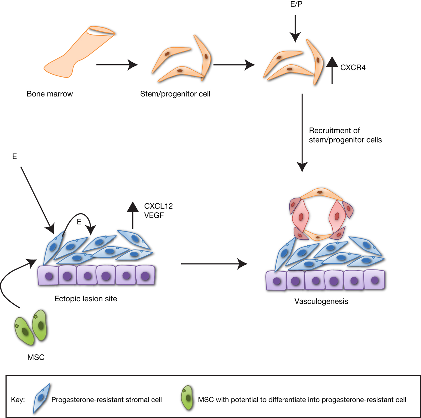
Response of EnSCs to oestrogen and progesterone differs in endometriosis patients when compared with normal. For example, the expression of EnSCs marker, Musashi1 (MSI1) in the ectopic endometrium does not correlate with the oestrogen and progesterone levels unlike observed in normal endometrium (Ref. Reference Chen127). It is shown that the recruitment of stem cells to the ectopic lesions involves oestrogen signalling (Figure 2) which is mediated through ER. Bazedoxifene (ER modulator) when administered with conjugated oestrogens demonstrates this by reduction in the stem cell recruitment to the endometriosis lesions, thus causing regression of lesions (Ref. Reference Sakr128). The physiological levels of oestrogen and progesterone also enhance the migration of BMdSCs to endometrium by increasing the expression of CXCL12 (C–X–C motif chemokine 12) in endometrial stromal cells and CXCR4 (C–X–C chemokine receptor type 4) in BMdSCs enabling chemoattraction of BMdSCs to the ectopic lesion site. The recruitment of circulating BMdSCs to the ectopic lesion in endometriosis might be induced by local oestrogen synthesised in these lesions (Ref. Reference Wang129). Oestrogen has been shown to enhance proliferation and migration of BMdSCs in an ER-dependent manner and increase the VEGF level in stromal cells of ectopic lesions leading to vascularisation. Progesterone, in turn, reduce microvessel density by down-regulating VEGF A. Hypoestrogenism given as therapy attenuate the growth of the lesion by inhibiting migration of BMdSCs to lesion site and making it available for recruitment to uterus (Ref. Reference Ersoy130). Another study (Ref. Reference Rudzitis-Auth131) has also shown that oestrogen induces more EPCs to incorporate into endometriotic lesion vasculature but does not increase the number of circulating EPCs. The uterine natural killer cell progenitors, which are significant for the inflammatory response in endometriosis, fail to develop normally as stem cell factor (SCF) essential for this development is diminished in endometriosis patients. Oestrogen and progesterone treatments fail to enhance SCF production in patients unlike in fertile women (Ref. Reference Thiruchelvam132) possibly owing to progesterone resistance in stromal cells of patients. Altogether, oestrogen deprivation as a long-term treatment regimen in endometriosis has superiority over progestin therapy due to the formers ability to reduce stem cell recruitment to lesions (Ref. Reference Ersoy130).
Recruitment of bone marrow-derived stem cells (BmdSCs) to ectopic endometriotic lesions is induced by oestrogen and progesterone. Oestrogen and progesterone (E/P) induce the expression of CXCR4 in BmdSCs. In endometriosis patients, stromal cells inherit progesterone resistance from their precursor mesenchymal stem cells (MSC) and create a high oestrogen environment at lesion site owing to local oestrogen synthesis. Oestrogen increases the expression of VEGF and CXCL12 in the stromal cells enabling chemo-attraction of BmdSCs for vasculogenesis to the lesion site, thus impairing their recruitment to uterine endometrium.

De-railed hormonal and stem cell signalling in endometriosis
The ability of niche cells to control the fate of EnSCs depends on the major signalling pathways involving Wnt, Notch, Hh and oestrogen–progesterone signalling. This niche signalling is deregulated in the disease condition.
Wnt signalling
The Wnt signalling, crucial for the stromal–epithelial cross-talk in the endometrium (Ref. Reference Zhang133) is aberrantly activated in endometriosis (Ref. Reference Santamaria123). PKF 115-854, an inhibitor of Wnt/β-catenin pathway is found to inhibit proliferation, migration and invasion of both epithelial and stromal cells obtained from endometriotic endometrium of patients (Ref. Reference Matsuzaki and Darcha134) suggesting the significance of Wnt pathway in disease progression. In endometriosis patients, WNT2, WNT7A (Ref. Reference Gaetje135) and β-catenin are highly expressed in ectopic compared with eutopic endometrium (Ref. Reference Xiong136). Musashi1 mediated activation of Wnt/β-catenin pathway and the positive correlation between EnSCs and β-catenin observed in endometriosis (Ref. Reference Yu137) is indicative of the involvement of this pathway in EnSCs of endometriosis. The aberrant activation of Wnt/β-catenin signalling enhances cell proliferation, migration and invasion in endometriosis, have been reviewed in recent articles (Refs Reference Matsuzaki138, Reference Kiewisz139).
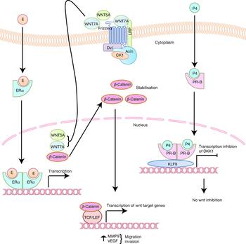
The elevated expression of Wnt proteins in the endometriotic tissues (Ref. Reference Gaetje135) might be because of reduced progesterone signalling as the Wnt inhibitor, DKK1 is found to be downregulated in endometriosis (Ref. Reference Brueggmann140). The hormone dependency of each component of Wnt signalling in endometriosis need to be investigated to get a clear picture of the molecular events leading to alteration of this pathway in endometriosis. Recent studies (Refs Reference Brueggmann140, Reference Brueggmann141, Reference Matsuzaki142) have initiated analyzing the expression pattern of Wnt pathway genes in a menstrual cycle phase-specific manner which might provide indirect evidence for hormone dependency of Wnt genes in endometrium. WNT7A and WNT5A are shown to have a cycle dependent expression (Ref. Reference Brueggmann140) whereas WNT4 is down-regulated in both eutopic and ectopic endometrium of endometriosis patients independent of cycle phase (Ref. Reference Liang143). Studies to elucidate the importance of KLF9 (Kruppel like factor 9), a PGR interacting protein, in endometriosis has revealed the PGR-KLF9 regulation of Wnt signalling (Ref. Reference Pabona144). KLF9 and PGR co-regulate DKK1 by co-recruiting to DKK1 promoter to down regulate the gene. In eutopic endometrium of endometriosis patients, DKK1 is found to be down-regulated along with PGR-B, KLF9 and WNT2 (Ref. Reference Pabona144). Wnt signalling in endometriosis endometrium is activated by oestrogen as it binds to the ERE of β-catenin promoter and induces its transcription (Ref. Reference Zhang145). The activation of Wnt/β-catenin signalling by oestrogen induces high expression of MMP9 resulting in enhanced migration and invasion of endometrial cells and promotes VEGF gene expression (Figure 3) aiding in the pathogenesis of endometriosis (Ref. Reference Zhang145).
Oestrogen-progesterone regulation of Wnt signalling in endometriosis. Oestrogen (E) induces expression of Wnt ligands, WNT5A and WNT7A as well as Wnt effector, β-catenin, in ERα-dependent manner. Progesterone (P4) binds to progesterone receptor-B (PR-B) and recruits transcriptional co-activator, KLF9 to the promoter of Wnt inhibitor, DKK1 and down regulate the gene. Binding of Wnt ligands to the receptor (Frizzled) activates the Wnt signalling and accumulation of β-catenin in the nucleus resulting in transcription of Wnt target genes like MMP9 and VEGF, associated with migration and invasion. P4-mediated inhibition of Wnt signalling which is present in the normal endometrium, is impaired in endometriosis.

Hh signalling
Hh signalling activation indirectly leads to EMT (epithelial mesenchymal transition), which is important for the development and progression of endometriosis, (Ref. Reference Albertsen and Ward146) through activation of FGF (fibroblast growth factor) as well as cross-talk with notch and TGF-β signalling cascades (Ref. Reference Katoh and Katoh147). It is important to note here that despite oestrogen and progesterone signalling impairment owing to altered expression of PGR, ESR1 and their target genes, endometriotic lesion development do not appear until the Hh pathway is altered (Ref. Reference Heard148). The endometrial expression of IHH is reduced in endometriosis (Ref. Reference Smith149) which can be attributed to the tissue resistance to progesterone (Ref. Reference Burney150) as the down-regulation is more evident in the secretory phase of patients in comparison with normal (Ref. Reference Smith149). Nr2f2, which is a downstream target of Ihh, is also down-regulated in endometriosis (Ref. Reference Lin151) and this might enhance oestrogen-induced proliferation in endometrium. However, the endeavour to understand the significance of KLF9, which is a PGR co-regulator (Ref. Reference Zhang152) and a regulator of ERα signalling (Ref. Reference Pabona153), in the pathophysiology of endometriosis using Klf9 null mice has shown that Shh-mediated Hh signalling is activated in the endometriotic lesions causing more proliferation and less apoptosis. Deregulation of oestrogen and progesterone signalling is also observed in the mice, however, the cause–effect relationship cannot be concluded from the study (Ref. Reference Heard154). Further studies are needed to understand the Hh signalling alterations in endometriosis like (1) Hh pathway regulation by KLF9, whether it is dependent or independent of oestrogen and progesterone (2) status of Ihh targets like Nr2f2 and its influence on oestrogen signalling, etc. In the Klf9 null mice Shh, Gli1, Smo and Stil are up-regulated resulting in Hh pathway activation (Ref. Reference Heard154) while in SF1 (Steroidogenic Factor 1) overexpressed mice model of endometriosis Ihh, Gli1, Gli2 and Ptch1 are down-regulated suppressing Hh signalling (Ref. Reference Vasquez155) suggesting existence of multiple regulatory axis for Hh signalling in endometriosis. The significance of Hh signalling in ectopic lesion development in endometriosis needs further in depth investigations to better comprehend its pathophysiology.
Notch signalling
NOTCH1 and NOTCH2 mutations are seen in ectopic endometriotic lesions and atypical endometriosis and these mutations are proposed to be critical for predisposing the endometriotic lesions to malignancy (Ref. Reference Er156). In endometriosis, NOTCH1 is also found to co-localise with MSI1 in endometrium (Ref. Reference Götte120) suggesting that the notch pathway alteration might influence the EnSCs. A Recent study (Ref. Reference He157) has shown that down-regulation of NOTCH1 decreases the in vitro clonogenicity and migratory capacity of EnSCs isolated from eutopic endometrium of endometriosis patients. Their data also provided evidence for reduction of endometriosis lesion growth upon Notch1 down-regulation in an in vivo model. However, another study (Ref. Reference Su158) has shown that Notch signalling components, receptors-NOTCH1 and NOTCH4, ligands- JAG2 and DLL4 and target genes – HES5 and HEY1 are all down-regulated in eutopic endometrium of endometriosis patients indicating down-regulation of the pathway specifically in stromal cells, impairing decidualisation. This is carried out by decreasing expression of NOTCH1-regulated FOXO-1-responsive genes. Progesterone resistance in endometriosis is speculated to be a reason for decreased notch signalling as progesterone enhances cleavage of NOTCH1 (Ref. Reference Su158). A study using Klf9 null mice revealed that notch signalling is activated in the ectopic lesions with an up-regulation of Jag2 and increased expression of NICD in stromal cells. Though the PGR level is found to decrease and ESR2/ESR1 increase in the lesion, the study did not address the possibility of hormonal regulation of notch signalling in endometriosis (Ref. Reference Heard154). The aberrant activation of Notch1 could lead to hypermethylation of PGR in endometriosis (Refs Reference Mikhailik101, Reference Wu159). Available data pertaining to activation status and expression pattern of Notch signalling components is inconclusive as the human data suggest a down regulation while mouse data an up-regulation of pathway components. The lesion, menstrual cycle phase and cell type specific expression pattern analysis need to be performed to produce more accurate data in this aspect. The oestrogen and progesterone influence on notch signalling in the context of endometriosis require elaborate investigation as this pathway is critical in the malignant transformation of endometriotic lesions.
Conclusion and future perspective
In endometriosis, the EnSCs are dysfunctional which may lead to alterations in their response to the hormonal cues ultimately disrupting endometrial homeostasis by affecting cell proliferation and differentiation. In addition, the stem cell niche of ectopic endometrium differs from eutopic endometrium as it enhances migration of the circulating BMdSCs to the site of lesion where these cells assist in lesion development. The major therapeutic intervention in endometriosis is by the inhibition of hormonal signalling, which only acts as a deterrent to the progress of the disease by selective targeting of the niche signals. This may cause a symptom-free but not disease-free survival as the dysfunctional stem cells remain intact. Recurrence of the disease may be attributed to stem cells post-hypoestrogenism inducing therapy since these cells can survive a long period of oestrogen absence. In addition stem cells in eutopic endometrium of endometriosis patients are shown to differentiate into less hormone-responsive cells unlike their normal counterparts (Ref. Reference Barragan125). It has also been shown that the deregulation of hormonal signalling alone cannot initiate lesion development but alterations in the stem cell-associated pathways such as Wnt, Hh and notch are a prerequisite for it. However, available expression data of these pathway components across the endometrium are inconsistent owing to variation in source of endometriotic samples, menstrual cycle phase during sample collection and lesion type. Recently researchers have commenced to address this by performing the expression analysis using a cycle phase-lesion type-specific approach.
Mice have been extensively used for endometrial stem cell as well as endometriosis research because of the complexity associated with usage of human samples. Though we have tried to incorporate maximum data from the studies on humans, data derived from mice models remains indispensable for this review as the data explaining action of hormones on endometrial stem cells are mainly obtained from latter. There exist both similarities and differences between mice and humans in terms of endometrial homeostasis. Both species undergo growth and regression of endometrium in response to hormones and harbour stem cells in endometrium to support endometrial regeneration, reviewed in (Ref. Reference Gargett12). However, humans undergo menstrual cycle which lasts for 28 days while in mice estrous cycle lasts for 4–5 days. Menstruation that is, the shedding of functionalis layer and regeneration from basalis happens in humans while in mice re-absorption of endometrium occurs. This would result in difference in the growth kinetics of cells (Ref. Reference Kurita160) and mechanism of endometrial re-growth in both species (Ref. Reference Groothuis2). Hence extrapolation of data obtained from mice studies to humans should be done with caution and validation of these findings in humans is crucial.
A major hurdle in study of human endometrial stem cells is the inability to maintain and expand these cells in culture, as these cells often undergo differentiation forfeiting their stem cell status (Ref. Reference Barragan125). This has lead researchers to depend on model organisms mainly mice, but the extrapolation of research outcome in these model organisms to humans has its own limitations as discussed. The recent development of organoid culture from endometrial samples by Turco et al. (Ref. Reference Turco161) and Boretto et al. (Ref. Reference Boretto162) has made it possible to maintain and expand human endometrial stem cells in culture in a near physiological state. This, along with the identification of markers for endometrial stem cells (Refs Reference Masuda48, Reference Valentijn51, Reference Nguyen52) would accelerate endometrial stem cell research in humans. As these organoid cultures are shown to be responsive to oestrogen and progesterone, this would greatly help us to understand the hormonal influence on stem cells, mechanism of stem cell proliferation and stem cell signalling in human. In addition, development of organoid cultures from endometriosis and normal endometrium may help us to perform comparative analysis of stem cell function in diseased versus normal.
Various questions need to be answered before we could unravel the aetiology of this complex disease like (1) How the Wnt, Hh and Notch signalling differ in the EnSCs of endometriosis patients compared to normal counterparts and its contribution to disease development and progression? (2) How the inter-relationship between these pathways and oestrogen–progesterone signalling is altered in endometriosis? (3) Is there a dose-dependent effect of hormones on these pathways, as the local oestrogen level increases in ectopic endometrium? (4) What is the cause-effect relationship between EnSC dysfunction and hormonal imbalance in endometriosis? The major roadblock in this research area is the lack of conclusive markers for endometrial epithelial progenitor cells and because of which researchers employ different strategies for identification of the stem/progenitor cell population leading to inconsistency in the data generated. The proposal of having a common ancestor for both MSC and epithelial progenitor cells arising from VSEL still needs to be ascertained by extensive experimental validation. However, from the existing data, we suggest, that re-examining the hormonal signalling in normal as well as diseased endometrium with an exclusive focus on stem cells, will enable us to design novel combinatorial therapy, which can be more effective than routine therapeutic strategies currently being employed.
Acknowledgements
We thank Mr. Tapas Pradhan and Mr. Samu John, Rajiv Gandhi Centre for Biotechnology for their critical comments on the manuscript.
Conflict of interest
The authors declare that they have no conflict of interest and there was no funding source for this work.
Further reading, resources and contacts
Endometriosis Research Center is a nonprofit foundation to promote public awareness about endometriosis. The organisation is actively involved in independent and collaborative research initiatives on endometriosis and critical evaluation of research outcomes.
endometriosis.org is a global forum which provide resources including clinical and scientific research updates about endometriosis and self-help books for women suffering from the disease as well as support to women through national support organisations situated all over the world.
https://endometriosisnews.com/
This is a news and information site providing information on clinical trials in endometriosis research.
http://www.endometrialdatabase.com/edb/
Endometrial database consists of published scientific articles in the field of endometrial research including endometriosis.