Introduction
The belief that the emotional state of the pregnant woman affects the development of the fetus is ancient and found in all cultures (Ferreira, Reference Ferreira1965). Animal research shows that exposure to prenatal stress is related to lower fetal and birth weight of the offspring (Pinto & Shetty, Reference Pinto and Shetty1995; Lesage et al. Reference Lesage, Del-Favero, Leonhardt, Louvart, Maccari, Vieau and Darnaudery2004). In humans, maternal prenatal depression, anxiety and stress are associated with higher rates of spontaneous abortion and pre-eclampsia (Kurki et al. Reference Kurki, Hiilesmaa, Raitasalo, Mattila and Ylikorkala2000; Nakano et al. Reference Nakano, Oshima, Sugiura-Ogasawara, Aoki, Kitamura and Furukawa2004). Moreover, maternal psychological distress in pregnancy is related to an increased risk of preterm delivery (Hedegaard et al. Reference Hedegaard, Henriksen, Sabroe and Secher1993; Rondo et al. Reference Rondo, Ferreira, Nogueira, Ribeiro, Lobert and Artes2003; Mancuso et al. Reference Mancuso, Schetter, Rini, Roesch and Hobel2004). Earlier research investigating the relation between maternal psychological distress and lower birth weight was inconsistent. Although some studies reported that maternal psychological distress is negatively related to birth weight (Lou et al. Reference Lou, Hansen, Nordentoft, Pryds, Jensen, Nim and Hemmingsen1994; Rondo et al. Reference Rondo, Ferreira, Nogueira, Ribeiro, Lobert and Artes2003; Rahman et al. Reference Rahman, Bunn, Lovel and Creed2007), other studies observed no (independent) relation between maternal psychological distress and low birth weight (Nordentoft et al. Reference Nordentoft, Lou, Hansen, Nim, Pryds, Rubin and Hemmingsen1996; Andersson et al. Reference Andersson, Sundstrom-Poromaa, Wulff, Astrom and Bixo2004; Evans et al. Reference Evans, Heron, Patel and Wiles2007).
Previous studies have investigated the influences of maternal prenatal distress on birth outcomes, such as birth weight. Birth outcomes are only crude summary measures of intrauterine growth and cannot provide information on the growth of the fetal head, abdomen, femur and body across different time periods in pregnancy. Furthermore, individuals can reach the same birth weight by different fetal growth trajectories (Bloomfield et al. Reference Bloomfield, Oliver and Harding2006). Therefore, in the current population-based cohort study, we examined the effect of maternal distress during pregnancy not only on birth weight but also on repeatedly measured fetal growth parameters such as head and abdominal circumference and femur length in mid- and late pregnancy. Furthermore, we also studied the ratio of abdominal and head circumference, which assesses levels of symmetry of fetal growth and is an indicator of brain sparing. We hypothesized that maternal distress in pregnancy negatively affects fetal size and growth from mid-pregnancy onwards.
Method
Design
This study was embedded in the Generation R Study, a population-based cohort study from fetal life onwards in Rotterdam, The Netherlands. The Generation R Study has previously been described in detail (Jaddoe et al. Reference Jaddoe, Mackenbach, Moll, Steegers, Tiemeier, Verhulst, Witteman and Hofman2006). The cohort includes 9778 mothers and their children that were born between April 2002 and January 2006. Assessments in pregnant women consisted of physical examinations, fetal ultrasounds, biological samples and questionnaires.
The study has been approved by the Medical Ethics Committee of the Erasmus Medical Centre, Rotterdam (no. MEC 198.782/2001/31). Written informed consent was obtained from all pregnant women.
Population for analysis
Of the total cohort of 9778 mothers, 8880 (91%) were enrolled in pregnancy (Jaddoe et al. Reference Jaddoe, Mackenbach, Moll, Steegers, Tiemeier, Verhulst, Witteman and Hofman2006). In this study, 104 fetal deaths and 93 mothers with twin pregnancies were excluded because growth potentials of fetuses in multiple pregnancies are not comparable with those of fetuses in singleton pregnancies. For mothers with multiple pregnancies, data on their second (n=500) or third (n=8) pregnancy enrolled in the study were excluded to avoid effects of paired data. The remaining 8130 mothers were eligible. There were 45 losses to follow-up during pregnancy. In 22.2% (n=1806) of the eligible mothers, no information on any of the three types of maternal distress was available. For 11 mothers there were no data on fetal ultrasound. Of the remaining 6313 (77.7%) mothers, 5976 mothers (94.7%) had two ultrasound assessments in mid- and late pregnancy and 337 (5.3%) mothers attended only one ultrasound assessment.
Maternal psychological distress in pregnancy
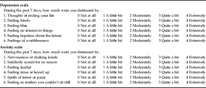
Information on maternal distress was obtained by postal questionnaires that were returned at, on average, 20.6 (s.d.=1.2) weeks of gestation. Anxious and depressive symptoms were assessed with the Brief Symptom Inventory (BSI), a validated self-report questionnaire with 53 items (De Beurs, Reference De Beurs2004). These items define a spectrum of psychiatric symptoms in the preceding 7 days. For this study, the six-item anxiety scale and the six-item depression scale were used (Table 1). Each item was rated on five-point uni-dimensional scales ranging from ‘0’ (not at all) to ‘4’ (extremely). Total scores for each scale were calculated by summing the item scores (range: 0–4) and dividing by the number of endorsed items. Following the BSI manual instructions (De Beurs, Reference De Beurs2004) we allowed one missing item per scale to minimize selective non-response. For depressive symptoms, 1.6% (n=100) of the participating mothers only filled in five of the six items. For anxious symptoms, one item was missing in 1.9% (n=120) of the mothers. The internal consistencies were α=0.80 for the depression scale and α=0.75 for the anxiety scale. Mothers scoring in the top 15% of the anxiety or depression scale scores of the BSI were considered to have anxious or depressive symptoms. The applied top 15% cut-offs were 0.50 for depressive symptoms and 0.66 for anxious symptoms, and lie within the range used to describe ‘above average’ scores, i.e. scores >0.33 and <0.67, on both the depression and the anxiety scale of the BSI in the Dutch norm population (De Beurs, Reference De Beurs2004). An earlier study used a very similar percentile cut-off to define increased antenatal anxiety using the Crown–Crisp Index (Birtchnell et al. Reference Birtchnell, Evans and Kennard1988; O'Connor et al. Reference O'Connor, Heron and Glover2002).
Listing of items included in the depression and anxiety scale of the Brief Symptom Inventory

Within a Generation R subgroup of 917 women, we tested the BSI's ability to identify clinical depression and anxiety using the applied cut-off scores. Data on clinical depression and anxiety during the last year were obtained with the Composite International Diagnostic Interview (CIDI). The CIDI is a structured interview based on DSM-IV criteria. Good reliability and validity have been reported (Andrews & Peters, Reference Andrews and Peters1998). A home interview was conducted during pregnancy by research assistants. The cut-offs for each scale had low positive predictive values for depressive (6.8%) and anxious (10.4%) disorders, but they were very good at assessing that a person is not depressed or anxious (negative predictive value=99.2% or negative predictive value=99.3%, respectively). However, if the prevalence is as low as in this subgroup, i.e. <2% for clinical depression and anxiety, the positive predictive value will not be close to 1 even if sensitivity and specificity are high. Inevitably most people with positive test results will be false positives (Altman & Bland, Reference Altman and Bland1994). Therefore, we also calculated the positive likelihood ratio (LR+) of the top 15% cut-offs for depressive (LR+=5.62) and anxious symptoms (LR+=9.68), which accounts for the prevalence. This demonstrated moderate quality of the cut-offs as indicators of certainty of diagnosis.
Family stress was assessed by the 7th subscale General Functioning (GF) of the Family Assessment Device (Byles et al. Reference Byles, Byrne, Boyle and Offord1988). GF is a validated 12-item measure of family health. The item scores were summed and divided by 12, yielding a total score from 1 to 4. We allowed 25% of the 12 GF items to be missing, which was the case in 4.2% (n=263) of the participating mothers in whom weighted sum-scores were calculated. A GF score >2.17 (cut-off) denotes unhealthy family functioning. In this study, just as in the Ontario Child Health Study, 10% of the families scored above this cut-off (Byles et al. Reference Byles, Byrne, Boyle and Offord1988). The internal consistency of GF was α=0.90.
Fetal ultrasound measurements and birth weight
Trained sonographers conducted fetal ultrasound examinations at the visits to the research centres in early (gestational age <18 weeks), mid-pregnancy (gestational age 18–25 weeks) and late pregnancy (gestational age ⩾25 weeks). These examinations were used for establishing gestational age and assessing fetal growth characteristics. Gestational age was established by the fetal ultrasound assessments since women do not remember the exact date of their last menstrual period or have irregular menstrual cycles (Altman & Chitty, Reference Altman and Chitty1997).
Online measurements included head and abdominal circumference, and femur length in mid- and late pregnancy that were all measured to the nearest millimetre using standardized techniques. The intra- and inter-observer reliability of fetal biometry in early pregnancy within the Generation R Study was high. Intra-observer intraclass correlation coefficients based on relative agreement varied from 0.982 to 0.995, inter-observer intraclass correlation coefficients varied from 0.982 to 0.988, with coefficients of variation between 2.2% and 5.9% (Verburg et al. Reference Verburg, Mulder, Hofman, Jaddoe, Witteman and Steegers2008a). The ratio of abdominal and head circumference, which was calculated by dividing abdominal circumference through head circumference, measures symmetry of fetal growth and indicates brain sparing. Estimated fetal weight was calculated using the formula by Hadlock et al. (Reference Hadlock, Harrist, Carpenter, Deter and Park1984) including head and abdominal circumference, and femur length. This formula by Hadlock et al. (Reference Hadlock, Harrist, Carpenter, Deter and Park1984) is frequently used in research and applied within Dutch medical practice. Before 18 weeks of gestation an accurate estimation of fetal weight cannot be achieved (Hadlock et al. Reference Hadlock, Harrist, Carpenter, Deter and Park1984). Gestational age-adjusted standard deviation scores of estimated fetal weight were constructed using reference growth curves from the total Generation R Study population (Verburg et al. Reference Verburg, Steegers, De Ridder, Snijders, Smith, Hofman, Moll, Jaddoe and Witteman2008b). Birth weight was obtained from medical records completed by midwives and gynaecologists.
Covariates
Information on maternal age, pre-pregnancy body mass index, educational level, ethnicity and parity (0, or ⩾1) was obtained by questionnaire at enrolment. Following the definition of Statistics Netherlands we divided education into five categories: primary education (no education, primary school), secondary education 1st phase (lower vocational training or ⩽3 years secondary school), secondary education 2nd phase (>3 years secondary school, intermediate vocational training), higher education 1st phase (higher vocational training) and higher education 2nd phase (university degree). Ethnicity of the mother was based on the country of birth of herself and her parents. Maternal height was measured during the first visit to the research centre. Information about maternal prenatal smoking and alcohol use was obtained by questionnaires in early, mid- and late pregnancy. Based on these questionnaires maternal smoking or alcohol use were categorized into ‘no’, ‘until pregnancy was known’ and ‘continued during pregnancy’ as described previously (Roza et al. Reference Roza, Verburg, Jaddoe, Hofman, Mackenbach, Steegers, Witteman, Verhulst and Tiemeier2007). Fetal sex and information on gestational diabetes, pre-eclampsia, and maternal hypertension during pregnancy were obtained from medical records.
Statistical analysis
To examine whether non-response was selective, we compared core data of pregnant women with information on psychological distress and fetal ultrasound assessments with eligible women not included because of missing data on one or the other assessment.
Multiple linear regression was used to examine the associations of maternal distress with absolute measures of fetal size in mid- and late pregnancy and birth weight. To investigate whether the wide range of gestational ages, in which fetal size was assessed, influenced our results we reran analysis using gestational age-adjusted standard deviation scores of estimated fetal weight as outcome measures. All models were controlled for maternal education and known determinants of fetal development, i.e. maternal height, age, body mass index, ethnicity, prenatal smoking, parity, gestational diabetes, pre-eclampsia, hypertension and fetal sex (Kramer, Reference Kramer1987). Models including absolute measures of fetal size were additionally controlled for gestational age. Furthermore, all analyses were also adjusted for maternal anxious symptoms or for family stress to determine whether a type of maternal distress was independently related to fetal size. To avoid collinearity and over-adjustment, maternal anxious and depressive symptoms were not included in the same model. Anxiety and depression as measured by the BSI were highly co-morbid (correlation: r=0.7, p<0.001). Maternal prenatal alcohol use did not significantly improve the models and was therefore not included in the analysis. On average, data were incomplete in 3.5% (range: 0.0–15.7%) of the confounders. To avoid the bias of a complete case analysis we accounted for missing information on confounders by using a missing dummy category for categorical variables or imputing the mean or median. The number of missing data per covariate is shown in Table 2. Using a categorical distinction of a top 15% cut-off for anxious and depressive symptoms and the established cut-off of a GF score >2.17 for family stress (Byles et al. Reference Byles, Byrne, Boyle and Offord1988), we established dichotomized main determinants that were used in our primary analyses.
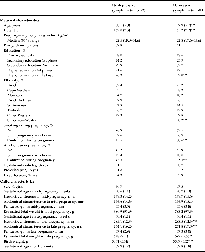
Maternal and child characteristics by level of depressive symptomsFootnote a

Values are given as mean (standard deviation) unless otherwise indicated.
a Independent t tests were used for continuous normal distributed variables, χ2 tests were used for categorical variables and Mann–Whitney U tests for continuous non-normal distributed variables. Data were missing on height (n=16), pre-pregnancy body mass index (n=989), parity (n=37), education (n=319), ethnicity (n=232), alcohol use during pregnancy (n=380), gestational diabetes (n=225), pre-eclampsia (n=225) and hypertension during pregnancy (n=224).
* p<0.05, ** p<0.01, *** p<0.001.
The associations of maternal distress with repeatedly measured parameters of fetal growth were analysed using longitudinal multilevel analysis to account for the dependency between measurements in the same subject. As fetal growth trajectories follow a non-linear pattern we used fractional polynomials of gestational age to model fetal growth. Fractional polynomials account for non-linearity and offer greater flexibility in curve shape than conventional polynomials (Royston & Altman, Reference Royston and Altman1994; Royston et al. Reference Royston, Ambler and Sauerbrei1999). We fitted an additive regression model to estimate fetal growth; the resulting variable was a sum of transformations of gestational age. A fractional polynomial of second-degree in a variable x (in this case gestational age) is a linear combination of power transformations of the form: β0+β1xp1+β2xp2. The transformation functions were chosen from first- or second-degree powers among from P=(−2, −1, −0.5, 0, 0.5, 1, 2, 3). The best-fitting model was chosen by comparing the deviance difference of the respective fractional polynomial regression model with the straight line model using approximate χ2 tests with significance level set at 0.1 (Royston et al. Reference Royston, Ambler and Sauerbrei1999). Random effects for both intercept and gestational age were included. Then, type of maternal distress was brought into the model as the main determinant. The interaction term of maternal distress with gestational age was included in the model to compare the slope of the curves between the different categories of affective symptoms and family stress. We tested whether this interaction term resulted in a significant improvement by comparing the −2 log likelihood of the model with the interaction term with the −2 log likelihood of the model without the interaction term. The following models were used:
Head circumference=β0+(β1×maternal distress)+(β2×gestational age)+(β3×gestational age2)+[β4×gestational age2×ln(gestational age)]+(β5×maternal distress×gestational age).
Ratio of abdominal and head circumference=β0+(β1×maternal distress)+(β2×gestational age)+[β3×ln(gestational age)]+(β4×gestational age−0.5)+(β5×maternal distress×gestational age).
Femur length=β0+(β1×maternal distress)+(β2×gestational age)+(β3×gestational age3)+(β4×maternal distress×gestational age).
Fetal weight gain=β0+(β1×maternal distress)+(β2×gestational age)+[β3×gestational age×ln(gestational age)]+(β4×maternal distress×gestational age).
The model of abdominal circumference was the same as that of head circumference. The model of fetal weight gain represents the increase in weight of the fetus from mid-pregnancy onwards and is based on estimated fetal weight in mid- and late pregnancy and birth weight. In these models, ‘β0+(β1×maternal distress)’ reflects the intercept and the terms including ‘βx×gestational age’ (or ‘βx×polynomials of gestational age’) reflect the slope of fetal growth per week. Terms including ‘βx×maternal distress×gestational age’ represent the differences in growth per week of the respective fetal body part (or in fetal weight gain) between the categories of maternal distress. Models were based on 11856 observations for head circumference, 11915 observations for abdominal circumference, 11570 observations for the ratio of abdominal and head circumference, 11925 observations for femur length and 18010 observations for fetal and birth weight. All models were controlled for potential confounders. Then, all models were additionally adjusted for maternal anxious symptoms or for family stress as well as for the respective interaction with gestational age to determine whether a type of maternal distress was independently related to fetal growth trajectories. SPSS for Windows version 15.0 (SPSS Inc., USA) and SAS version 9.1 (SAS Institute Inc., USA) including the Proc Mixed module for longitudinal multilevel analysis were used.
Non-response analysis
The non-response analysis showed that mothers included in the study were more likely to be Dutch [52.6% v. 30.6%, χ2(7)=327.9, p<0.001] and to be higher educated [higher education with a university degree 23.7% v. 13.3%, χ2(4)=266.3, p<0.001] than non-responders. Children of mothers in the study had a higher birth weight [3416 (s.d.=556) g v. 3343 (s.d.=581) g, t=4.83, p<0.001] and gestational age at birth [39.9 (s.d.=1.8) weeks v. 39.6 (s.d.=2.3) weeks, t=5.99, p<0.001].
Results
Table 2 presents maternal and child characteristics of mothers with and without depressive symptoms during pregnancy. Mothers reporting depressive symptoms were younger, less tall, had higher rates of education, were less often Dutch, and continued smoking during pregnancy more often than mothers not reporting depressive symptoms. Children of mothers with depressive symptoms during pregnancy had lower fetal weight in late pregnancy and lower birth weight (Table 2). Distributions for mothers who reported anxious symptoms (n=937) or family stress (n=625) and who did not report anxious symptoms (n=5376) or family stress (n=5688) were similar (data not shown).
Family stress in pregnancy was moderately correlated with both anxiety (r=0.3, p<0.001) and depression (r=0.4, p<0.001). Head and abdominal circumference, and femur length were all highly correlated in late pregnancy (r=0.6, p<0.001). Correlations between these ultrasound measurements in mid-pregnancy were similar (data not shown).
Table 3 shows that maternal distress was not related to estimated fetal weight in mid-pregnancy. In contrast, a crude analysis demonstrated that all types of maternal distress were negatively associated with fetal weight in late pregnancy (data not shown). However, only anxious symptoms were negatively linked to estimated fetal weight in late pregnancy after controlling for potential confounders (Table 3). Almost identical results were found when we used standard deviation scores of estimated fetal weight as outcome. Anxious symptoms were negatively related to standard deviation scores of estimated fetal weight in late pregnancy [β=−0.09, 95% confidence interval (CI) −0.17 to −0.02, p=0.013] but not in mid-pregnancy (β=−0.06, 95% CI −0.14 to 0.02, p=0.123). The other forms of maternal distress were not related to standard deviation scores of estimated fetal weight in mid- and late pregnancy after adjustment for potential confounders (data not shown). Similarly, all forms of maternal distress were negatively related to birth weight before adjustments were made (data not shown). After controlling for potential confounders, only anxious symptoms were associated with lower birth weight (Table 3).
Associations of maternal distress during pregnancy with fetal size in mid- and late pregnancy and size at birthFootnote a

Values are given as β coefficient (95% confidence interval).
a Adjusted for gestational age in mid- or late pregnancy or at birth, fetal sex, maternal age, height, body mass index, education, ethnicity, smoking during pregnancy, parity, gestational diabetes, hypertension in pregnancy, pre-eclampsia and for maternal anxious symptoms in pregnancy in the case of family stress or for family stress in the case of maternal anxious/depressive symptoms.
b β Coefficients represent the differences in fetal or birth weight between high levels of maternal distress and low levels of maternal distress.
* p<0.05.
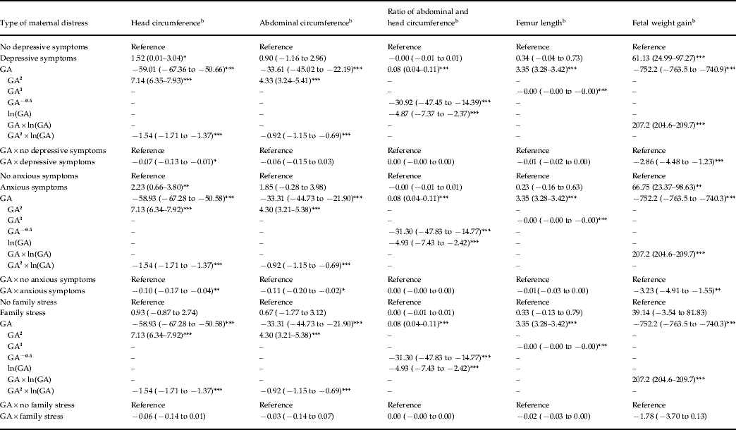
Table 4 presents the adjusted associations between maternal distress and repeatedly measured fetal growth characteristics. The effect estimates of the different forms of maternal distress, the respective slope based on fractional polynomials of gestational age and the interaction terms of maternal distress with gestational age are shown. The main effects of maternal distress on the fetal growth characteristics cannot be interpreted because the interaction effects were included in the models. Anxious symptoms were negatively associated with growth trajectories of the fetal head and abdomen and with fetal weight gain but not with growth patterns of the femur or asymmetric growth. Depressive symptoms had a negative association with fetal head growth and fetal weight gain but not with growth of the femur, abdomen or asymmetric growth. Family stress was not related to any parameter of fetal growth. However, when the association between family stress and the different fetal growth characteristics was not additionally adjusted for anxious symptoms but only for the other confounders we did find significant associations. Family stress was negatively linked to fetal head growth (β=−0.09 mm/week, 95% CI −0.16 to −0.01, p=0.024) and fetal weight gain (β=−2.53 g/week, 95% CI −4.41 to −0.13, p=0.009). To illustrate the non-linear pattern of the modelled fetal growth trajectories, Fig. 1 presents patterns of weight gain of fetuses of mothers with and without anxious symptoms during pregnancy.
Maternal anxious symptoms during pregnancy and fetal weight gain. Values are weight gain patterns of fetuses of mothers with ( ) and without (·······) anxious symptoms during pregnancy based on linear mixed models that were adjusted for gestational age, fetal sex, maternal age, height, body mass index, education, ethnicity, smoking during pregnancy, parity, gestational diabetes, hypertension in pregnancy and pre-eclampsia.

Associations of maternal depressive and anxious symptoms and family stress in pregnancy with fetal growthFootnote a

GA, Gestational age.
Values are given as β coefficient (95% confidence interval).
a Models were constructed using fractional polynomials for gestational age and adjusted for fetal sex, maternal age, height, body mass index, education, ethnicity, smoking during pregnancy, parity, gestational diabetes, hypertension in pregnancy and pre-eclampsia.
b β Coefficients are relative to the respective group of no maternal distress during pregnancy.
* p<0.05, ** p<0.01, *** p<0.001.
To place the magnitude of observed effects on the rate of fetal weight gain we also investigated the association of maternal prenatal smoking with fetal weight gain. Maternal smoking during pregnancy was linked to a 7.33 g (95% CI −8.84 to −5.82, p<0.001) lower fetal weight gain per week after control for potential confounders. In comparison with, for example, the negative effect of maternal anxious symptoms on fetal weight gain (i.e. β=−3.23 g/week, 95% CI −4.91 to −1.55, p=0.002), the negative effect of maternal smoking on fetal weight gain was 2.3 times higher.
Discussion
In this study we showed that affective symptoms during pregnancy were negatively associated with growth trajectories of, in particular, fetal head and abdominal circumference. Furthermore, children of mothers with anxious or depressive symptoms had reduced fetal weight gain during pregnancy. Only maternal anxious symptoms during pregnancy were related to lower birth weight.
So far, studies relating maternal psychological distress to lower birth weight have shown inconsistent findings. While some studies found no (independent) association between maternal psychological distress and low birth weight (Nordentoft et al. Reference Nordentoft, Lou, Hansen, Nim, Pryds, Rubin and Hemmingsen1996; Andersson et al. Reference Andersson, Sundstrom-Poromaa, Wulff, Astrom and Bixo2004; Evans et al. Reference Evans, Heron, Patel and Wiles2007), our results as regards maternal anxious symptoms in pregnancy are in line with the positive findings from earlier studies of birth weight (Lou et al. Reference Lou, Hansen, Nordentoft, Pryds, Jensen, Nim and Hemmingsen1994; Rondo et al. Reference Rondo, Ferreira, Nogueira, Ribeiro, Lobert and Artes2003; Rahman et al. Reference Rahman, Bunn, Lovel and Creed2007). However, birth weight is only a summative measure of a long, rapid and non-linear period of intrauterine growth. While undergoing fetal growth restriction due to environmental influences an individual fetus may still reach a normal birth weight because of his/her high genetic growth potential. Nevertheless, fetal growth restriction may affect fetal physiology and lifetime health (Hanson, Reference Hanson2002).
Only a single cross-sectional study reported an association between maternal psychological distress and fetal size in mid-pregnancy, indexed by fetal weight (Diego et al. Reference Diego, Jones, Field, Hernandez-Reif, Schanberg, Kuhn and Gonzalez-Garcia2006). This study was based on a small sample (n=98) with measurements in mid-pregnancy only and an incomplete control for confounders.
In our study, maternal distress was related to fetal size in late pregnancy and at birth but not to fetal size in mid-pregnancy, which suggests that influences of maternal distress on fetal growth are strongest in the last trimester of pregnancy. This is not surprising because fetal growth prior to 20 weeks is predominantly determined by genetic predisposition, whereas growth in the third trimester is more likely to be related to intrauterine environment. The finding may also reflect that effects of maternal distress on fetal growth are cumulative and easier to detect in the last trimester of pregnancy because of the increasing discriminative power of the measurements.
Maternal distress was associated with reduced fetal weight gain, and growth of the fetal head and abdomen but not with growth of the femur. Probably, maternal distress affects development of central organs more than that of distal body parts and bone structure.
Family stress was not related to fetal growth independently of maternal anxious symptoms. Arguably, we over-corrected our analyses by also adjusting for anxious symptoms. It is possible that aspects of family stress, such as lack of trust in family members, is one cause of anxious symptoms in pregnant women.
Our findings support the notion that maternal distress affects fetal head growth. As head circumference correlates with brain volume (Cooke et al. Reference Cooke, Lucas, Yudkin and Pryse-Davies1977), fetal head growth can be interpreted as an indicator of fetal brain development. Earlier studies reported a relation of maternal distress in pregnancy with childhood behavioural problems and poorer growth in infancy (O'Connor et al. Reference O'Connor, Heron and Glover2002; Rahman et al. Reference Rahman, Iqbal, Bunn, Lovel and Harrington2004). Moreover, previous research showed that intrauterine growth restriction indexed by birth length is associated with childhood behavioural problems and that head circumference at birth predicts cognitive functioning in childhood (Gale et al. Reference Gale, O'Callaghan, Bredow and Martyn2006; Wiles et al. Reference Wiles, Peters, Heron, Gunnell, Emond and Lewis2006). Possibly, fetal head growth is an intermediate in the relation of maternal psychological distress during pregnancy and subsequent child development.
Whereas our results showed that maternal distress is negatively related to several indicators of growth we observed no association with asymmetric fetal growth. This suggests that maternal distress during pregnancy leads to generally reduced fetal growth patterns but not to asymmetric growth restriction. Furthermore, these findings imply that the fetal brain is not spared when the fetus is exposed to maternal psychological distress.
Several mechanisms have been put forward to explain the association between maternal distress in pregnancy and fetal growth. Human and animal research suggests that maternal stress and distress during pregnancy leads to an elevated maternal hypothalamus–pituitary–adrenal (HPA) axis activity, which causes an increased release of glucocorticoids (Huizink et al. Reference Huizink, Mulder and Buitelaar2004; Mancuso et al. Reference Mancuso, Schetter, Rini, Roesch and Hobel2004; Diego et al. Reference Diego, Jones, Field, Hernandez-Reif, Schanberg, Kuhn and Gonzalez-Garcia2006), that in turn negatively affect fetal development (Mancuso et al. Reference Mancuso, Schetter, Rini, Roesch and Hobel2004; Diego et al. Reference Diego, Jones, Field, Hernandez-Reif, Schanberg, Kuhn and Gonzalez-Garcia2006). Maternal stress hormones may be transduced to the fetus by transplacental transport and by stress-induced release of placental hormones that enter the fetal circulation (Huizink et al. Reference Huizink, Mulder and Buitelaar2004). It was shown that maternal cortisol levels are strongly correlated with fetal levels, although fetal concentrations are lower compared with maternal concentrations (Gitau et al. Reference Gitau, Cameron, Fisk and Glover1998). Glucocorticoids are involved in fetal tissue proliferation and differentiation and are growth inhibiting (Fowden & Forhead, Reference Fowden and Forhead2004; Huizink et al. Reference Huizink, Mulder and Buitelaar2004). It is also possible that the association between maternal distress and fetal growth might be partly accounted for by a general reduced food intake of the mother or by a low intake of essential fatty acids or vitamins, such as folic acid or vitamin B12.
Our results might also be explained by an underlying common genetic factor affecting both maternal distress and fetal growth. Although we controlled for genetic effects on fetal growth by adjusting for maternal height and pre-pregnancy body mass index, residual genetic influences are likely.
The main strength of this large prospective population-based cohort study was that the repeated fetal ultrasound assessments were combined with information on birth weight, so that we were able to assess fetal growth from mid-pregnancy until birth. In addition, we controlled for many confounders known to affect fetal development.
Several potential limitations must be considered. As maternal psychological distress was only assessed at 20 weeks of pregnancy, we do not know whether maternal affective symptoms and family stress varied in intensity or were persistent throughout pregnancy. Second, the anxiety and depression scale of the BSI were strongly correlated. It seems plausible that these scales measure very similar concepts. This reflects the co-morbidity between anxiety and depression, which has frequently been reported (Beekman et al. Reference Beekman, de Beurs, van Balkom, Deeg, van Dyck and van Tilburg2000). We could not disentangle whether maternal anxiety and depression have independent effects on fetal growth, because of collinearity and possible over-adjustment in our analysis. Moreover, we were not able to control for antidepressant drug use during pregnancy. A recent population-based study (n=29005) of Dutch pregnant women showed, however, that only 1.8–2% took antidepressants at some point during pregnancy (Ververs et al. Reference Ververs, Kaasenbrood, Visser, Schobben, de Jong-van den Berg and Egberts2006). Our data also do not allow us to determine which physiological mechanisms may account for the findings of this study. As data on maternal distress were more complete in Dutch and higher-educated mothers whose children had a higher birth weight, we cannot rule out selection effects on fetal growth trajectories. Finally, while the size of the association between maternal psychological distress and fetal growth was small, such effects may be important in public health terms. The relations between maternal psychological distress and outcomes were evident within the normal range of maternal distress and fetal growth. Possibly the observed effects would have been larger if more individuals with higher rates of maternal distress and lower rates of fetal growth had been studied.
In conclusion, maternal psychological distress during pregnancy affects fetal development. Future research should address mechanisms underlying the relation between maternal psychological distress and fetal growth, e.g. dysregulation of the HPA axis, and long-term effects on child development. Furthermore, our findings highlight the importance of distress in pregnant women because this may affect the fetus. Information about distress can easily be obtained by questionnaires. Pregnant women at elevated risk could then be invited to participate, for example, in stress-reduction programmes.
Acknowledgements
The Generation R Study is conducted by the Erasmus Medical Centre (Rotterdam, The Netherlands) in close collaboration with the School of Law and Faculty of Social Sciences of the Erasmus University Rotterdam, the Municipal Health Service Rotterdam area, the Rotterdam Homecare Foundation and the Stichting Trombosedienst & Artsenlaboratorium Rijnmond (STAR), Rotterdam. We gratefully acknowledge the contribution of participating mothers and their children, general practitioners, hospitals, midwives and pharmacies in Rotterdam. The first phase of the Generation R Study was made possible by financial support from the Erasmus Medical Centre, Rotterdam, the Erasmus University Rotterdam and the Netherlands Organization for Health Research and Development (Zon Mw).
Declaration of Interest
None.