Anomalous systemic arterial supply to the normal basal segments of the lung without pulmonary sequestration, partial anomalous origin of a branch pulmonary artery from the descending aorta (aPADAO), is a rare congenital anomaly. Reference Bhalla, Gupta, Mukund, Kumar and Gupta1–Reference Gümüştaş, Akça, Ciftçi, Topçu and Akgül4 It can be considered a part of the broad spectrum of pulmonary sequestration and classified as Pryce type I sequestration. Reference Bhalla, Gupta, Mukund, Kumar and Gupta1–Reference Kim, Kim, Ha, Bae and Park5 However, the involved lung segments have a normal bronchial distribution, which could be distinguished from intralobar pulmonary sequestration. It is also important to differentiate aPADAO from other mimickers. Careful interpretation of chest computed tomography angiograph is important in the differential diagnosis and making a correct diagnosis. Reference Bhalla, Gupta, Mukund, Kumar and Gupta1,Reference Kim, Kim, Ha, Bae and Park5 Herein, we present two cases of anomalous systemic arterial supply to the pulmonary region, namely aPADAO and intralobar pulmonary sequestration, which were both successfully treated by transarterial embolisation using the Amplatzer Vascular Plug.
Case report
Patient 1
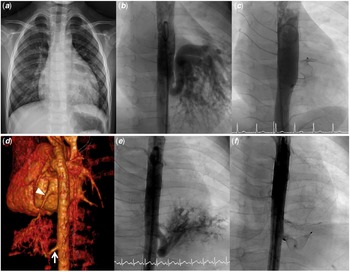
A 4-year-old boy, 15 kg and 105 cm, with a birth history of G2P1A1, gestational age of 38 weeks, and birth body weight of 3150 gm, was referred to our hospital for suboptimal weight gain and heart murmur. He had been admitted to a tertiary medical centre due to left lower lobe pneumonia 1 month before visiting our hospital. During hospitalisation, physical examination revealed normal breath sounds, regular heart beats, and a grade 3/6 systolic murmur over the left lower sternal border. The standard 12-lead electrocardiogram showed normal sinus rhythm and left ventricular hypertrophy. Chest X-ray revealed enlarged cardiac silhouette and retrocardiac opacity with increased vascularity over the left lower lung (Fig 1a). Transthoracic echocardiography showed normal left ventricular systolic function, cardiomegaly with left atrial and ventricular dilation, and abnormal vascularity located towards the posterior of the left atrium. Chest computed tomography angiography revealed a tortuous anomalous artery arising from the descending aorta and supplying the left lower pulmonary region. The pulmonary venous returns were normal with drainage to the left atrium. The pulmonary bronchial tree was connected normally and lung parenchyma was normal. Anomalous systemic arterial supply to normal basal segment of the left lower lung (aPADAO) was diagnosed. Cardiac catheterisation was performed under heavy sedation for transcatheter closure of this anomalous systemic arterial supply. Selective descending aortogram showed a feeding artery (internal diameter, approx. 0.7 cm; length, approx. 4 cm) from the descending aorta to the left lower lung (Fig 1b), and the levophase angiogram revealed normal pulmonary venous return to the left atrium. Selective left pulmonary arteriography showed no blood supply to the left lower lung. Then, transarterial embolisation was performed using a 12 mm Amplatzer Vascular Plug II over the feeding artery (Fig 1c). There were no complications after the embolisation, and he was discharged 2 days after the procedure. A follow-up chest computed tomography angiography showed no pulmonary infarction 6 months later.
Chest X-ray showed cardiomegaly and retrocardiac opacity ( a ). Aortogram revealed a feeding artery from the descending aorta to the left lower lung, and no residual shunt after transarterial embolisation ( b , c ). Chest computed tomography angiography showed intralobar pulmonary sequestration over the left lower lung with a feeding artery from the descending aorta (arrow) and venous drainage via the pulmonary vein to the left atrium (arrowhead) ( d ). Aortogram revealed a feeding artery from the descending aorta to the left lower lung, and no residual shunt after transarterial embolisation ( e , f ).

Patient 2
A 4-month-old female infant was referred by a paediatric surgeon because of suspected pulmonary sequestration prenatally. Chest computed tomography angiography after birth showed intralobar pulmonary sequestration over the left lower lobe of the lung (Fig 1d). Cardiac catheterisation was performed under heavy sedation for transcatheter closure of this anomalous systemic arterial supply. Selective descending aortogram showed a feeding artery (internal diameter, approx. 0.5 cm; length, approx. 1.7 cm) from the descending aorta to the left lower lung (Fig 1e), and the levophase angiogram revealed normal pulmonary venous return to the left atrium. Then, transcatheter embolisation was performed using an 8 mm Amplatzer Vascular Plug IV over the feeding artery (Fig 1f). There were no complications after the embolisation, and she was discharged the next day after the procedure.
Discussion
Anomalous systemic arterial supply to the normal basal segments of the lung without sequestration (aPADAO) is a rare congenital anomaly. Reference Bhalla, Gupta, Mukund, Kumar and Gupta1–Reference Gümüştaş, Akça, Ciftçi, Topçu and Akgül4 This anomaly is characterised as an anomalous systemic artery arising from the descending aorta supplying the basal segments of the lung which lacks a pulmonary arterial supply, the segments of the lung present a normal bronchial system distribution as well as normal pulmonary parenchyma, and the drainage and return of pulmonary veins are normal. Reference Singhi, Nicholson, Francis, Kumar and Hawker2–Reference Gümüştaş, Akça, Ciftçi, Topçu and Akgül4,Reference Wu, Yu, Zhang and Deng6 The cause of systemic arterial supply to the normal lung is unknown. Reference Gümüştaş, Akça, Ciftçi, Topçu and Akgül4,Reference Kim, Kim, Ha, Bae and Park5 It was previously classified as Pryce type I sequestration. Reference Bhalla, Gupta, Mukund, Kumar and Gupta1–Reference Kim, Kim, Ha, Bae and Park5 There is no consensus regarding the classification and definition of pulmonary sequestration. Because it has a normal bronchial connection which distinguishes it from classic bronchopulmonary sequestration, its classification in the broad spectrum of sequestration disorders remains controversial. Reference Singhi, Nicholson, Francis, Kumar and Hawker2 Despite some differences, it is probably closely related to intralobar pulmonary sequestration in many respects. Nowadays, most authors consider it to be a type of sequestration complex or a part of the broad spectrum of pulmonary sequestration. Reference Singhi, Nicholson, Francis, Kumar and Hawker2
In patients with aPADAO, basal segments especially in the left lower lung are most often involved and may be asymptomatic with a slight male predominance. Reference Bhalla, Gupta, Mukund, Kumar and Gupta1,Reference Singhi, Nicholson, Francis, Kumar and Hawker2,Reference Gümüştaş, Akça, Ciftçi, Topçu and Akgül4,Reference Kim, Kim, Ha, Bae and Park5 The signs and symptoms of this disease can be exertional dyspnoea, a lower thoracic murmur, recurrent infection, haemoptysis, and congestive heart failure due to left-sided cardiac overload. Reference Bhalla, Gupta, Mukund, Kumar and Gupta1–Reference Sugihara, Murata and Uchiyama3,Reference Kim, Kim, Ha, Bae and Park5 Mitsunori Higuchi et al. reported that the symptoms include haemoptysis (37.9%), abnormal shadow of chest X-ray (15.5%), heart murmur (10.3%), exertional dyspnoea (10.3%), and most commonly involve the left lung (91.4%). Of particular note, 81.3% of the reported cases were East Asian patients. Reference Higuchi, Chida, Muto, Fukuhara, Suzuki and Gotoh7 In case 1, left lower heart murmur, a retrocardiac opacity over the left lower lung on the chest X-ray was also noted. Therefore, an abnormal shadow on the chest X-ray could be a hint of an anomalous systemic arterial supply to the lung which may be misdiagnosed as pneumonia or other diseases. Caution is thus warranted when related clinical symptoms are encountered and a careful differential diagnosis should be performed.
It is important to differentiate anomalous systemic arterial supply to normal basal segment of the lung (aPADAO) from other mimickers, such as classic pulmonary sequestration, congenital descending aorta to pulmonary vein fistula, scimitar syndrome, and major aortopulmonary collateral artery, and so on. In this report, we present two cases of anomalous systemic arterial supply to the left lower lobe of the lung with normal pulmonary venous return. One was a patient with aPADAO who had normal basal segments of the left lower lung with normal bronchial connection and pulmonary parenchyma, and the other had intralobar pulmonary sequestration without a normal bronchial connection.
Pulmonary sequestration, which is divided into extralobar or intralobar, is characterised by abnormal lung tissue formation separate from the tracheobronchial tree. This abnormal tissue has systemic arterial supply from the aorta, but without a normal pulmonary artery supply, and can have multiple internal calcifications. Both intralobar and extralobar sequestrations tend to have arterial supply via an anomalous branch off the aorta. Intralobar pulmonary sequestration often has venous drainage via the pulmonary veins to the left atrium, while the extralobar pulmonary sequestration drains via systemic veins into the right atrium. Extralobar pulmonary sequestration is further differentiated from intralobar pulmonary sequestration by the presence of its own pleura. Congenital descending aorta to pulmonary vein fistula is characterised by a descending aorta to pulmonary vein fistula with enlarged pulmonary veins, normal pulmonary artery distribution, and no pulmonary parenchymal abnormalities to suggest sequestration. Scimitar syndrome falls under the category of congenital anomalous pulmonary venous return abnormalities. Scimitar syndrome is seen almost exclusively in the right lung and is characterised by enlarged abnormal pulmonary venous drainage into the systemic system, hypoplastic right lung, dextrocardia, hypoplasia of the right pulmonary artery, and abnormal systemic arterial supply. Reference Gümüştaş, Akça, Ciftçi, Topçu and Akgül4,Reference Kunwar, Paddalwar and Ghogare8 Isolated major aortopulmonary collateral artery is an anomalous blood vessel arising from the descending aorta and connects with pulmonary artery. It serves as an additive source of blood supply to the lung. There is no congenital heart disease, and pulmonary arteries as well as systemic and pulmonary venous drainage are normal. Isolated major aortopulmonary collateral artery regressed spontaneously in the majority of cases. Reference Kunwar, Paddalwar and Ghogare8
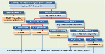
According to above description, we set up a diagnostic algorithm of anomalous systemic arterial supply to the pulmonary region (Fig 2). Once a case of anomalous systemic arterial supply to the pulmonary region is found, the first step is to check whether or not there is congenital heart disease and significantly decreased or absent pulmonary flow. If the pulmonary flow is significantly decreased or absent, diseases of pulmonary atresia or tetralogy of Fallot with major aortopulmonary collateral artery, systemic artery to pulmonary artery fistulas or scimitar syndrome, and so on, should be considered. If the pulmonary flow is normal or near normal, the second step is to check the pulmonary veins. If there is pulmonary venous dilatation with pulmonary vein and a descending aorta connection, a diagnosis of congenital descending aorta to pulmonary vein fistula is confirmed. If there is abnormal pulmonary venous return to systemic vein, extralobar pulmonary sequestration should be considered and could be confirmed by an abnormal bronchial connection. For step 3, if the pulmonary venous return is normal, the bronchial distribution should be checked. If the involved lung segments do not have a normal bronchial connection, the diagnosis of intralobar pulmonary sequestration is confirmed. Then, in step 4, if the pulmonary venous return and bronchial distribution are both normal, the involved segments of the lung with single or dual blood supply should be checked. aPADAO only has blood supply from the aorta to the involved segments. In contrast, isolated major aortopulmonary collateral artery has a dual blood supply from the aorta and pulmonary artery. Because chest computed tomography angiography can provide visual information about the condition of the bronchial system and pulmonary parenchyma, it can replace bronchography for accurate evaluation of the bronchial system and is the only diagnostic method that can provide evidence of an anomalous systemic artery, absence of pulmonary arterial supply, pulmonary venous return, and normal bronchial system, as well as normal pulmonary parenchyma simultaneously. Careful interpretation of chest computed tomography angiography is important in the differential diagnosis and for making a correct diagnosis. Reference Bhalla, Gupta, Mukund, Kumar and Gupta1,Reference Kim, Kim, Ha, Bae and Park5
The diagnostic algorithm of anomalous systemic arterial supply to the pulmonary region. AAOPA = anomalous origin of a branch pulmonary artery from the ascending aorta; aPADAO = anomalous systemic arterial supply to the normal basal segments of the lung; AO = aorta; cDAPVF = congenital descending aorta to pulmonary vein fistula; CHD = congenital heart disease; ELPS = extralobar pulmonary sequestration; ILPS = intralobar pulmonary sequestration; MAPCA = major aortopulmonary collateral artery; PA = pulmonary artery; PAVSD = pulmonary atresia with ventricular septal defect; PV = pulmonary vein; TOF = tetralogy of Fallot.

Because of its potential risk for haemoptysis, congestive heart failure, and recurrent infection, treatment is also recommended for asymptomatic patients. Reference Sugihara, Murata and Uchiyama3–Reference Kim, Kim, Ha, Bae and Park5 Previously, treatment for aPADAO was resection of the affected lung with ligation of the anomalous artery or lung-conserving surgery with re-implantation of the anomalous systemic artery to the pulmonary artery (unifocalisation). Reference Sugihara, Murata and Uchiyama3,Reference Kim, Kim, Ha, Bae and Park5 Furthermore, previous treatment for intralobar pulmonary sequestration was surgical ligation of the anomalous artery with lobectomy or segmentectomy. In particular cases of normal pulmonary artery with dual blood supply of lung tissue from both systemic and pulmonary artery, simple anomalous systemic artery ligation without lung resection would suffice. Reference Bhalla, Gupta, Mukund, Kumar and Gupta1 In recent years, interruption of flow through the anomalous artery (simple ligation or transarterial embolisation) without lung resection has been reported as an alternative method for aPADAO and intralobar pulmonary sequestration, and may bring about satisfactory results. Reference Sugihara, Murata and Uchiyama3–Reference Kim, Kim, Ha, Bae and Park5 Although simple ligation or transarterial embolisation could cause pulmonary infarction in cases of single blood supply solely from systemic artery, in most previous cases there were no severe complications because of the abundant collateral circulation from the bronchial, intercostal, and the other nearby arteries. Reference Sugihara, Murata and Uchiyama3 Therefore, according to recent reports, lung resection may be safely deferred when the diagnosis of aPADAO is well confirmed. Reference Singhi, Nicholson, Francis, Kumar and Hawker2–Reference Gümüştaş, Akça, Ciftçi, Topçu and Akgül4 Recently, transarterial embolisation with coil or vascular plug has been mainly attempted to reduce complications and risks of surgery as it is a minimally invasive technique. Reference Singhi, Nicholson, Francis, Kumar and Hawker2–Reference Kim, Kim, Ha, Bae and Park5 We used a vascular plug to occlude the anomalous systemic artery as in other reported cases, and no complications occurred after embolisation.
Conclusion
The clinical symptoms and signs, especially chest X-ray, should be evaluated carefully to avoid misdiagnosis of anomalous systemic arterial supply to the pulmonary region. A correct diagnosis can be achieved using the diagnostic algorithm and interpretation of chest computed tomography angiography step by step. Transarterial embolisation is a minimally invasive and apparently safe therapeutic option for reducing complications and risks of surgery.
Acknowledgements
None.
Financial support
This research received no specific grant from any funding agency, commercial or not-for-profit sectors.
Conflicts of interest
None.