Introduction
The fruticose lichens include some of the most complex vegetative structures built by micro-organisms. With physical attachment to their substratum limited to one or a few fixation points, these lichens are free to grow in three spatial dimensions as they build light-intercepting surfaces for their photosynthetic symbionts. Fruticose thalli may be ascending, bushy, or pendulous, with a great variety of morphologies, branching patterns and tissue types. Although their visibility and plant-like forms have attracted the attention of biologists, remarkably little is known about how such thalli grow. For all but two species it is unclear whether growth in length is distributed throughout the thallus or localized mainly in specific zones such as apical regions, as typically occurs in plants. Observations of anatomy and structure in a number of foliose and fruticose lichens have suggested a meristem-like role for thallus apices as active regions of morphogenesis, cell division, and tissue differentiation (Boissière Reference Boissière1979; Greenhalgh & Anglesea Reference Greenhalgh and Anglesea1979; Anglesea et al. Reference Anglesea, Greenhalgh and Veltkamp1983; Henssen & Dobelmann Reference Henssen and Dobelmann1987; Sanders Reference Sanders1993; Hill Reference Hill1994; Honegger Reference Honegger and Nash2008). Correlating these features with actual patterns of thallus growth, however, requires observations carried out in the field.
Several studies with foliose lichens have shown measurable growth to be restricted mainly to the apices or margins of lobes (Hale Reference Hale1970; Fisher & Proctor Reference Fisher and Proctor1978; Armstrong & Smith Reference Armstrong and Smith1998). Since foliose thalli are typically fixed to the substratum by rhizines, limitation of growth mainly to the free margins is not particularly surprising. Fruticose ‘beard’ lichens, however, have much less contact with their substratum. In the two species thus far investigated, growth appears to occur diffusely throughout the thallus, rather than just apically/marginally as observed in crustose and foliose thalli. The distinctive reticulate thallus of Ramalina menziesii Taylor arises from buds that form perforations and expand into nets (Cramer Reference Cramer1891; Lutz Reference Lutz1894; Peirce Reference Peirce1898; Larson Reference Larson1983). For each net, histogenesis of thallus tissue layers and formation of perforations are localized at the apical margin, while expansion of the reticulate tissue occurs diffusely throughout the thallus (Sanders Reference Sanders1989). In Usnea longissima Ach. the thallus consists of very long, cylindrical axes with short lateral branches. Growth measurement studies carried out in the field have shown that elongation occurs throughout thallus axes (Rolstad & Rolstad Reference Rolstad and Rolstad2008). Stratification of the symbionts into lichen tissues occurs at the apices of U. longissima; the cortex formed there matures and is eventually destroyed by diffuse expansion of the underlying medullary cord (Sanders & de los Ríos Reference Sanders and de los Ríos2012). Although they differ substantially in morphology, both of these ‘beard lichens’ are characterized by apical zones of histogenesis where the symbionts become organized into distinct tissues, while overall growth of these tissues continues diffusely throughout the thallus. Such non-localized expansion has often been referred to as ‘intercalary growth’, a term avoided here because it implies a defined zone of growth intercalated between regions where growth has ceased (for discussion of terminology, see Sanders & de los Ríos Reference Sanders and de los Ríos2012).
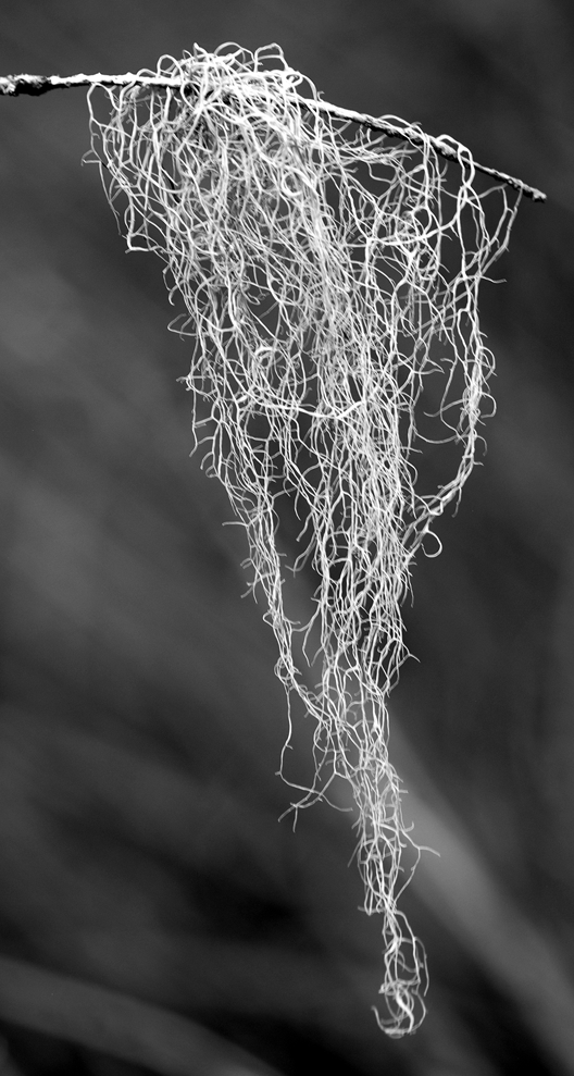
In theory, a structure growing over its entire length has the potential to increase exponentially over time, since the amount of tissue contributing to growth is itself ever-increasing. Amounts of elongation proportional to initial length were observed in thallus nets of Ramalina menziesii (Sanders Reference Sanders1989), and in segments of Usnea longissima (Rolstad & Rolstad Reference Rolstad and Rolstad2008; Jansson et al. Reference Jansson, Palmqvist and Esseen2009). The relatively high rates of elongation measured in these lichens (Herre Reference Herre1904; Keon & Muir Reference Keon and Muir2002) may also be reflected in substantial biomass accumulation rates (Boucher & Nash Reference Boucher and Nash1990), suggesting that the capacity for growth throughout mature tissue might have important implications for productivity. Furthermore, the expansion of mature, thick-walled fungal tissue raises questions about the anatomical events involved; in plants, the formation of thick secondary walls is usually incompatible with continued tissue expansion. It is therefore of interest to know whether diffuse growth is an exceptional feature of Ramalina menziesii and Usnea longissima, or whether it occurs commonly in fruticose beard lichens. Here we examine growth and branch formation in Ramalina usnea (L.) R. Howe (Fig. 1), a long, pendulous lichen that occurs widely in southern Florida, the Caribbean, and coastal Central and South America (Rundel Reference Rundel1978).
Ramalina usnea, small thallus on Taxodium branchlet.

Methods
Growth studies were carried out in a remnant of wetland at the southern edge of the FGCU campus in south-western Florida. Sixteen thalli of Ramalina usnea growing on Taxodium were chosen and their substratum branchlets broken off with the lichen attached. Thalli consisted of abundantly branched axes that were often tangled around their supports (the term axis is used here to refer to the morphological unit of thallus construction; there is no medullary cord, which is sometimes designated by this term in the genus Usnea). One to two segments of variable length on each thallus were chosen for measurement (26 in total), based mainly on the feasibility of attaching stable markers to the delicate thallus axes. Thus, broader and sturdier axis regions >0·5 mm wide were preferentially selected, presumably representing older thallus tissue that could not be traced back to its original apex. Marking and measuring was carried out with the aid of a dissecting microscope. Two spaced branching points along each selected axis were chosen to delimit a measurable segment of that axis. The segments were labelled by tying coloured thread at the chosen branch points, looping and knotting the thread around both main and lateral axes so that the thread would not slip from the branch position marked (Fig. 4A). Distance along the axis segment was measured from the points corresponding to the mid-width of the two marked side branches diverging from that axis. After measurement, thalli were returned to the field and the branches reattached near to their original position using nylon sewing monofilament. Marking and initial measurement of thalli were carried out on 13 and 21 May 2013; those recovered were re-measured on 27 December 2013 and again on 8 May 2014. The amounts of elongation were recorded and divided by the respective number of days to facilitate comparison between differing interval lengths. All measurements were made to the nearest millimetre by both authors independently and then compared to assess consistency. Most duplicate measurements differed by 0 or 1 mm; in very few cases they differed by 2 mm. Where different by 1 mm, the lower of the two values was used; where different by 2 mm, the mean was used. Average per-day increase in length was plotted against original length for each segment measured. Trend lines were generated (with y-intercepts=0) and Pearson product-moment correlation coefficients (r) were calculated using Microsoft Excel. A t-test for significance of linear correlation was performed with Bonferroni correction, using α=0·0033 and df=n − 2.
Hand-cut sections were made with fragments of razor blade held with a hemostat, and observed and photographed with an Olympus BX-51 compound microscope. Voucher specimens are deposited at UC (1965877).
Results
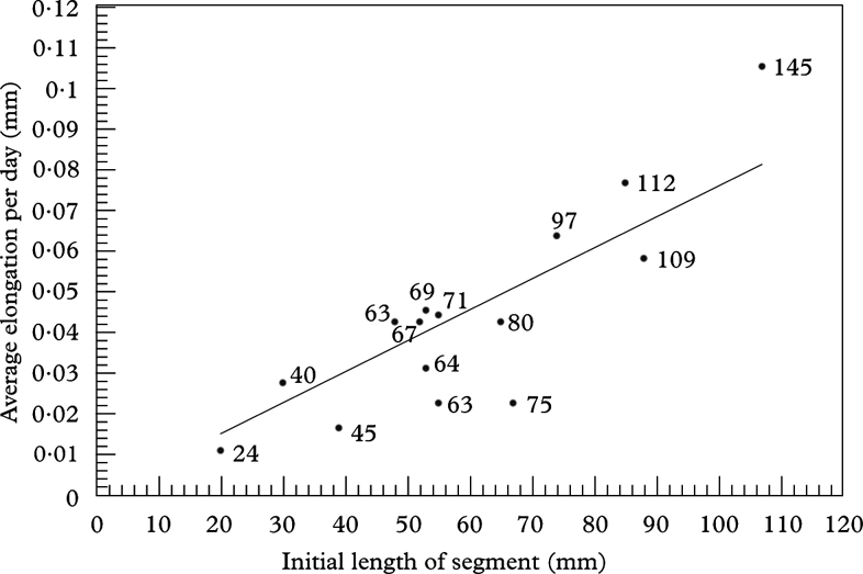
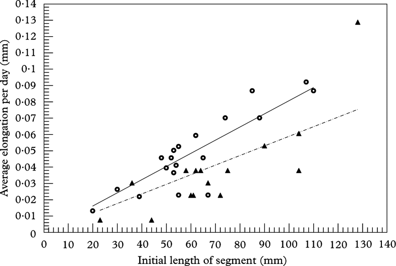
Nineteen of the 26 marked thallus segments were recovered and re-measured after seven months; fifteen of these persisted to the final measurement after approximately one year. All thalli recovered showed elongation of measured segments. Generally, the long segments grew much more than short ones; although there was a substantial degree of variability, the amount of length increase was roughly proportional to the initial length of the segment (Fig. 2). There was some indication of greater average growth rates during the summer-autumn interval (which includes the season of greatest rainfall in south-west Florida) compared to that of winter-spring (Fig. 3), although the reverse was observed in a few segments.
Plot of elongation rates as a function of initial length for the segments persisting through the full study period (May 2013 to May 2014). Growth is expressed on an average per-day basis to standardize for the slightly different starting dates (13 and 21 May 2013). The number beside each data point gives the final length of the segment in mm. t 15=5·379; P<0·005.

Relationships between average segment elongation rates per day and initial lengths for the two measurement intervals, May 2013 to 27 December 2013 (circles; solid line) and 27 December 2013 to 8 May 2014 (triangles; broken line). t 17, interval 1=7·337; t 15, interval 2=4·416; P<0·005.

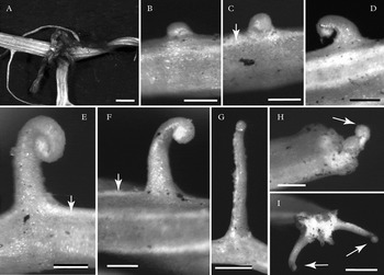
New thallus axes originated as branch primordia that first appeared as minute convex swellings on the edges of parent axes (Fig. 4B), most commonly along the longitudinal white striations corresponding to discontinuities (pseudocyphellae) in the cortex (Fig. 4C, E–F). With further expansion, one edge of the convex dome separated from the surface to reveal an inrolled structure, which was maintained as the branch elongated (Fig. 4C–G). Branch primordia also formed commonly along broken ends of thallus axes (Fig. 4H & I). Examination of numerous apices showed no indication of branching by means of furcation or division of the apex itself.
Formation of new branch axes in Ramalina usnea. A, branch point labelled with knotted thread; B, emerging bud primordium, side view; C & D, bud with inrolled margin emergent, front view and side view, respectively. Note origin from linear pseudocyphella (arrow); E–G, expanding buds maintaining inrolled apical margin. Arrows, linear pseudocyphellae; H & I, new buds (arrows) formed on broken surface of damaged tissue. Scales: A=1 mm; B & G=0·2 mm; C, D–F, H=0·1 mm; I=0·5 mm.

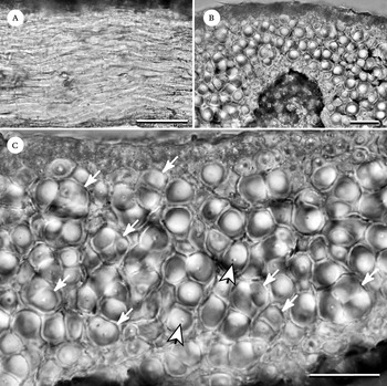
The thallus possessed a cortex of densely packed, elongate hypha-like cells oriented longitudinally in parallel with the thallus axis (Fig. 5A). The cortex was of irregular thickness, with thinner areas where the algal layer and medullar space penetrate upward (Fig. 5B). Cortical cell walls were extremely thick, occupying considerably more volume than the cell lumen (Fig. 5C). Cells in transverse section appeared to be grouped into bundles (Fig. 5C), although optical sectioning showed that along their length, cells pass into and out of such groupings with neighbouring cells.
Hand-cut sections of thallus of Ramalina usnea. A, longitudinal section showing hypha-like fungal cells in longitudinal orientation; B, transverse section of cortex showing irregular thickness, with algal cells (a) at perimeter of medullary region extending upward; C, transverse section detail showing thick-walled cells of cortex seemingly grouped in bundles (arrows) in this plane of section. Note relatively tiny proportion of cross-sectional area occupied by cell lumen (arrowheads). Scales: A=50 µm; B & C=20 µm.

Discussion
Since thallus segments chosen for study were broader portions of axes several times wider and thicker than apical regions, the growth observed represents non-apical elongation of axis regions many centimetres distant from any original apex. Because of frequent fragmentation and new branch formation, the original apices of the measured regions would not be identifiable even in the unlikely event that they were still present on the thallus. We were unable to mark and measure the fine young branches with intact apices and therefore cannot compare their contribution to axis growth with that of the broader tissue segments measured here. However, observations of branch formation (Fig. 4B–G) leave no doubt that growth occurs in apical and subapical regions; indeed, the much greater abundance of fine axes produced by new apices implies that their contribution to growth is no less significant than that documented in the broader thallus regions. We therefore conclude from the rough proportionality of increase in length to the original length of the measured segments that elongation is not localized to any specific zone, but occurs instead throughout the thallus. Longer tissue segments will thereby have more tissue contributing to further growth. Growth is not necessarily distributed uniformly, however, and variation between comparable segments is likely to reflect fine-scale heterogeneities in the microenvironmental conditions that individual thalli and individual thallus branches experience.
In formation and growth of new thallus units, Ramalina usnea shows several broad similarities to the Californian lace lichen R. menziesii. In both lichens, new thallus units arise from buds that emerge from mature tissue, frequently along pseudocyphellae or breakage points. The bud is circinate (inrolled) at the apex, which results from more accelerated cortical development on the convex surface (Sanders Reference Sanders1989). Thallus components are therefore actually dorsiventral in their ontogeny, although this is evident only in reference to an intact apex. Mature, organized tissue continues to expand in both Ramalina species, as well as in Usnea longissima (Rolstad & Rolstad Reference Rolstad and Rolstad2008), giving overall expansion rates that are proportional to length. The extremely fine younger portions of Ramalina usnea break easily, with new branch primordia often organized at the broken ends (Fig. 4H & I), as seen in the fine, coastal forms of Ramalina menziesii. A thallus axis may thereby be built from several branch buds arising in tandem after repeated breaks, forming a kind of sympodium (Sanders Reference Sanders1992). Ramalina usnea may closely resemble coastal forms of R. menziesii, whose fine nets often break apart and give the impression of branches; misidentified specimens of those forms appear to be the basis for reports extending the range of R. usnea to California (e.g., Hale Reference Hale1979; Brodo et al. Reference Brodo, Sharnoff and Sharnoff2001), where this taxon does not actually occur (Rundel Reference Rundel1978; Kashiwadani & Nash Reference Kashiwadani, Nash, Nash, Ryan, Diederich, Gries and Bungartz2004).
But Ramalina usnea clearly differs morphologically from R. menziesii in that its branch units do not develop into nets. Although some sporadic reticulations may occasionally be observed in R. usnea, there is no indication of any programmed perforation process at the apex like that characteristic of R. menziesii; this feature can always be used to distinguish the latter from the former. There was also no evidence, at least in the population examined, that the apices of R. usnea ever divide or furcate as commonly occurs in R. menziesii (Sanders Reference Sanders1989). The much narrower apices of Ramalina usnea (<0·10 mm) do not appear to undergo the widening that furcation typically accompanies. Rundel (Reference Rundel1978), however, described the thallus of R. usnea as dichotomously branched. This may have been meant to refer merely to the overall appearance of the branches, as parent axis and branch are often deformed to make a Y-shaped junction. It is also conceivable that some of the more broadly laciniate populations included in the material Rundel (Reference Rundel1978) studied do in fact have apical branching not seen in south-west Florida material. Where two new branches form at the wound surface of a broken thallus axis, a misleading appearance of dichotomy may also be created (Fig. 4I).
Mature cortical tissue provides the structural framework in Ramalina; its ability to continue growth, at least in R. usnea and R. menziesii, raises questions about how this is accomplished at the cellular level. Because the cells are oriented longitudinally, their massively thickened walls must allow for extension throughout their length; hypha-like tip growth cannot account for the expansion of the cortex as a whole. Their secondarily thickened walls must therefore function quite differently from those of plants, which generally do not permit further cell expansion. Previous ultrastructural study of the cortex in Ramalina menziesii revealed cell walls composed of distinct layers that accumulate and deform with age. Younger branch cells growing intrusively into the older wall layers of adjacent cells were recognizable in transverse section by their fewer wall layers and their interruption through the concentric arrangement of the older cell's wall layers. The outermost wall layers of an older cell containing additional cells growing through its wall material delimited the apparent bundle of cells visible with light microscopy (Sanders & Ascaso Reference Sanders and Ascaso1995). A similar arrangement was observed in the central cord tissue of Usnea longissima (Sanders & de los Ríos Reference Sanders and de los Ríos2012). Although the nature and functional properties of these wall layers remain to be explored, evidence of their continued production and accumulation over time has clear significance for cells that are continuously elongating. While Ramalina usnea has not been investigated ultrastructurally, the bundled appearance of the thick-walled cortical cells in transverse section (Fig. 5C) suggests a comparable pattern of ongoing wall layer deposition. Some other species of the genus also appear to have similar anatomical features (e.g., Kashiwadani Reference Kashiwadani1990). Though the fruticose lichens are plant-like in morphology and apical localization of histogenesis, their fungal tissues appear to grow in ways that are neither like plants nor like conventional fungal hyphae. These growth processes merit closer study at all levels of organization.
The manuscript benefitted from critical review by two anonymous referees. Dr Galen Papkov kindly provided guidance with the statistics.