Introduction
Assisted reproduction techniques (ART) are procedures usually accepted for the treatment of infertility. The success rate of in vitro fertilization (IVF) and embryo transfer depends on some factors including maternal age. In female reproduction, fertility begins to decline significantly after age 37 (Piette et al., Reference Piette, De Mouzon, Bachelot and Spira1990; Oktay et al., Reference Oktay, Turan, Bedoschi, Pacheco and Moy2015). This variation can be accompanied by a change in the oocyte microenvironment. Several reports have demonstrated that the follicular environment and follicular cell function are altered according to female age (Pacella et al., Reference Pacella, Zander-Fox, Armstrong and Lane2012). Human follicular fluid (HFF) forms the microenvironment for developing oocytes and has an important influence on embryo development (Palhares et al., Reference Palhares, Romão, Da Broi, Martins and Jordão2012). Its content includes granulosa cells and leukocytes and various mediators including growth factors, reactive oxygen species (ROS) and antioxidants (Attaran et al., Reference Attaran, Pasqualotto, Falcone, Goldberg, Miller, Agarwal and Sharma2000; Carbone et al., Reference Carbone, Tatone, Monache, Marci, Caserta, Colonna and Amicarelli2003). ROS are involved as second messengers that are capable of modulating the expression of genes that control physiological processes of gametes and embryos (Hensley et al., Reference Hensley, Robinson, Gabbita, Salsman and Floyd2000; Dröge, Reference Dröge2002) and they are suggested to be involved in the process of ovarian ageing (Agarwal et al., Reference Agarwal, Gupta and Sharma2005). However, the exact function of ROS in FF remains unknown (Oyawoye et al., Reference Oyawoye, Abdel Gadir, Garner, Constantinovici, Perrett and Hardiman2003; Pasqualotto et al., Reference Pasqualotto, Agarwal, Sharma, Izzo, Pinotti, Joshi and Rose2004). Despite their physiological condition, the peroxidative damage caused by ROS is associated with the process of ageing. Various reports have shown that oxidative stress (OS) is one of the major causes of age-related cellular and molecular damage (Harman, Reference Harman1984). Therefore, OS might be responsible for the reduced reproductive potential and oocyte–embryo quality associated with ageing (Liu et al., Reference Liu, Trimarchi and Keefe2000).
At the same time, FF has its antioxidant system that includes superoxide dismutase (SOD), catalase (CAT), glutathione (GSH), glutathione reductase (GR) (Carbone et al., Reference Carbone, Tatone, Monache, Marci, Caserta, Colonna and Amicarelli2003; Lambrinoudaki et al., Reference Lambrinoudaki, Augoulea, Christodoulakos, Economou, Kaparos, Kontoravdis and Creatsas2009). SOD reduces superoxide anion to hydrogen peroxide and molecular oxygen (Zelko et al., Reference Zelko, Mariani and Folz2002). The removal of hydrogen peroxide is catalyzed by CAT, which reduces hydrogen peroxide to water (Fridovich, Reference Fridovich1986). Therefore, the SOD and CAT serve, in tandem, as front-line antioxidant defences (Scandalios, Reference Scandalios2005). The relationship between ageing and antioxidant enzyme function continues to be a contentious area. Recently various studies have demonstrated that ROS scavenging efficiency in the follicular fluid (FF) undergoes a significant decrease during ageing as a result of changes in some antioxidant enzymatic activities (Carbone et al., Reference Carbone, Tatone, Monache, Marci, Caserta, Colonna and Amicarelli2003). This condition might be a result of changes in the microenvironment of the aged ovary. The aim of this work was to investigate the correlation between age and lipid peroxidation (MDA) and antioxidant enzyme activities and also determine the OS status in patients of advanced age undergoing in IVF.
Materials and methods
This was a prospective study, approved by Anfa Fertility Center and Physiopathology Molecular Genetics and Biotechnology Laboratory of University Hassan II, Casablanca (Morocco) informing the selected patients with consent, including 201 infertile women under IVF treatment whose age was between 24 and 45 years old, which included 111 patients with primary infertility and 90 patients with secondary infertility. This study excluded patients with polycystic ovary syndrome, with endometriosis and smokers. All patients were stimulated with an antagonist protocol using follicle stimulating hormone (FSH; Orgalutran 0.25 and Gonal-F). Further FSH administration (Gonal-F; Serono Laboratories, Saint Cloud, France) was started by daily subcutaneously injection (150–225 IU/day or ¼ 300 IU/day). The FSH dose was based on the woman’s age, antral follicle count (AFC) on days 2–3 of the cycles, and anti-Müllerian hormone (AMH) concentration that was maintained constant for 5 days and it was adjusted according to usual parameters of follicle growth determined using ultrasound monitoring. A potent, third-generation gonadotrophin-releasing hormone (GnRH) antagonist, Ganirelix (Orgalutran VR, MSD Schering-Plough, France) was injected subcutaneously once daily starting on day 6 of FSH administration. A subcutaneous injection of human chorionic gonadotrophin (HCG; Gonadotrophins Chorioniques Ovitrelle VR, Merck Serono) was performed when triggering criteria were reached ≥3 follicles of 17 mm.
Follicular fluid samples were obtained from mature follicles at the time of oocyte retrieval separated using a Ficoll-based protocol (3 ml), as described by Ferrero et al. (Reference Ferrero, Delgado-Rosas, Garcia-Pascual, Monterde, Zimmermann, Simón and Gómez2012). After centrifugation at 400 g for 30–40 min, three layers could be distinguished: a top layer containing the FF, a bottom layer containing erythrocytes and, in the middle, a ring-like layer containing the cumulus–granulosa cells. The only the top layer (purified FF) was collected, centrifuged at 500 g, filtered and kept at 20°C until use. The protein content was measured according to Bradford (Reference Bradford1976) using bovine serum albumin as standard.
Lipid peroxidation was estimated by thiobarbituric acid reactive substance (TBARS) reaction with malondialdehyde (MDA) as described by Samokyszyn and Marnett (Reference Samokyszyn and Marnett1990). Briefly, 100 µl of purified FF were added to 10% trichloroacetic (TCA) and 0.67% thiobarbituric acid (TBA). The MDA level was expressed as μmol/µg protein. The MDA level was expressed.
SOD activity was measured according to Paoletti et al. (Reference Paoletti, Aldinucci, Mocali and Caparrini1986). The reaction medium contained 5 mM EDTA, 2.5 mM MnCl, 0.27 mM NADH, 3.9 mM 2-mercaptoethanol, 50 mM phosphate buffer pH 7 and 50 μl of the FF. The enzyme activity was expressed as micromoles (μmol) of NADH consumed/min/µg of protein.
To estimate the GR activity, the method of Di Ilio et al. (Reference Di Ilio, Polidoro, Arduini, Muccini and Federici1983) was used. The reaction medium was composed of 1 mM EDTA, 25 μM GSSG, 1.5 μM nicotinamide adenine dinucleotide phosphate (NADPH), 50 m phosphate buffer pH 7.4 and 50 μl of FF and the absorbance was read at 340 nm after the addition of 1.6 μM of NADPH. Enzyme activity was expressed in millimoles (mmol) of NADPH consumed/min/µg of protein.
Catalase activity was measured according to Aebi (Reference Aebi1984). The reaction medium was composed of 50 mM phosphate buffer, pH 7 and 30 μl of FF. Specific activity was given as micromoles of consumed H2O2/min/µg of protein.
Glutathione (GSH) was determined according to the method of Weckbecker and Cory (Reference Weckbecker and Cory1988). This essay was based on the measurement of optical absorbance of 2-nitro-5-mercapturic acid. The latter resulted from the reduction of 5.5-dithio-bis-2-nitrobenzoic acid (Ellman’s reagent) by groups (–SH) in glutathione. The GSH concentration was given as nmol/µg of protein.
Statistical analysis
Results were expressed as the mean ± standard deviation (SD). We applied Pearson’s linear correlation coefficients to estimate correlations between different parameters, using Statistical Package for the Social Sciences (SPSS) software. Statistical significance was defined for P-values < 0.05. Receiver operating characteristic (ROC) curves were generated to determine age cutoff points that matched a higher MDA level with respect to the greatest sensitivity and specificity values.
Results
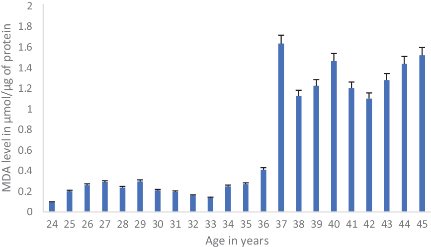
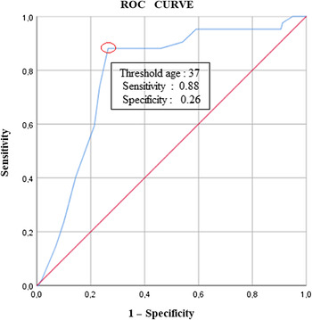
This study aimed to determine the threshold age for increasing the MDA (lipid peroxidation marker) level. The age cutoff point allowed us to divide our samples into two groups that aimed at correlation statistical analysis. Evolution data of MDA level in FF as a function of women’s age between 24 and 45 years are shown in Figure 1. We noted a very significant increase in the MDA level from the age of 37 years. The Youden’s index on the ROC curve with maximal sensitivity (0.88) and specificity (0.26) showed that the threshold age for higher MDA rates was 37 years (Figure 2). This age cutoff of 37 years old allowed us to differentiate the study into two age groups. Group I included patients whose age was less than 37 years, and group II included patients whose age was equal or greater than 37 years. As shown in Table 1, statistical analysis revealed a positive correlation between MDA and maternal age. Indeed, the MDA level was significantly higher in group II compared with group I (P < 0.0001). In the same way, we noted a positive correlation between GR, GSH and maternal age. The GR enzyme activity and GSH level were significantly higher in group II compared with group I (respectively, P < 0.04 and P < 0.0001). While the results indicated that maternal age was inversely correlated with SOD and CAT activities.
Evolution of the malondialdehyde (MDA) level in follicular fluid as function women’s ages between 24 and 45 years. Values are in means ± standard deviation (SD).

Receiver operating characteristic (ROC) curves of maternal age that show the threshold age for higher malondialdehyde rates. Note red line, reference line; blue line, patient age between 24 and 45 years.

Lipid peroxidation and antioxidant activities in follicular fluid from women undergoing IVF before or after 37 years of age

Discussion
In medically assisted procreation processes, a women’s age and embryo quality were the important factors leading to the establishment of pregnancy (IVF) (Wiener-Megnazi et al., Reference Wiener-Megnazi, Vardi, Lissak, Shnizer, Reznick, Ishai and Dirnfeld2004). Furthermore, female reproductive ageing assumes that the decline in both the quantity and quality of the oocyte/follicle determines an age-dependent loss of female fertility (Ottolenghi et al., Reference Ottolenghi, Uda, Hamatani, Crisponi, Garcia, Ko and Forabosco2004). Ovarian oxidative balance seems to be critical for oocyte quality in advanced maternal age patients (Pella et al., Reference Pella, Suárez-Cunza, Orihuela, Escudero, Pérez, García and Romero2020), although the primary cause of ovarian ageing involves the accumulation of damage by OS, at least in part, with an increasingly compromised microcirculation around the leading follicle (Friedman et al., Reference Friedman, Danforth, Herbosa-Encarnacion, Arbogast, Alak and Seifer1997; Van Blerkom et al., Reference Van Blerkom, Antczak and Schrader1997). Based on this assumption, that the most relevant idea is that age-related OS can cause infertility, it is important to know age when the state of OS begins to change.
In the present study, we evaluated the antioxidant and lipid peroxidation status with advancing age. As far as we know, in this study for the first time, it was determined that 37 years was the threshold age for increase in MDA (lipid peroxidation marker) levels. The results showed a positive correlation between MDA and age. These results were consistent with various studies that have noted an increased OS with age (Borowiecka et al., Reference Borowiecka, Wojsiat, Polac, Radwan, Radwan and Zbikowska2012). It is well known that germ cell membranes are particularly vulnerable to attack by ROS, as they are very rich in polyunsaturated fatty acids (Zeron et al., Reference Zeron, Ocheretny, Kedar, Borochov, Sklan and Arav2001; Lenzi et al., Reference Lenzi, Gandini, Lombardo, Picardo, Maresca, Panfili and Dondero2002). Furthermore, one cause of failed IVF in older women was correlated with ROS level in FF (Wiener-Megnazi et al., Reference Wiener-Megnazi, Vardi, Lissak, Shnizer, Reznick, Ishai and Dirnfeld2004). By contrast, we noted in our study that there was a negative correlation between SOD and CAT and age. These intriguing observations agreed with several studies that suggested that, during the process of ovarian ageing and the consequent follicular depletion and decrease oocyte quality, there was a gradual increase in follicular ROS level. The latter could result from either an increase in ROS production or a decrease in ROS degradation by antioxidant (Tatone et al., Reference Tatone, Carbone, Falone, Aimola, Giardinelli, Caserta and Amicarelli2006; Elizur et al., Reference Elizur, Lebovitz, Orvieto, Dor and Zan-Bar2014) especially SOD and CAT that are present as the front-line antioxidant defence (Scandalios, Reference Scandalios2005; Wdowiak, Reference Wdowiak2015; Ighodaro and Akinloye, Reference Ighodaro and Akinloye2018). For instance, the impairment of antioxidant enzymatic defence results in ovarian ageing caused by higher oxidative stress (OS) damage (Aitken and Clarkson, Reference Aitken and Clarkson1988).
Our results also showed a positive correlation between GR and GSH activity with age. In this sense, various studies have shown that the advanced age of women increased apoptosis of granulosa cells (GCs; Sadraie et al., Reference Sadraie, Saito, Kaneko, Saito and Hiroi2000). According to several reports, GSH acts as antiapoptotic (Han et al., Reference Han, Song, Yu and Chen2017), this explains the increased levels of GSH in the FF. Indeed, GR reduces oxidized glutathione (GSSG) back to glutathione (GSH) using reduced NADPH as the electron donor reductant, and which clarified the increase levels of GR in the FF. In addition, GSH is the most abundant cellular thiol and provides the major antioxidant defence mechanism in all mammalian cells by neutralizing toxic peroxides. Therefore, adequate antioxidant concentrations may be essential for protecting oocytes and follicles from excess ROS. However, the relationship between the effects of ROS and activities of antioxidants on female reproduction has not yet been elucidated in detail. Interestingly, decreased levels of SOD and CAT, and increased levels of GSH, have been found to compensate for the increased ROS levels (Şimşek et al., Reference Şimşek, Nazirolu, Şimşek, Cay, Aksakal and Kumru1998; Burton et al., Reference Burton, Hempstock and Jauniaux2003). We observed that reproductive ageing was accompanied by a change in the antioxidant enzymatic pattern in FF that impaired ROS scavenging efficiency. Moreover, various reports have suggested that the coordinate action of antioxidant enzymes, with non-enzymatic antioxidants, may provide maximum protection against ROS and their byproducts (Carbone et al., Reference Carbone, Tatone, Monache, Marci, Caserta, Colonna and Amicarelli2003). Various studies have suggested that antioxidants levels in FF can predict fertilization potential (Paszkowski et al., Reference Paszkowski, Traub, Robinson and McMaster1995; Oyawoye et al., Reference Oyawoye, Abdel Gadir, Garner, Constantinovici, Perrett and Hardiman2003) and poor oocyte and embryo quality in women who are undergoing IVF (Saikat et al., Reference Saikat, Narendra, Ratna, Baidyanath and Koel2010). These reports offer further evidence that ROS play a role in female reproductive function (Oyawoye et al., Reference Oyawoye, Abdel Gadir, Garner, Constantinovici, Perrett and Hardiman2003).
Conflict of interest
The authors declare that they have no conflict of interest.
Financial support
This research received no specific grant from any funding agency, commercial or not-for- profit sectors.
Ethical standards
Not applicable