Introduction
Glomus jugulare tumours are highly vascular, with intense contrast enhancement and a characteristic ‘salt and pepper’ appearance due to multiple small flow voids seen on magnetic resonance imaging (MRI). To our knowledge, we present the first case of a glomus jugulare tumour associated with a posterior fossa cyst.
Case report
A 75-year-old woman presented with a one-year history of left-sided, pulsatile tinnitus. This was not associated with any other symptoms, including hearing loss or disequilibrium. She had no past medical history of note.
On examination of the left ear, a small, purple mass was visible through the postero-inferior quadrant of the tympanic membrane. Tuning fork tests were normal, neurological examination was unremarkable, and no bruit was audible around the mastoid occipital bone or neck.
The patient subsequently underwent MRI scanning. Axial and coronal T1-weighted images were obtained through the internal auditory meatus, as well as axial T2 and T1 post-contrast slices of the whole brain. An example is shown in Figure 1, demonstrating an axial T2-weighted scan through the posterior fossa at the level of the internal auditory meatus.
Axial T2-weighted magnetic resonance imaging scan through the posterior fossa at the level of internal auditory meatus.

In view of the patient's age and lack of symptoms, a conservative approach was proposed and accepted by the patient. The patient was followed up in the out-patients clinic, with a plan to keep her symptoms under review and to obtain further serial scans at intervals (initially annually).
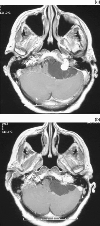
One year after initial presentation, the patient complained of sudden onset hearing loss on the left side, with progressive balance problems. Examination findings demonstrated no change in the appearance of the tympanic membrane, although a House–Brackmann grade II facial palsy was noted and pure tone audiometry demonstrated a ‘dead’ left ear. All other cranial nerves were intact. Repeated MRI scanning demonstrated no change in the size of the tumour but showed a substantial increase in the size of the cyst, leading to considerable distortion of the medulla. Examples of these axial T1-weighted scans with gadolinium enhancement are shown in Figure 2. Figure 2(a) shows an enhancing tumour mass extending from the left jugular foramen into the posterior fossa. Figure 2(b) shows the cyst as it appears in a contrast-enhanced, T1-weighted image. The cyst fluid in this image is of a slightly higher signal intensity than cerebrospinal fluid (CSF), compared with the T2-weighted scan in Figure 1, in which the cyst has a similar signal intensity to CSF. This may indicate the presence of protein within the cyst fluid. The cyst is clearly extra-axial and associated with the tumour. There is no enhancement of the cyst wall.
Axial T1-weighted magnetic resonance imaging (MRI) scans with gadolinium enhancement. (a) An enhancing tumour mass can be seen extending from the left jugular foramen into the posterior fossa. (b) Contrast-enhanced, T1-weighted MRI image showing the cyst.

A neurosurgery and oncology opinion was sought, and treatment options were thought to comprise surgery, radiotherapy or embolisation. Both radiotherapy and embolisation were thought unlikely to have any direct effect on the cyst itself, and would only ‘treat’ the solid component of the tumour. After consultation with the patient, a left posterior craniotomy was performed, with opening and drainage of the cyst. A sample of the cyst wall was sent for histological analysis, which demonstrated an appearance consistent with an arachnoid cyst. After drainage of the cyst, a highly vascular lesion was identified in the cerebellopontine angle.
Post-operatively, the patient made a good recovery, with resolution of the facial palsy and improvement of balance; however, there was no change in the pure tone audiogram. As it was felt that there was a risk of cyst recurrence, follow up was arranged at three-monthly intervals. At the time of writing, two years post-operatively, the patient was well. Further MRI scans at six months and at one and two years post-operatively demonstrated no significant change in the solid component of the lesion and no recurrence of the cyst. At the time of writing, the patient was being managed by a conservative approach of continued observation and serial imaging, given the risk of recurrence of the cyst.
Discussion
Rosenwasser first described removal of glomus jugulare tumours in 1945.Reference Rosenwasser1 Advances in neuroimaging, skull base techniques, embolisation, anaesthesia and post-operative care have improved both our ability to excise these tumours, and patients' outcomes.Reference Fisch, Fagan and Valvanis2–Reference Woods, Strasnick and Jackson5 A subgroup of complex glomus jugulare tumours includes: multiple, giant or neuropeptide-secreting lesions; those associated with other lesions, such as dural arterio-venous malformation or an adrenal tumour; and tumours in which there has been previous treatment with adverse outcome.Reference Al-Mefty6 We performed a literature search using Medline through PubMed (1950–2007) and Ovid (1958–2007), searching for similar cases using the search term ‘glomus jugulare’. To our knowledge, the current report represents the first published case of a glomus jugulare tumour associated with a posterior fossa cyst. We believe that this entity should be included in the subgroup of complex glomus jugulare tumours.
• A subgroup of complex glomus jugulare tumours exists
• This subgroup includes: multiple, giant or neuropeptide-secreting lesions; those associated with other lesions, such as dural arterio-venous malformation or an adrenal tumour; and tumours in which there has been previous treatment with adverse outcome
• This paper describes a case of a glomus jugulare tumour associated with a posterior fossa cyst; this entity should be included in this subgroup of complex glomus jugulare tumours
Arachnoid cysts may occur in association with cerebellopontine angle tumours, including acoustic neuromas, and it is therefore not unknown to have coexisting pathologies.Reference Schuhmann, Tatagiba, Hader, Brandis and Samii7 It has been postulated that arachnoid cysts arise as a result of: splitting of the arachnoid membrane by a focal increase in pulsation of the CSF; infectious events; or haemorrhage into the subarachnoid space.Reference Handley, Graham, Daspit and Spetzler8, Reference Lalwani9 In our patient, it may therefore be possible that a haemorrhage occurred from the glomus tumour, which resulted ultimately in formation of the cyst. Further acute haemorrhages could result in an enlargement of the cyst, which may also explain the sudden onset in deterioration of symptoms one year after presentation.
In children, growth of arachnoid cysts tends to occur with age. However in adulthood, de novo occurrence of large arachnoid cysts is uncommon; therefore, when discovered, such cysts are usually small and cause minor or no symptoms. Furthermore, in adults, the presentation and growth of these cysts are more akin to those of solid tumours such as acoustic neuromas.Reference Alaani, Hogg, Siddiq, Chavda and Irving10
In the management of other cerebellopontine angle lesions, it has been stated that a ‘watch and wait’ policy would appear to be a valid initial option in cases in which there is no significant compromise of local neural or vascular structures, no severe symptoms, and no suspected or proven rapid growth.Reference Alaani, Hogg, Siddiq, Chavda and Irving10, Reference Samii, Carvalho, Schuhmann and Mathies11 We are therefore satisfied that our initial watch and wait management plan was appropriate, especially given the potential morbidity of surgery and the resolution of the patient's symptoms following eventual drainage of the cyst. In future, it may become possible to drain such cysts endoscopically; this is minimally invasive, carries a lower morbidity and has had good results in the management of arachnoid cysts.Reference Schroeder, Gaab and Niendore12