A double outlet right ventricle is a complex CHD (CCHD) with an incidence of 0.03 a 0.14/1000 alive newborns. This cardiopathy forms part of a variable group of congenital cardiopathies that have in common the type of ventricular-arterial connection, in which more than 50% of the semilunar valves originate in the right ventricle.Reference Tynan, Becker, Macartney, Jiménez, Shinebourne and Anderson1 In these cases, ventricular septal defects work as the only output from left to right ventricle. The ventricular septal defects can be sub-aortic forming part of a Fallot tetralogy spectrum, double related, non-related or sub-pulmonar (Taussig-Bing), which can also generate pulmonary stenosis (sub-valvular, valvular or supra-valvular). In this cardiopathy, both ventricular outlets have two variants anterior–posterior and side by side.Reference De la Cruz, Cayre, Arista, Sadowinski and Serrano2
Many authors have suggested that double outlet right ventricle originates by a specific morphologic and haemodynamic pattern of embryonic heart in XIII and XIV Streeter horizons, in which the right ventricle has two outputs.Reference Goor and Edwards3–Reference De Vries and Saunders5
Muñoz et al have proposed a genetic aetiology within different anatomic types in this cardiopathy depending on the rotations of the truncus arteriosus, if the rotation is inferior to 180°, it affects the infundibulum and the aortic-pulmonary septum that divides both arteries. This theory is based on the anatomical study of both outputs and the relation between these large vessels.Reference Muñoz-Castellanos and Kuri-Nivon6
Echocardiography is a non-invasive diagnostic technique useful in CHD, including double outlet right ventricle. The double outlet right ventricle should be repaired between 3 and 6 months of age or even earlier, when the patient presents signs of heart failure.Reference Kliegman, Stanton, St. Geme and Schor7,Reference Jone, VonAlvensleben, Burkett, Darst, Collins, Miyamoto, Hay, Levin, Deterding and Abzug8
Pregnancy in patients with CHD conditions their circulatory system to a volume overload, establishing a high risk for either the mother or the fetus. Within cardiac complications, patients may develop pregnancy induced-hypertension, preeclampsia, eclampsia, haemolysis, HELLP syndrome, thrombo-embolic syndromes, premature membrane rupture, premature labour and postpartum haemorrhage.Reference Drenthen, Pieper and Roos-Hesselink9 Pregnancy patients with congenital cardiopathy should undergo a fetal echocardiogram between 18 and 22 weeks of gestational age.Reference Chakraborty, Gorla and Swaminathan10
The objective of this study is to present the case of a 19-year-old woman with a double outlet of the right ventricle who had 2 pregnancies.
Case presentation
In a 19-year-old female patient, a heart murmur was detected at the age of 1 year, and at 10 years, she was proposed for cardiac surgery, but her family rejected surgical treatment because of financial resources. The patient developed cardiac symptoms, such as cyanosis and dyspnoea on exertion, until the age of 15 years.
At 18 years, she had her first pregnancy, which resulted in an abortion at 12 weeks of gestational age. In her second pregnancy, the patient remained asymptomatic until 12 weeks of gestational age, when she presented dyspnoea of moderate efforts and orthopnoea of two pillows, which led her to a private cardiologist, who referred her to our institution. On physical examination, she had grade I jugular plethora, bilateral thrill and palpable apex at 6th left intercostal space, left anterior axillary line, systolic–diastolic murmur III/IV in the upper right sternal border radiating to subscapular right area, holosystolic murmur III/IV that radiates to mesocardium, holosystolic murmur in lower right sternal border II/IV and a single second heart sound and extremities with acropachy and 2-second-capillary flow.
Laboratory data with haemoglobin of 16.3 mg/dL, haematocrit of 46.8%. Electrocardiogram in sinus rhythm with a heart rate of 80 bpm, QRS complex axis −60° and right ventricular enlargement.
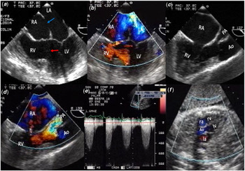
The echocardiogram showed situs solitus, double outlet right ventricle, 29 mm ostium secundum atrial septal defect, 30 mm subaortic ventricular septal defect, subvalvular and valvular severe pulmonary stenosis, with a peak gradient of 111mmHg, anterior aorta and posterior pulmonary artery, dilated coronary sinus secondary to persistence of left superior vena cava, no valvular aortic stenosis and mild mitral and tricuspid regurgitation (Fig. 1, Supplementary videos 1 and 2).
Echocardiographic study. (a) Bidimensional transoesophageal image at 0a. The blue arrow points to the ostium secundum atrial septal defect and the red arrow the ventricular septal defect. (b) Bidimensional and colour flow image at 0° with shunt from right to left at the level of atrial septal defect and from left to right at the level of ventricular septal defect. (c) Bidimensional transoesophageal study at 137° showing the double outlet right ventricle with anterior aorta and posterior pulmonary artery with subvalvular and valvular stenosis. (d) With colour flow, a subvalvular and valvular turbulence is observed. (e) Transthoracic bidimensional and continuous Doppler in 5-chamber view with severe subvalvular and valvular pulmonary stenosis with peak gradient of 95 mmHg. (f) Fetal echocardiography showing normal 5-chamber view.

Cardiac catheterisation demonstrated low oxygen saturation in left ventricle and aorta compared to pulmonary vein. Free wall right ventricular hypertrophy with a small cavity, valvular and sub-valvular pulmonary stenosis with peak gradient of 95 mmHg and normal coronary arteries.
The patient had a vaginal delivery without complications; the newborn was a male with a birth weight of 2100 g. Afterwards, the patient continued her follow-up for 4 years in the adult congenital cardiopathies department, remaining asymptomatic and without deterioration of her ventricular systolic function with respect to pregnancy.
Newborn control transthoracic echocardiogram showed no congenital cardiac abnormalities.
Discussion
The double outlet right ventricle represents a rare entity within CCHD. In literature, there are few cases of successful pregnancies of patients with this cardiopathy. Our patient has the diagnosis of double outlet right ventricle, and during her first pregnancy, she had an abortion at 12 weeks of gestational age, but her second pregnancy had a successful resolution.
In pregnancies of patients with CCHD, arrhythmias are a strong predictor of maternal complications; meanwhile, pulmonary regurgitation is a strong predictor of fetal and neonatal complications. Factors that may lead to bad outcomes for women are tricuspid regurgitation, the use of anticoagulants, anti-platelet drugs and being New York Heart Association class III or IV.
There have been case reports of pregnancies that have reached full-term deliveries. Wang et al reported a case series of 21 patients that had an adequate delivery without surgical management (one of them in our country), with a better prognosis with New York Heart Association class I and II. These authors found that to have a favourable delivery, pregnant patients need to have an adequate ventricular function and no pulmonary hypertension.Reference Jone, VonAlvensleben, Burkett, Darst, Collins, Miyamoto, Hay, Levin, Deterding and Abzug8 Pulmonary hypertension may lead to polycythemia, low oxygen saturation and right-to-left shunt. These conditions can exacerbate hypoxemia and increase complications towards delivery.Reference Wang, Luo, Xin and Yu11,Reference Dubois, Belkacem, Berl, Dailland and Carli12 However, as occurred in our patient, it is very important to note that pulmonary stenosis has a protective effect because it allows oxygenated blood to pass from the left atrium to the aorta, and it can also improve pulmonary venous return to systemic circulation with greater oxygen saturation, which is related to fewer complications during pregnancy in these patients.Reference Meyer, Jongbloed and Ho13–Reference Steinberg and Dantzker17
Ammash and Warnes mention that a person with adequate pulmonary circulation may reach the 6th decade without the need of surgical treatment. Although only 1% of pregnant patients present a CHD, cardiac disease is the main cause of non-obstetric death in these patients.Reference Drenthen, Pieper and Roos-Hesselink9,Reference Chakraborty, Gorla and Swaminathan10
The case reported by Gu et al in 2016 is similar to our case because their patient had a subaortic ventricular septal defect diagnosed in the adulthood and also she had an abortion.Reference Gu, Cai, Liu and Lv18
Drenthen et al demonstrated that female patients with CCHD who received palliative heart surgery or those without any surgical management, therefore, 11% of cases develop heart failure and supraventricular tachyarrhythmias. In pregnant patients with atrial arrhythmias, the use of beta-blockers and cardioversion, if necessary, is recommended to return a sinus rhythm and decrease the risk for heart failure. Within the obstetric complications of pregnancy, hypertension was not prevalent in pregnant patients with CHD compared with those who had none. In fetal complications, there has been found a higher prevalence (16%) of preterm labour.
Based on this research, patients with congenital cardiopathies should be assessed and warned about the risk they are exposed to with pregnancy, especially those with oxygen saturations lower than 85%, pulmonary hypertension, cyanosis or heart failure.Reference Drenthen, Pieper and Roos-Hesselink9,Reference Wang, Luo, Xin and Yu11,Reference Dubois, Belkacem, Berl, Dailland and Carli12,Reference Yucel and DeFaria Yeh19
In pregnant patients with CCHD, a strict periodic follow-up of their gestation and a multi-disciplinary strategy prior to delivery should be done.
For the pregnancy resolution, vaginal delivery and epidural anaesthesia are preferred, C-section is used in case of severe ventricular dysfunction, New York Heart Association class III or IV, aortic dissection, use of Warfarin, Eisenmenger and Marfan syndrome with aortic diameter >4.5 cm.Reference Wang, Luo, Xin and Yu11,Reference Gu, Cai, Liu and Lv18,Reference Yucel and DeFaria Yeh19
Conclusion
Our patient had a successful resolution of her second pregnancy and anatomical characteristics of her subaortic ventricular septal defect. Pulmonary and systemic circulations had a haemodynamic balance, until volume overload made symptoms of heart failure apparent.Reference Yucel and DeFaria Yeh19
Acknowledgements
None.
Financial Support
None.
Conflict of Interest
No conflict of interest between the authors.
Ethical Standards
The authors assert that all procedures contributing to this work comply with the ethical standards of the relevant national guidelines on human experimentation and with the Helsinki Declaration of 1975, as revised in 2008, and has been approved by our institutional committees.
Supplementary Material
To view supplementary material for this article, please visit https://doi.org/10.1017/S1047951120000566