Coronary artery fistula is an abnormal connection between a coronary artery and any cardiac chamber or great vessel. Angiography remains the gold standard for diagnosing the disease. In routine angiocardiography, it is found in up to 0.2% of cases.Reference Mangukia 1 It may occur isolated or along with congenital heart disease.
Hemangiomas are the most common vascular tumours in childhood. Most are asymptomatic and disappear spontaneously, but depending on the location they may disrupt vital functions or cause disfigurement. In recent years, the treatment with propranolol hydrochloride, a non-selective blocker, has been observed to produce dramatic involution of infantile hemangiomas.Reference Hogeling, Adams and Wargon 2
We report a case of multiple micro-fistula emptied into the left ventricle that was treated successfully with propranolol.
Case report
We report a case of a female neonate who was admitted for evaluation due to murmur. The examination disclosed a grade II/VI murmur, non-continuous in the left sternal border. Her physical appearance was abnormal. She had a slightly coarse facial appearance, a left preauricular pit, a rigid right helix, ascending palpebral fissures, deep-set eyes, strabismus, depressed nasal bridge, anteverted nose, and retrognathia. She also had hypoplastic nipples, bilateral single transverse palmar sulcus, and anterior anus. The electrocardiogram was normal without changes on ST segment.
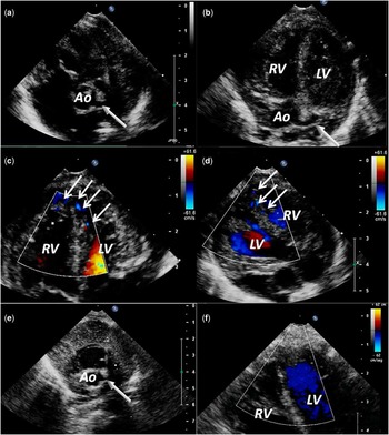
The two-dimensional transthoracic echocardiography revealed dilated left and right coronary arteries and the colour Doppler showed multiple linear colour-flow signals perpendicular to the epicardium draining in the left ventricular chamber; pulse-wave Doppler further identified a diastolic flow pattern towards the left ventricular cavity suggestive of multiple coronary artery–left ventricular micro-fistulas (Fig 1). The left ventricular ejection fraction was normal. She also had a small ventricular septal defect in the middle of the septum.
( a – d ) Transthoracic parasternal long-axis, short-axis, and apical four-chamber view showing dilated left and right coronary arteries (white arrows) and the colour Doppler showing multiple linear colour-flow signals at the apical and medium interventricular septum draining in the left ventricular chamber (head arrows). Those multiple diastolic flows reflect the presence of a micro-fistula between the coronary system and the left ventricular cavity. ( e – f ) Transthoracic short-axis and apical-four chamber views after β-blocker treatment. The left coronary artery has a normal calibre (white solid arrow) and colour Doppler shows the disappearance of multiple linear colour-flow signals at the apical and medium interventricular septum. Ao=aorta; LV=left ventricle; RV=right ventricle.

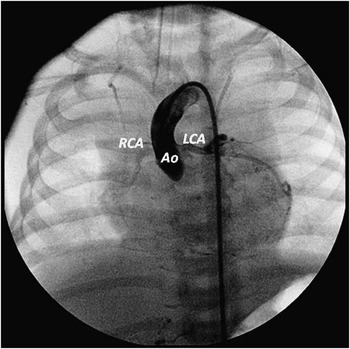
A cardiac catheterisation and coronary arteriography showed multiple micro-fistulas from the left and right coronary system. All three arteries communicated with the left ventricle cavity through many small diffuse fistulas, resulting in complete left ventricle contrast opacification. She also had an aberrant right subclavian artery (Fig 2).
Coronary arteriography showing dilated left and right coronary arteries and many small diffuse fistulas, from the left coronary system resulting in a left ventricle contrast opacification. The patient has an aberrant right subclavian artery (not shown). Ao=aorta; LCA=left coronary artery; RCA=right coronary artery.

Karyotype showed an abnormal chromosome 11 with an extra material at the end of the long arm. The array-comparative genomic hybridisation technique was performed and showed two alterations: Dup 3q26.1q29 of 32 megabases and Del 11q25 of 520 kilobases.
The patient was placed on medical therapy with propranolol with steps increase from 0.5 mg/kg/day to 2 mg/kg/day. No side effects were seen. After 6 months, the echocardiography was normal and the micro-fistulas had disappeared.
Discussion
Coronary artery fistula is a rare malformation. In all, 60% of these fistulas originate from the right coronary artery and mostly drain into the right heart chambers. Fistulas may be isolated, but between 20% and 45% are associated with other congenital heart disease – tetralogy of Fallot, atrial and ventricular septal defects, patent ductus arteriosus, and pulmonary atresia. Fistulas draining into the left ventricle are uncommon and its rare association with apical myocardial hypertrophy has been reported.Reference Dresios, Apostolakis and Tzortzis 3
Congenital fistulas are more common than acquired fistulas and they may represent a persistence of embryogenic myocardial sinusoids that arise from the endothelial protusions into the trabecular spaces. Normally, myocardial sinusoids become narrowed and persist as thebesian vessels in adults. If there is any interference in the normal development of these vessels, fistulous communication persists between coronary arteries and the cardiac chamber.Reference Mangukia 1
The natural history is marked by progressive dilatation of the coronary fistulas, accompanied by worsening symptoms. Most infants and children are asymptomatic, but if surgical correction is not indicated early complications occur, usually over 20 years of age with fatigue, dyspnoea, angina, and congestive heart failure. Spontaneous closure is very uncommon.Reference Mangukia 1
The two-dimensional transthoracic echocardiography may establish the diagnosis demonstrating the origin and drainage site, but catheterisation with coronary angiography remains the gold standard for the definitive diagnosis and in most cases the treatment. In our case, transthoracic colour Doppler echocardiography revealed multiple linear colour flows from the epicardial surface into the left ventricle cavity during diastole. Those multiple diastolic flows reflect the presence of a micro-fistula between the epicardial coronary system and the left ventricular cavity.
Surgical treatment is recommended in large, haemodynamic significant fistulas, even if they are asymptomatic. The treatment options include surgical ligation or transcatheter interventions. The surgical closure is until now the most effective treatment, but recent results of both techniques indicate a good prognosis.Reference Mangukia 1 When fistulas are diffuse, intervention might become impossible and treatment with calcium channel blockers or β-blockers should be considered in those symptomatic patients.Reference Duckworth, Mukharji and Vetrovec 4 In our case, our patient presented diffuse coronary fistulas. Owing to the fact that it was not considered for intervention and we assumed the diffuse coronary fistulas as a vascular malformation, we began propranolol therapy at 2 mg/kg/day like infantile hemangiomas. The result was the disappearance of the fistulas within six months. In the follow-up, recurrence of the fistulas was not observed.
In the last few years, newer treatment options are available that seem to have fewer side effects than another treatment options such as steroids, vincristine, interferon, or cyclophosphamida. Propranolol, a β-blocker, has been observed to produce dramatic involution of infantile hemangiomas. Its exact mechanism of action is still unknown, but some hypotheses include decreasing arterial flow, inducing endothelial cell apoptosis, and inhibition of angiogenesis.Reference Storch and Hoeger 5
The malformative syndrome described in our patient has characteristic features and a slight language delay with normal growth. Deletion of chromosome 11 is very small and does not include genetic sequences. The genetic basis of the picture is due to 3q-11q translocation and is mainly related to the partial trisomy of a large fragment of chromosome 3. The array-comparative genomic hybridisation technique is a technique used in genetic diagnosis, which allows us to analyse the entire genome of an individual for alterations of gain or loss of genetic material.Reference Malan and Romana 6 We did not find in the literature this association between this chromosomal abnormality and the presence of coronary fistulas.
We suggest propanolol as a new option of treatment of multiple and diffuse coronary artery fistulas, even in asymptomatic patients to try to avoid complications.Further studies are needed to support this hypothesis, as there is not enough experience.
Acknowledgement
None.
Financial Support
This research received no specific grant from any funding agency, commercial or not-for-profit sectors.
Conflicts of Interest
None.