

INTRODUCTION
The geographical ranges of Gambierdiscus species are mostly confined to the circumtropical shallow coastal and reef habitats (e.g. Lewis, Reference Lewis2001; GEOHAB, Reference Berdalet, Tester and Zingone2012; Parsons et al., Reference Parsons, Aligizaki, Dechraoui Bottein, Fraga, Morton, Penna and Rhodes2012) mainly coinciding with global distribution of ciguatera fish poisoning (CFP) between 35° north and 35° south latitudes, particularly in the Pacific and West Indian Oceans and the Caribbean Sea (e.g. Lehane & Lewis, Reference Lehane and Lewis2000; Lewis, Reference Lewis2001; Pérez-Arellano et al., Reference Pérez-Arellano, Luzardo, Perez Brito, Hernández Cabrera, Zumbado, Carranza, Angel-Moreno, Dickey and Boada2005; Tester et al., Reference Tester, Feldman, Nau, Faust, Litaker and Lang2009, Reference Tester, Feldman, Nau, Kibler and Litaker2010). Some of the Gambierdiscus species have been confirmed to produce maitotoxins and ciguatoxins, being the primary causative organism of CFP (e.g. GEOHAB, Reference Berdalet, Tester and Zingone2012 and references therein).
Until recently, scarce records of Gambierdiscus in the Indian Ocean were restricted to the western tropical area, where this genus has been detected in reef lagoons of the Mayotte, Mauritius and Réunion Islands (Grzebyk et al., Reference Grzebyk, Berland, Thomassin, Bosi and Arnoux1994; Turquet et al., Reference Turquet, Quod, Ten-Hage, Dahalani, Wendling, Hallegraeff, Blackburn, Bolch and Lewis2001; Hurbungs et al., Reference Hurbungs, Jayabalan, Chineah, Lalouette and Bachraz2002; Litaker et al., Reference Litaker, Vandersea, Faust, Kibler, Chinain, Holmes, Holland and Tester2009; Parsons et al., Reference Parsons, Aligizaki, Dechraoui Bottein, Fraga, Morton, Penna and Rhodes2012). Gambierdiscus toxicus Adachi & Fukuyo was the only species recorded from the Indian Ocean (Litaker et al., Reference Litaker, Vandersea, Faust, Kibler, Chinain, Holmes, Holland and Tester2009). However to date, five Gambierdiscus species have been reported from the coastal waters of Pakistan in the northern Indian Ocean, such as G. toxicus, G. belizeanus Faust, G. polynesiensis Chinain & Faust, G. australes Chinain & Faust and G. cf. yasumotoi Holmes (Munir et al., Reference Munir, Siddiqui and Morton2011). Then, Gambierdiscus sp. has been found in the central Red Sea (Catania, Reference Catania2012), but this ciguatoxic genus has never been reported from the Sea of Oman and the Arabian Gulf previously.
Despite great diversity of the dinoflagellate assemblage inhabiting the coral sands of the Gulf of Aqaba, northern Red Sea (Saburova & Polikarpov, unpublished), published data on benthic dinoflagellates from this area are totally lacking. Very recently, epiphytic dinoflagellate assemblage has been preliminarily described for the first time for the central Red Sea off the Saudi Arabia coast, where Gambierdiscus species was detected (Catania, Reference Catania2012). The diversity of benthic dinoflagellates in Kuwait's coastal area (north-western Arabian Gulf) has been recently documented (Saburova et al., Reference Saburova, Al-Yamani, Polikarpov, Krupp, Musselman, Kotb and Weidig2009; Al-Yamani & Saburova, Reference Al-Yamani and Saburova2010), with emphasis on sand-dwelling intertidal species. However, the occurrence of Gambierdiscus species has not been previously noted for Kuwait.
Recently, regular benthic surveys at southern Kuwait's shores and occasional sampling of the bottom sediments within the coral reef at the Gulf of Aqaba have revealed the presence of Gambierdiscus species in the composition of the benthic dinoflagellate assemblages for the above two sampled localities. The objective of this study was to examine newly found Gambierdiscus species from two localities in the marginal seas of the northern Indian Ocean to better document the global distribution of this ciguatera-causing genus. In this paper, morphological details of G. yasumotoi and G. cf. belizeanus, and their occurrence among different localities and substrates are presented.
MATERIALS AND METHODS
Sampling sites
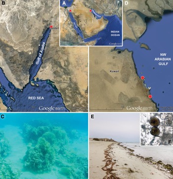
Studies on benthic dinoflagellates were carried out in the Arabian (Persian) Gulf and in the Red Sea that constitute the marginal seas of the Indian Ocean basin enclosed by arid lands (Figure 1A). These coastal biomes are also known as the Red Sea–Arabian Gulf Biogeographic Province (Longhurst, Reference Longhurst2007).
Area of investigations: (A) satellite image (Google Earth) showing the sampling area locations in marginal seas of the Indian Ocean; (B) sampling site overlaid on satellite image (Google Earth) of the northern Red Sea in the Gulf of Aqaba; (C) inshore coral reef in the Gulf of Aqaba along Jordanian coast, where samples were collected; (D) sampling sites overlaid on satellite image (Google Earth) of north-western Arabian Gulf, Kuwait's marine area; (E) southern Kuwait's shore, where samples were collected and Gambierdiscus yasumotoi was detected (F).

In the Arabian Gulf, two sampling sites were located on the southern coast of Kuwait at 28°37′–28°53′N 48°16′–48°23′E (Figure 1D). Kuwait's marine area stretches for about 180 km, occupying the north-western corner of the Arabian Gulf. The hypersaline, shallow and sub-tropical Kuwait marine environment provides a broad range of benthic marine habitats varying from the extensive mudflats in the north to numerous sandy beaches and beach rocks extending down to the Saudi Arabia border in the south. Coral reefs occur mainly within the southern waters of Kuwait as numerous inshore patch reefs, however, fringing reefs are found around offshore islands. In Kuwait's marine environment, beds of Sargassum grow in a discontinuous linear strip parallel to the coast where appropriate substrates exist (Al-Yamani et al., Reference Al-Yamani, Bishop, Ramadhan, Al-Husaini and Al-Ghadban2004).
The sampling site in the Red Sea was located at the northern end of the Gulf of Aqaba in the Jordanian coastal area at 29°25′58″N 34°58′26″E (Figure 1B). The Gulf of Aqaba, approximately 180 km long and on average 8 km wide, is a marine environment that experiences extremes of temperature and exceedingly low levels of precipitation. The Jordanian coastline has a length of about 27 km with a discontinuous series of fringing reefs of 13 km length, interrupted by bays, which are mostly covered with seagrass meadows (UNEP/IUCN, 1988).
Sampling
At the southern Kuwait coastal area, the bottom sediments and both floating and attached to the bottom macroalgae were sampled within the inshore Qit'at Julai'ah coral reef at depths of 1–3 m, and to the south of the first sampling site in small semi-protected shallow lagoons and on the sea shore in November 2012 and March 2013 (Figure 1D, E). The seawater temperature range was 23.5–25°C and the salinity range was 41.2–42.4, when sampling was carried out in November 2012. In March 2013, the seawater temperature ranged from 21 to 22.3°C and the salinity range was 42.1–42.8.
In Jordan, the bottom sandy sediments, macroalgae and near-bottom water were collected on the shallow slope of the inshore coral reef (Figure 1C) within the swimming zone of the Aqaba Marine Park at depths of 1.5–2 m in October 2010. Samples were collected at a seawater temperature range of 24–25°C and at salinity of 40.
The surface layer (0–2 cm) of the sandy sediments was sampled within coral reefs using a plastic bottle directly from the sea bottom by snorkelling. The floating macroalgae were handpicked in near-shore waters, and the growing macroalgae were detached gently from the substrate manually. The samples were collected together with the surrounding seawater, and kept in the dark in ambient temperature until samples were transferred to the laboratory.
Samples processing
The dinoflagellate cells were separated from the sandy sediment by extraction using the frozen seawater method (Uhlig, Reference Uhlig1964) with a 110 µm mesh size. Macroalgae samples were shaken vigorously in 300–500 ml of seawater to detach the epiphytic dinoflagellates. The suspension was then passed over 250 µm sieve to remove large particles. The macroalgae were dried using absorbent paper and then weighed.
From each sample 50 to 100 ml aliquots were placed into cell culture flasks, enriched with equal volume of sterile Guillard's F/2 medium without silicates (Sigma-Aldrich), and exposed in the incubator FytoScope Chamber FS130 RGBIR MINI (Photon Systems Instruments, Czech Republic) at 24.5°C under 100 µmol photons m−2 s−1 of light and 12:12 h light:dark photoperiod for several weeks. If adequate densities of Gambierdiscus cells were found during incubation, these aliquots were used to establish cell cultures (only carried out for Kuwait's samples). Alternatively, 50 ml aliquots of each sample were preserved by 4% Lugol's solution and utilized for examination by epifluorescence or scanning electron microscopy (SEM), and for cells enumeration.
The material was preliminarily viewed alive with a Leica DMIL inverted microscope at 35× to 200× magnifications. For detailed observation, cells were isolated by micropipetting in preparation for high-magnification photomicroscopy. Cells were examined using a Leica DMLM microscope (Leica, Wetzlar, Germany) connected to a Leica DFC 320 digital camera at 630× to 1000× magnification. Some morphometric features were observed under epifluorescence microscopy after staining with Calcofluor White (Sigma Chemical Co.) according to the method of Fritz & Triemer (Reference Fritz and Triemer1985), and SEM was employed for detailed observations of the thecal surface.
For SEM, mixed samples of extracted cells after preservation with Lugol's iodide, were filter mounted by a Swinnex filter-holder, rinsed twice with distilled water and subsequently dehydrated in a series of increasing ethanol concentrations (15, 30, 50, 70, 90, 95 and 100%). The filters were critical point dried, scutter-coated with gold-palladium and examined either by Tescan Vega-3, or by JEOL JCM-5700 CarryScope scanning electron microscope.
Morphometric measurements were made either from the calibrated digital light microscopy images using Leica Application Suite v. 3.7 software (Leica Microsystems Ltd, Switzerland) or were calculated from scanning electron micrographs. The SEM photographs were presented on a black background using Adobe Photoshop CS2, v. 9.0.2 (Adobe Systems, San Jose, CA, USA). The thecal plates are named according to Kofoid (Reference Kofoid1909).
RESULTS
A Gambierdiscus species with globular morphology was found in samples from Kuwait. Examination of the samples collected in the Gulf of Aqaba revealed the co-occurrence of both anterio-posteriorly compressed and globular Gambierdiscus cells in the sandy bottom sediments from shallow coral reef.
Species identifications were based on observations of the morphological characters using light, epifluorescence, and scanning electron microscopy. Both alive and preserved cells from field samples and material from the established cultures (only carried out for the Kuwait samples) were examined.
Based on the main diagnostic features of the genus Gambierdiscus (Chinain et al., Reference Chinain, Germain, Deparis, Pauillac and Legrand1999; Richlen et al., Reference Richlen, Morton and Barber2008; Litaker et al., Reference Litaker, Vandersea, Faust, Kibler, Chinain, Holmes, Holland and Tester2009) including cell shape and dimensions, thecal surface structure, and the size and shape of the apical pore complex (Po), the second apical plate (2'), and posterior intercalary plate (1p), and following the key to the Gambierdiscus species (Litaker et al., Reference Litaker, Vandersea, Faust, Kibler, Chinain, Holmes, Holland and Tester2009), the globular species from both the Kuwait and Jordanian samples was identified as G. yasumotoi, whereas the anterio-posteriorly compressed species from the Gulf of Aqaba was presumably assigned to G. belizeanus.
SYSTEMATICS
Class DINOPHYCEAE West & Fritch, 1927
Order GONYAULACALES Lindemann, 1928
Family GONIODOMACEAE Lindemann, 1928
Genus Gambierdiscus Adachi & Fukuyo, 1979
Gambierdiscus yasumotoi Holmes, Reference Holmes1998
(Figures 2–5)
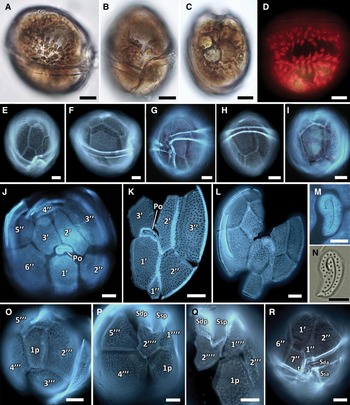
Light micrographs of Gambierdiscus yasumotoi recorded from the southern Kuwait area in November 2012: (A–C) living cell in lateral (A), ventral (B), and antapical (C) views; (D) dorsal view showing chloroplasts distribution; (E–I) cells shown from various perspectives; (J, K) squashed theca in apical view showing epithecal plate pattern; (L) squashed theca in apical view showing aberrant epithecal plate pattern in cultural cell; (M, N) apical pore plate; (O–Q) antapical view showing hypothecal plate pattern; (R) ventral view showing epithecal and sulcal plate pattern. A–C, N, BF; D, chlorophyll epifluorescence; E–M, O–R, epifluorescence images of calcofluor-stained cells. Scale bars: A–L, O–R, 10 µm; M, N, 5 µm. Abbreviations used in Figures 2–7: Po, apical pore plate; 1–3', apical plates; 1–6”, precingular plates; 1–5'”, postcingular plates, 1–2””, antapical plates; 1p, posterior intercalary plate; Sda, sulcal right anterior plate; Ssa, sulcal left anterior plate; t, transition plate; Sdp, sulcal right posterior plate; Ssp, sulcal left posterior plate; BF, brightfield; DF, darkfield.

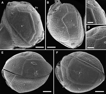
Scanning electron micrographs of Gambierdiscus yasumotoi recorded from the southern Kuwait area in November 2012: (A) epitheca in right lateral view showing epithecal plate pattern; (B) hypotheca in left lateral view showing hypothecal plate pattern; (C, D) detail of apical pore; (E, F) oblique left lateral views. Scale bars: A, B, E, F, 10 µm; C, D, 5 µm.

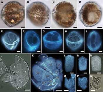
Light micrographs of Gambierdiscus yasumotoi recorded from Jordanian coastal area in October 2010: (A–D) living cells in lateral view; (E–I) cells shown from various perspectives; (J) squashed theca in apical view showing epithecal plate pattern; (K) squashed theca in right lateral view showing plate pattern; (L–N) 1p plates; (O–Q) 2”” plates. A–D, BF; J, Q, DF; E–I, K–P, epifluorescence images of calcofluor-stained cells. Scale bars: A-K, 10 µm; L-Q, 5 µm.

Scanning electron micrographs of Gambierdiscus yasumotoi recorded from Jordanian coastal area in October 2010: (A) epitheca in right lateral view showing epithecal plate pattern; (B) right lateral view showing plate pattern; (C) detail of apical pore; (D, E) cells in left (D) and right (E) lateral view; (F) detail of sulcal area. Scale bars: A, B, D–F, 10 µm; C, 5 µm.

REFERENCES
Holmes, Reference Holmes1998; Litaker et al., Reference Litaker, Vandersea, Faust, Kibler, Chinain, Holmes, Holland and Tester2009: figures 22–24 and 60–66.
DESCRIPTION
Cells were globular in shape in lateral view, oblong in apical/antapical view and slightly laterally flattened, with dome-shaped epitheca and truncate–conical hypotheca (Figures 2A–C, E–I; 3E, F; 4A–I & 5A, B, D, E). In the Kuwait samples and cultures, the depth of cells (dorsoventral diameter) ranged from 54 to 73 µm (an average 62.9 ± 4.4, N = 30), cell width (transdiameter) ranged from 46 to 61 µm (54.1 ± 5.1, N = 9), with an average depth/weight (D/W) ratio of 1.28, and cell length was 49–70 µm (61.7 ± 6.2, N = 20). In the Jordanian population, cells had a depth of 54–71 µm (62.6 ± 6.1, N = 7), a width of 53–56 µm (54.7 ± 1.5, N = 3), with an average D/W ratio of 1.14, and a length of 55–72 µm (62.9 ± 6.8, N = 7). The pre-median cingulum was deeply excavated, descending, and was displaced by once its width (Figures 2A, B, E, G, R & 4E). The apical pore plate (Po) was elongated and teardrop shaped, 9.2–9.5 µm long, and 3.8–4.2 µm wide, with the large fish-hook shaped apical opening surrounded by 33–42 pores (an average 37.1 ± 3.6, N = 8) (Figures 2E, J–N; 3C–E; 4J & 5A, C). The posterior intercalary plate (1p) was pentagonal, narrow, 35.6–42.5 µm long, and 20.8–22 µm wide, with an average length/weight (L/W) ratio of 1.72 (Figures 2I, O; 3F & 4I, L–N). The second antapical plate 2”” was fork-shaped and rather large, with robust and bevelled fork edges (Figures 2P, Q; 3B; 4K, O–Q & 5F). Numerous small elongated golden-brown chloroplasts were peripherially arranged (Figures 2A, D & 4A, B). Nucleus was large, oblong and located in the right posterior cell part (Figure 4D). Several specimens with aberrant plate pattern were observed among cultural cells (Figure 2L).
Gambierdiscus cf. belizeanus Faust, Reference Faust1995
(Figures 6 & 7)
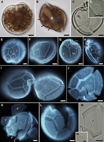
Light micrographs of Gambierdiscus cf. belizeanus recorded from Jordanian coastal area in October 2010: (A, B) living cell in apical (A) and lateral (B) views showing chloroplasts distribution, nucleus (marked by N), and red inclusion bodies (indicated by arrows); (C) squashed theca in antapical view showing areolated thecal surface; (D) detail of thecal surface; (E) cell in apical view showing epithecal plate pattern; (F) oblique apical view; (G) cell in antapical view showing hypothecal plate pattern; (H) epitheca in left lateral view; (I) total theca split into apical and antapical half; (J, K) squashed theca in apical view showing epithecal plate pattern; (L) squashed theca in antapical view; (M) hatchet shaped 2' plate associated with apical pore plate; (N) apical pore plate. A, B, BF; C, D, M, N, DF; E–L, epifluorescence images of calcofluor-stained cells. Scale bars: A–M, 10 µm; N, 2 µm.

Scanning electron micrographs of Gambierdiscus cf. belizeanus recorded from Jordanian coastal area in October 2010: (A, B, D) cell in apical (A) and oblique left lateral (B, D) views showing epithecal plate pattern; (C, F) detail of apical pore; (E) detail of thecal surface. Scale bars: A, B, D, 10 µm; C, E, 5 µm; F, 2 µm.

REFERENCES FOR GAMBIERDUSCUS BELIZEANUS
Faust, Reference Faust1995; Litaker et al., Reference Litaker, Vandersea, Faust, Kibler, Chinain, Holmes, Holland and Tester2009: figures 22–24, 68; Leaw et al., Reference Leaw, Lim, Tan, Tuan-Halim, Cheng, Ng and Usup2011; Munir et al., Reference Munir, Siddiqui and Morton2011.
DESCRIPTION
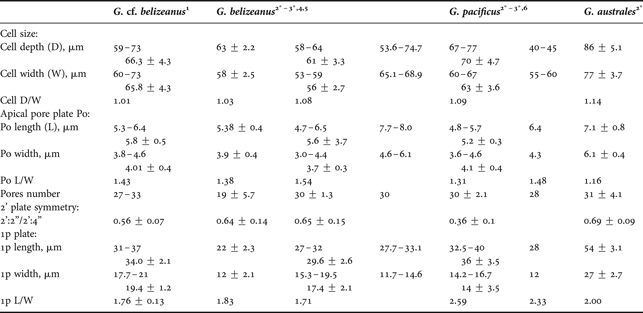
Cells were almost round in apical view, anterio-posteriorly compressed, and lenticular in lateral view (Figures 6A, B, E & 7A). The depth of cells (dorsoventral diameter) ranged from 59 to 73 µm (66.3 ± 4.3, N = 13), cell width (transdiameter) range was 60–73 µm (65.8 ± 4.3, N = 14), with an average D/W ratio of 1.01, and cell length was 47–51 µm. The thecal surface was moderately areolated; areolae were dense, and round to oblong (Figures 6C, D, J, M & 7E). Round pores were located within areolae. The apical pore plate (Po) was irregularly elongated, 5.3–6.4 µm long, and 3.8–4.6 µm wide, with average L/W ratio of 1.43. Po plate possessed the large fish-hook shaped apical opening surrounded by 27–33 pores (Figures 6M, N & 7A, C, F). The second apical plate (2') was the largest among apical series, hatchet shaped, with a ratio of the 2':2” to 2':4” suture lengths of 0.56 ± 0.07, N = 16 (Figures 6E, F, I–K, M & 7A, B, D). The posterior intercalary plate (1p) was pentagonal, narrow, with almost parallel sides, 31–37 µm long (34.0 ± 2.1, N = 13) and 17.7–21.6 µm wide (19.4 ± 1.2, N = 13), with an average L/W ratio of 1.76 ± 0.13, N = 13 (Figure 6C, G, I, L). Cells were photosynthetic, with numerous golden-brown drop-shaped chloroplasts radiating from the cell centre (Figure 6A, B). Nucleus was large, oblong and crescent-shaped, and located in the right posterior cell part (Figure 6B).
TAXONOMIC REMARKS
Our material agreed in size and shape with G. belizeanus, and shape, dimensions and symmetry of the epithecal plates were consistent with previous descriptions of this species (Faust, Reference Faust1995; Litaker et al., Reference Litaker, Vandersea, Faust, Kibler, Chinain, Holmes, Holland and Tester2009; Leaw et al., Reference Leaw, Lim, Tan, Tuan-Halim, Cheng, Ng and Usup2011; Munir et al., Reference Munir, Siddiqui and Morton2011). However, observed cells differed from those described in G. belizeanus in less areolated thecal surface and in size of the 1p plate.
Gambierdiscus species occurrence
In Kuwait (north-western Arabian Gulf), G. yasumotoi was the only Gambierdiscus species that was detected. It was found for the first time in late 2012 in the bottom sandy sediments of small patch inshore coral reef on the southern Kuwait shore (Figure 1D). The species was observed in low numbers in two samples out of the ten samples examined. Additionally, G. yasumotoi was detected in the epiphytic dinoflagellate assemblages southward of the first sampling site (Figure 1D–F). It was present in two samples of the 16 macroalgae samples analysed and was observed in low abundances on the floating brown macroalgae Padina tetrastomatica (6 cells g−1 wet weight) and on Sargassum oligocystum (4 cells g−1 wet weight). Gambierdiscus species occurred along with other epiphytic dinoflagellates of the genera Amphidinium Claperède & Lachmann, Coolia Meunier, Ostreopsis Schmidt, and Prorocentrum Ehrenberg.
During the early spring survey in March 2013 at the same sampling site, a more dense population of G. yasumotoi was recorded in a small and very shallow lagoon with limited connection with the open sea, and covered with a dense carpet of filamentous algae on the bottom. Gambierdiscus species was found epiphytically on filamentous green algae Chaetomorpha sp. Gambierdiscus yasumotoi prevailed numerically with the cells concentration varying from 83 to 171 cells g−1 wet weight (an average 116.7 ± 47.5), and contributed from 35 to 57% of the total epiphytic dinoflagellates assemblage. Co-occurring epiphytic dinoflagellates were represented by eight species, including rather abundant Coolia monotis Meunier, Amphidinium carterae Hulburt, and Peridinium quinquecorne Abé, and scanty number of Prorocentrum formosum Faust, P. rhathymum Loeblich, Sherley & Schmidt, P. tsawwassenense Hoppenrath & Leander, Adenoides eludens (Herdman) Balech, and Cabra matta Murray & Patterson.
In the Gulf of Aqaba (northern Red Sea, Jordan), co-occurrence of G. cf. belizeanus and G. yasumotoi was found in the bottom sandy sediments of a well-developed inshore coral reef (Figure 1C). Both species occurred commonly, being recorded in nine out of the twelve sediment samples collected, but only few cells of G. yasumotoi were observed in samples of near-bottom water, and both species were not found from the macroalgae samples.
DISCUSSION
This study presents the first preliminary report of the ciguatera-related genus Gambierdiscus in the northern parts of the Indian Ocean basin. Accurate taxonomic identification among Gambierdiscus species is difficult based on the morphological criteria because some species of this genus are morphologically highly uniform. Recently, the current taxonomic state of Gambierdiscus has been reviewed by Litaker et al. (Reference Litaker, Vandersea, Faust, Kibler, Chinain, Holmes, Holland and Tester2009), and reliable recognition among Gambierdiscus species based on their morphology has been demonstrated. Several morphological and morphometric features were selected as diagnostic characters to distinguish species from each other, including cell shape and size (for globular species), and shape, size and symmetry of the second apical plate (2') and posterior intercalary plate (1p), as well as the character of the thecal surface (for anterio-posteriorly compressed species) (Litaker et al., Reference Litaker, Vandersea, Faust, Kibler, Chinain, Holmes, Holland and Tester2009).
Based on the morphological characteristics of the cells, the globular Gambierdiscus species from the north-western Arabian Gulf as well as from the northern Red Sea is assigned to G. yasumotoi Holmes, Reference Holmes1998. Following the original description (Holmes, Reference Holmes1998) and identification key to the species of Gambierdiscus (Litaker et al., Reference Litaker, Vandersea, Faust, Kibler, Chinain, Holmes, Holland and Tester2009), the cell shape, dimensions, plate tabulation and structure of the 2”” from our material confirm the identification and allow to distinguish our findings from another globular Gambierdiscus species, G. ruetzleri (Litaker et al., Reference Litaker, Vandersea, Faust, Kibler, Chinain, Holmes, Holland and Tester2009).
Given the shape and size of the posterior intercalary plate (1p) in our anterio-posteriorly compressed Gambierdiscus species isolated from the bottom sediments of the Gulf of Aqaba in the Red Sea, it falls into the group of Gambierdiscus species with a narrow 1p, including G. australes, G. belizeanus and G. pacificus (Litaker et al., Reference Litaker, Vandersea, Faust, Kibler, Chinain, Holmes, Holland and Tester2009). Alternative character of the thecal surface (smooth or heavily areolated) allows delimiting between very similar morphologically G. belizeanus and G. pacificus, whereas the smooth-surfaced species (G. australes and G. pacificus) are further distinguished by shape of the second apical plate (Litaker et al., Reference Litaker, Vandersea, Faust, Kibler, Chinain, Holmes, Holland and Tester2009).
Our cells differ dramatically from G. australes in length of the 1p plate; however, our material has a morphological and morphometric characters intermediate between that of G. belizeanus and G. pacificus (Figures 6 & 7; Table 1). The dimensions of the 1p plate in specimens examined from the Red Sea (L = 34.0 ± 2.1, W = 19.4 ± 1.2 µm) match better with those described in G. pacificus (L = 36, W = 14 µm) than in G. belizeanus (L = 22, W = 12 µm), however, the proportion of this plate (L/W = 1.76) is much closer to those in G. belizeanus (1.83) than in G. pacificus (2.59) (Table 1; Litaker et al., Reference Litaker, Vandersea, Faust, Kibler, Chinain, Holmes, Holland and Tester2009). The thecal surface in our specimens is less deeply areolated compared to G. belizeanus, but differs from the smooth cell surface described in G. pacificus (Chinain et al., Reference Chinain, Germain, Deparis, Pauillac and Legrand1999) and considered as the main distinctive characteristic between G. belizeanus and G. pacificus (Litaker et al., Reference Litaker, Vandersea, Faust, Kibler, Chinain, Holmes, Holland and Tester2009). Moreover, the shape, dimensions and symmetry of the epithecal plates in our cells are consistent with those in G. belizeanus (Faust, Reference Faust1995; Litaker et al., Reference Litaker, Vandersea, Faust, Kibler, Chinain, Holmes, Holland and Tester2009) and differ from those in G. pacificus (Chinain et al., Reference Chinain, Germain, Deparis, Pauillac and Legrand1999; Litaker et al., Reference Litaker, Vandersea, Faust, Kibler, Chinain, Holmes, Holland and Tester2009). Here, we presumably ascribe the anterio-posteriorly compressed Gambierdiscus species from the Gulf of Aqaba to G. belizeanus due to its areolated theca, dimensions of the 1p plate and epithecal plate pattern. Further molecular identification of this species is requested to clarify this issue.
Morphometric characters for Gambierdiscus species with narrow posterior intercalary plate (1p) as measured in this and previous studies (the range and/or the average sizes ±1 standard deviation).

1, this study; 2, Litaker et al., Reference Litaker, Vandersea, Faust, Kibler, Chinain, Holmes, Holland and Tester2009; 3, Chinain et al., Reference Chinain, Germain, Deparis, Pauillac and Legrand1999; 4, Leaw et al., Reference Leaw, Lim, Tan, Tuan-Halim, Cheng, Ng and Usup2011; 5, Munir et al., Reference Munir, Siddiqui and Morton2011; 6, Ho Van The & Nguyen Thi Mai Anh, 2010; *, species identification was confirmed by molecular analysis.
In this study G. cf. belizeanus was observed from the sediment samples exclusively, that confirms the sand-dwelling character of this species (Faust, Reference Faust1995). In contrast, G. yasumotoi was found widely distributed among various substrates, being observed in the bottom sediments, surrounded water, and as an epiphyte on macroalgae.
Gambierdiscus belizeanus has been originally described from the coastal area of Belize in the Caribbean Sea (Faust, Reference Faust1995). Currently, this species is known to occur in the Atlantic, Pacific and Indian Oceans and can be considered as circumtropically distributed (Leaw et al., Reference Leaw, Lim, Tan, Tuan-Halim, Cheng, Ng and Usup2011; Munir et al., Reference Munir, Siddiqui and Morton2011; GEOHAB, Reference Berdalet, Tester and Zingone2012; Parsons et al., Reference Parsons, Aligizaki, Dechraoui Bottein, Fraga, Morton, Penna and Rhodes2012), although the record of this species from the Atlantic Ocean has been the only record confirmed by molecular identification (Litaker et al., Reference Litaker, Vandersea, Faust, Kibler, Nau, Holland, Chinain, Holmes and Tester2010). Since the original description of G. yasumotoi from the macroalgae collected on the coral reef of Pulau Hantu Island (Singapore, South China Sea) (Holmes, Reference Holmes1998), its distribution has been referred to the tropical area of the western Pacific Ocean (Holmes et al., Reference Holmes, Lee, Teo, Khoo, Reguera, Blanco, Fernández and Wyatt1998; Litaker et al., Reference Litaker, Vandersea, Faust, Kibler, Nau, Holland, Chinain, Holmes and Tester2010; GEOHAB, Reference Berdalet, Tester and Zingone2012; Parsons et al., Reference Parsons, Aligizaki, Dechraoui Bottein, Fraga, Morton, Penna and Rhodes2012). However, recently the globular Gambierdiscus species morphologically close to G. yasumotoi has been reported from coastal waters of Pakistan in a sub-tropical area of the north-western Indian Ocean (Munir et al., Reference Munir, Siddiqui and Morton2011). Gambierdiscus yasumotoi has been reported to be toxic, being a producer of the maitotoxin-like compound that is lethal to mice (Holmes, Reference Holmes1998), and its toxic strains have been isolated from Indonesian waters (Parsons et al., Reference Parsons, Aligizaki, Dechraoui Bottein, Fraga, Morton, Penna and Rhodes2012). Recently, the ciguatoxicity has been shown in a Caribbean isolate of G. belizeanus (Chinain et al., Reference Chinain, Darius, Ung, Cruchet, Wang, Ponton, Laurent and Pauillac2010).
The marginal area of the north-western Indian Ocean including the Arabian Gulf and the Red Sea appears to be free of ciguatera (Albinali, Reference Albinali2011; Catania, Reference Catania2012). Gambierdiscus species have never been recorded previously among other potentially ciguatera-related benthic dinoflagellates either in Kuwait's coastal area (Al-Yamani & Saburova, Reference Al-Yamani and Saburova2010) or in the northern Red Sea, although the occurrence of anterio-posteriorly compressed Gambierdiscus species has been reported recently from the central Red Sea off the Saudi Arabia coast (Catania, Reference Catania2012). It is interesting to note that this Gambierdiscus species from the central Red Sea seems to be morphologically similar to those described from the northern Red Sea in this study in distinctly areolated thecal surface and in narrow 1p plate (Catania, Reference Catania2012).
The inshore and well-developed coral reefs in the Gulf of Aqaba in the northern Red Sea provide a perfect habitat for benthic dinoflagellates and match well with preferred habitats reported for Gambierdiscus (Litaker et al., Reference Litaker, Vandersea, Faust, Kibler, Nau, Holland, Chinain, Holmes and Tester2010 and references therein) with respect to annual water temperature, salinity, hydrology and available substrates to which cells can attach. The average monthly seawater temperature of the Gulf of Aqaba ranges from 21°C during February–March to about 26°C in August–September with the seasonal amplitude of about 5.5°C. The average value of salinity is close to 40.6 with unessential annual variations (Al-Rousan et al., Reference Al-Rousan, Felis, Manasrah and Al-Horani2007). It was not surprising to detect Gambierdiscus species among diverse and abundant benthic dinoflagellates assemblage inhabiting the coral reef sediments of the Gulf of Aqaba. If at least two Gambierdiscus species were revealed during our occasional survey within a very limited area, the true diversity of this genus in the Jordanian coastal area can be underestimated. In contrast to the central Red Sea, where negative correlation between salinity and abundance of Gambierdiscus has been shown within the salinity range of 28–39 (Catania, Reference Catania2012), high and stable salinity of the Jordanian coastal waters may support a more abundant and diverse assemblage of CFP-related dinoflagellates. The results of our pilot survey highlight the need for detailed studies to assess the species composition, abundance and annual pattern of potentially toxic benthic dinoflagellates throughout the northern Red Sea.
Kuwait's sub-tropical location means that it is warm enough to support tropical biota, but its high-latitude location results in marked amplitude of summer/winter temperature differences. The average annual seawater temperature in Kuwait's area is 23.8°C, varying seasonally from 14°C during winter up to 30.5°C in summer months. Kuwait's southern and offshore waters are generally warmer with an annual average temperature of 25.6°C (Al-Yamani et al., Reference Al-Yamani, Bishop, Ramadhan, Al-Husaini and Al-Ghadban2004; Sheppard et al., Reference Sheppard, Al-Husiani, Al-Jamali, Al-Yamani, Baldwin, Bishop, Benzoni, Dutrieux, Dulvy, Durvasula, Jones, Loughland, Medio, Nithyanandan, Pilling, Polikarpov, Price, Purkis, Riegl, Saburova, Namin, Taylor, Wilson and Zainal2010). The main parameters of the southern part of Kuwait's marine environment appear to be close to those that are considered as optimal habitat for Gambierdiscus (Litaker et al., Reference Litaker, Vandersea, Faust, Kibler, Nau, Holland, Chinain, Holmes and Tester2010). However, given the peculiarities of Kuwait's waters, it can be assumed that Kuwait's marine environment may sustain the growth of Gambierdiscus only seasonally, forcing these tropical dinoflagellates to survive near their maximum tolerance during temperature extremes. In this respect, it is important to note that year-round occurrence of Gambierdiscus with marked seasonal pattern has been revealed recently in the Japanese temperate coastal waters (Ishikawa et al., Reference Ishikawa, Takeichi, Sakamoto and Yamaguchi2011), where overwintering cells of Gambierdiscus sp. were found in low numbers at seawater temperatures below 10°C, and long-term survival of Gambierdiscus cells at 10°C were confirmed in laboratory experiments. Alternatively, the occurrence of Gambierdiscus in Kuwait's coastal waters may be interpreted by sporadic (or regular) recruitment from the warmer areas of the Arabian Gulf due to migration on the floating macroalgae.
The detection of Gambierdiscus species in the coastal areas of Kuwait and Jordan constitutes the first record of the potentially causative agent of ciguatera in the Arabian Gulf and in the northern Red Sea as well as the second and northernmost report of this genus in the Indian Ocean basin. In both cases, Kuwait and Jordan, cells of Gambierdiscus species were observed in much lower concentrations than background levels of Gambierdiscus in the Atlantic and Pacific Oceans that pose little or no risk of CFP (Litaker et al., Reference Litaker, Vandersea, Faust, Kibler, Nau, Holland, Chinain, Holmes and Tester2010). However, the presence of potentially toxigenic Gambierdiscus species in Kuwait's and the Jordanian marine environment may indicate the potency of these areas on the possible occurring of CFP, and underscores the need for further studies on ecology and toxicology of CFP-related dinoflagellates in the north-western Indian Ocean.
ACKNOWLEDGEMENTS
The authors thank Drs Santiago Fraga (Centro Oceanográfico de Vigo, IEO, Vigo, Spain) and Jacob Larsen (IOC Science and Communication Centre on Harmful Algae, University of Copenhagen, Denmark) for their valuable discussions, and Dr Tatiana Mikhailova (Komarov Botanical Institute, St Petersburg, Russia) for her help in the identification of macroalgae. We are grateful to the anonymous referees for their constructive comments. We also acknowledge the help of Mr Alok Nigam (Behbehani United General Trading Co., Microscopy Products, Kuwait) in providing the light microscopy technical service, and Ms Ahlam S. Al-Kadi and Mr Mohammed T. Rajab (Nanoscopy Science Center, Faculty of Science, Kuwait University, Kuwait) and Mr Viktor Karlov (Institute of Oceanology, Moscow, Russia) for their skilful technical assistance with SEM.
FINANCIAL SUPPORT
Funding support for study on Gambierdiscus in Kuwait was provided by the Kuwait Institute for Scientific Research.