Introduction
Kingfishers (Alcedinidae Rafinesque) are known to be definitive hosts of a wide range of digeneans that use fishes as second intermediate hosts (Van Haitsma, Reference Van Haitsma1925; Hoffman, Reference Hoffman1956; Boyd & Fry, Reference Boyd and Fry1971; Merino et al., Reference Merino, Martínez, Alcántara, Navarro, Mas-coma and Rodríguez-Caabeiro2003; Muzzall et al., Reference Muzzall, Cook and Sweet2011). Among these digeneans, members of the Diplostomidae Poirier, 1886 are most common worldwide, including in the New World (Hoffman, Reference Hoffman1956; Muzzall et al., Reference Muzzall, Cook and Sweet2011; López-Jiménez et al., Reference López-Jiménez, Pérez-Ponce de León and García-Varela2018; Achatz et al., Reference Achatz, Curran, Patitucci, Fecchio and Tkach2019a, Reference Achatz, Pulis, Fecchio, Schlosser and Tkachb, Reference Achatz, Bell, Melo, Fecchio and Tkach2021a, Reference Achatz, Chermak and Martensb). Members of nine diplostomid genera are known to parasitize kingfishers (Dubois, Reference Dubois1968; Niewiadomska, Reference Niewiadomska, Gibson, Jones and Bray2002; Achatz et al., Reference Achatz, Bell, Melo, Fecchio and Tkach2021a, Reference Achatz, Chermak and Martensb); recent studies on diplostomids of kingfishers have revealed that they are certainly more diverse than previously known. This particularly concerns the genera Crassiphiala Van Haitsma, Reference Van Haitsma1925 and Uvulifer Yamaguti, 1934 (López-Jiménez et al., Reference López-Jiménez, Pérez-Ponce de León and García-Varela2018; Achatz et al., Reference Achatz, Curran, Patitucci, Fecchio and Tkach2019a, Reference Achatz, Pulis, Fecchio, Schlosser and Tkachb, Reference Achatz, Bell, Melo, Fecchio and Tkach2021a, Reference Achatz, Chermak and Martensb). Members of both genera are known to encyst on the skin/fins of fishes and often cause ‘black spot disease’ in their fish second intermediate hosts (Van Haitsma, Reference Van Haitsma1925; Hunter, Reference Hunter1933; Hoffman, Reference Hoffman1956). Their association with potential health concerns of fishes has led to interest in revealing the identities of these digeneans.
Herein, we obtained DNA sequences from seven diplostomid taxa infecting kingfishers, including Crassiphiala and Uvulifer spp., collected in North and South America as well as the Philippines. The newly generated partial sequences of the nuclear large ribosomal subunit (28S) gene were used to study the interrelationships of these diplostomids. We used sequences of the more variable mitochondrial cytochrome c oxidase subunit 1 (cox1) gene for differentiation among closely related species. We provide descriptions of a new diplostomid genus and three new species of diplostomids. Additionally, we provide an amended diagnosis of Crassiphiala and a description of Crassiphiala bulboglossa Van Haitsma, 1925, the type-species of the genus, using newly collected high-quality specimens. We have generated the first DNA sequence data of a member of Subuvulifer Dubois, 1952.
Materials and methods
Adult diplostomids were collected from the intestines of a belted kingfisher Megaceryle alcyon (Linnaeus) in North Dakota, USA, a ringed kingfisher Megaceryle torquata (Linnaeus) and a green kingfisher Chloroceryle americana (Gmelin) in Pantanal, Mato Grosso State, Brazil, M. torquata in Lábrea, State of Amazonas, Brazil and a white-throated kingfisher Halcyon smyrnensis (Linnaeus) from the Mindoro Island, Philippines. A single M. alcyon from North Dakota was collected using the federal collecting permit MB072162-0. A single M. torquata from Lábrea was collected as a part of the biodiversity survey funded by the National Science Foundation, USA, based on the collecting permit 37740-4 from the Ministry of the Environment (Ministério do Meio Ambiente), Brazil. Birds in Pantanal were collected based on the collecting permit 10698-1 and birds in the Philippines were obtained for parasitological examination from Dr Carl Oliveros as part of the biodiversity survey funded by the National Science Foundation.
Metacercariae of Uvulifer semicircumcisus Dubois et Rausch, 1950 were collected from the skin and fins of the Northern redbelly dace Chrosomus eos Cope in Minnesota, USA. The metacercariae were removed from the host and directly fixed in 70% ethanol.
Specimens for morphological study were stained with aqueous alum carmine and permanently mounted following the protocol of Lutz et al. (Reference Lutz, Tkach, Weckstein and Webster2017). Morphology was studied using an Olympus BX51 compound microscope (Tokyo, Japan) equipped with differential interference contrast optics and a digital imaging system; drawings were prepared with the aid of a drawing tube. All measurements are provided in micrometres. Type series and morphological vouchers of adult specimens are deposited in the Museu Paraense Emílio Goeldi (MPEG), Belém, Brazil and the collection of the H. W. Manter Laboratory, University of Nebraska, Lincoln, Nebraska, USA (table 1). Previously deposited vouchers identified as Crassiphiala spp. and sequenced by Achatz et al. (Reference Achatz, Pulis, Fecchio, Schlosser and Tkach2019b) were re-examined.
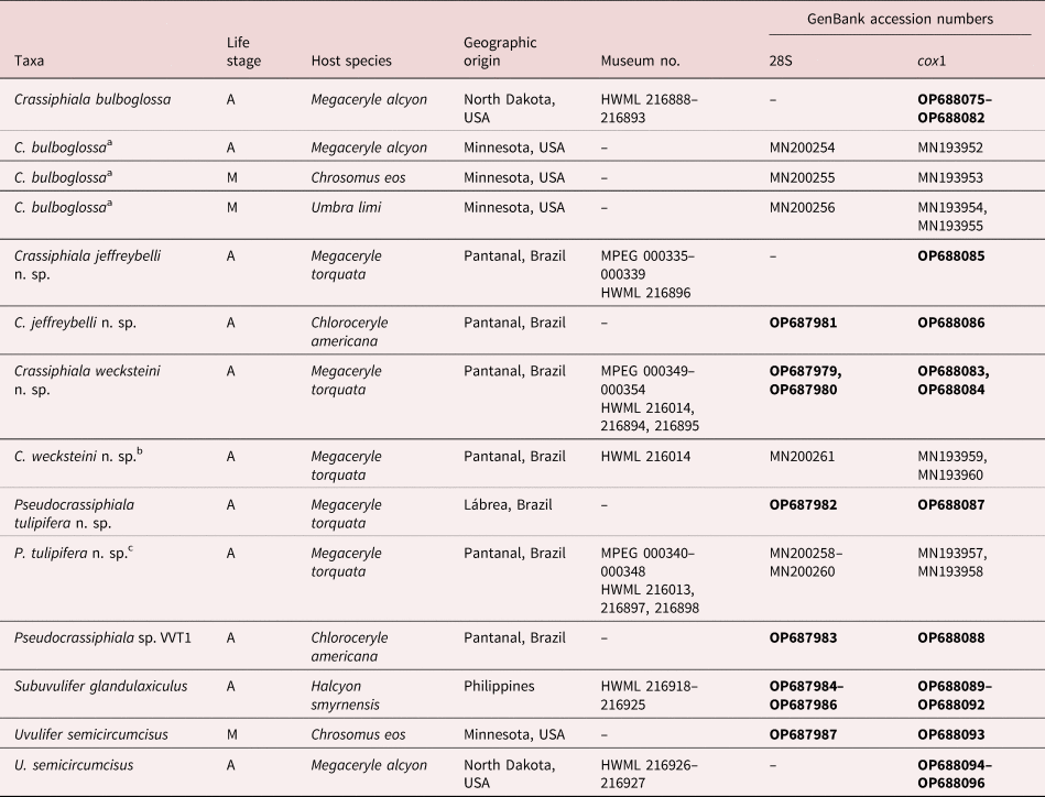
Hosts, GenBank accession numbers and museum accession numbers assigned by the Museu Paraense Emílio Goeldi (MPEG) and Harold W. Manter Laboratory (HWML) for diplostomids studied in present work.

Abbreviations for life stage: A, adult; C, cercaria; M, metacercaria. GenBank accession numbers for new sequences generated in the present study are in bold.
a Previously published as Crassiphiala sp. lineage 2 of Achatz et al. (Reference Achatz, Pulis, Fecchio, Schlosser and Tkach2019b).
b Previously published as Crassiphiala sp. lineage 5 of Achatz et al. (Reference Achatz, Pulis, Fecchio, Schlosser and Tkach2019b).
c Previously published as Crassiphiala sp. lineage 4 of Achatz et al. (Reference Achatz, Pulis, Fecchio, Schlosser and Tkach2019b).
The DNA extraction as well as the amplification and sequencing of partial 28S and cox1 genes were performed as previously described by Achatz et al. (Reference Achatz, Pulis, Fecchio, Schlosser and Tkach2019b, Reference Achatz, Martens and Kostadinova2022a). Newly generated contiguous sequences were assembled with the assistance of Sequencher version 4.2 software (GeneCodes Corp., Ann Arbor, Michigan, USA) and deposited in GenBank (table 1).
Phylogenetic relationships of the diplostomid taxa studied in the present work were estimated based on an alignment of partial 28S sequences. The alignment included newly generated sequences from members of Crassiphiala, Uvulifer, Subuvulifer and the new genus (table 1) and previously published sequences of 37 diplostomids and 14 strigeids. The alignment included representatives from all currently sequenced genera of the Diplostomidae and Strigeidae Railliet, 1919; we only included sequences that were at least 1,100 base pairs (bp) long to avoid significant loss of data. Only two representatives of the Proterodiplostomidae Dubois, 1936 were included because the monophyly of the family has been previously demonstrated (Tkach et al., Reference Tkach, Achatz, Pulis, Junker, Snyder, Bell, Halajian and Melo2020). Suchocyathocotyle crocodili (Yamaguti, 1954) was used as the outgroup for the analyses based on the study by Achatz et al. (Reference Achatz, Pulis, Junker, Binh, Snyder and Tkach2019c).
ClustalW implemented in MEGA7 was used to align newly generated and previously published sequences (Kumar et al., Reference Kumar, Stecher and Tamura2016). The alignment was trimmed to the length of the shortest sequence; ambiguous positions were excluded from each analysis.
The best-fitting nucleotide substitution models for the alignment were determined using MEGA7. The analysis used the general time-reversible model with estimates of invariant sites and gamma-distributed among-site variation (GTR + G + I) model. Bayesian Inference (BI) as implemented in MrBayes v3.2.6 software and Maximum Likelihood (ML) as implemented in MEGA7 were used to perform the phylogenetic analyses (Ronquist & Huelsenbeck, Reference Ronquist and Huelsenbeck2003; Kumar et al., Reference Kumar, Stecher and Tamura2016). The BI analysis was performed as follows: Markov chain Monte Carlo (MCMC) chains were run for 3,000,000 generations with sample frequency set at 1,000. This number of generations was considered sufficient as the standard deviation stabilized below 0.01. Log-likelihood scores were plotted and only the final 75% of trees were retained to produce the consensus trees. Nodal support of ML analysis was estimated by performing 1,000 bootstrap pseudoreplicates. Pairwise comparisons were performed using MEGA7.
Results
Molecular phylogenies
Upon trimming to the length of the shortest sequence, the alignment was 1,115 bp long; 33 nucleotide sites were excluded due to ambiguous homology. The strongly supported topologies were identical between the BI and ML analyses. The Diplostomidae and Strigeidae were clearly non-monophyletic throughout the basal polytomy (fig. 1); at the same time, the representatives of the Proterodiplostomidae formed a strongly supported clade (100% BI; 99% ML). These results are generally similar to those published and discussed in previous molecular phylogenetic studies (e.g. Blasco-Costa & Locke, Reference Blasco-Costa and Locke2017; Hernández-Mena et al., Reference Hernández-Mena, García-Varela and Pérez-Ponce de León2017; Achatz et al., Reference Achatz, Pulis, Fecchio, Schlosser and Tkach2019b, Reference Achatz, Pulis, Junker, Binh, Snyder and Tkachc, Reference Achatz, Bell, Melo, Fecchio and Tkach2021a, Reference Achatz, Chermak and Martensb, Reference Achatz, Martens and Kostadinova2022a, Reference Achatz, Martens, Kudlai, Junker, Boe and Tkachb; Tkach et al., Reference Tkach, Achatz, Pulis, Junker, Snyder, Bell, Halajian and Melo2020; Locke et al., Reference Locke, Drago and López-Hernández2021). Therefore, below we focus on details related to the clades that contain newly generated data.
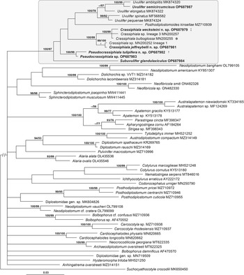
Phylogenetic interrelationships among 59 diplostomoideans based on Bayesian Inference (BI) and Maximum Likelihood (ML) analyses of partial 28S rDNA gene sequences. Topology from the BI analysis is provided. Bayesian inference posterior probability/ML bootstrap values are provided above internodes. The BI posterior probability values lower than 90% and ML bootstrap values lower than 50% are not shown. The new sequences generated in this study are in bold. The scale bar indicates the number of substitutions per site. The clade containing digeneans studied in the present work is in the shaded box. GenBank accession numbers are provided after names of taxa. *Previously published as Crassiphiala sp. lineage 2 of Achatz et al. (Reference Achatz, Pulis, Fecchio, Schlosser and Tkach2019b). ‡Previously published as Crassiphiala sp. lineage 5 of Achatz et al. (Reference Achatz, Pulis, Fecchio, Schlosser and Tkach2019b). †Previously published as Crassiphiala sp. lineage 4 of Achatz et al. (Reference Achatz, Pulis, Fecchio, Schlosser and Tkach2019b).

Members of Subuvulifer, Crassiphiala, Uvulifer, Posthodiplostomoides Williams, 1969 and the new genus formed a strongly supported clade (100% BI; 97% ML) in the basal polytomy of the Diplostomoidea consisting of five clades/branches with unresolved relationships, each representing a single genus (fig. 1). Subuvulifer and Posthodiplostomoides were represented in the tree by a single species each, Subuvulifer glandulaxiculus Pearson et Dubois, 1985 and Posthodiplostomoides kinsellae Achatz, Chermak, Martens, Pulis et Tkach, 2021. The clade containing two species of Pseudocrassiphiala n. gen. was strongly supported (100% BI; 99% ML).
The strongly supported (100% BI; 99% ML) clade containing Crassiphiala spp. (weak support in BI and ML) included two clusters: Crassiphiala jeffreybelli n. sp. + Crassiphiala sp. lineage 1 of Achatz et al. (Reference Achatz, Pulis, Fecchio, Schlosser and Tkach2019b) (99% BI; 100% ML) and C. bulboglossa + Crassiphiala wecksteini n. sp. + Crassiphiala sp. lineage 3 of Achatz et al. (Reference Achatz, Pulis, Fecchio, Schlosser and Tkach2019b) (100% BI; 99% ML).
Lastly, the clade of Uvulifer spp. included a well-supported (99% BI; 92% ML) grouping of Uvulifer pequenae Achatz, Curran, Patitucci, Fecchio et Tkach, 2019 + Uvulifer spinatus López-Jiménez, Pérez-Ponce de León et García-Varela, 2018 and a weakly supported clade (low BI support; 57% ML) of Uvulifer elongatus Dubois, 1988 + a strongly supported cluster (100% BI; 99% ML) of Uvulifer ambloplitis (Hughes, 1927) + U. semicircumcisus.
Descriptions of new taxa
At the time of publication of the previous diagnosis of Crassiphiala (Niewiadomska, Reference Niewiadomska, Gibson, Jones and Bray2002), the genus was considered monotypic. We provide a new diagnosis of the genus to accommodate the features of the new species described herein as well as Crassiphiala ceryliformis Vidyarthi, 1938. The latter species was originally placed in Crassiphiala and subsequently transferred into Uvulifer; we return it to Crassiphiala based on morphological evidence (see detailed discussion below).
Family Diplostomidae Poirier, 1886
Genus Crassiphiala Van Haitsma, 1925
Diagnosis. Body distinctly bipartite; prosoma generally flattened or with slight concavity, much shorter than cylindrical opisthosoma. Tegument unarmed. Oral sucker present; ventral sucker and pseudosuckers absent. Holdfast organ elliptical or bulbous, with median opening, may occupy entire width of prosoma. Pharynx present; caeca reach level of seminal vesicle. Testes two, tandem. Seminal vesicle compact, winding, with pouch-like expansion at proximal end. Ejaculatory pouch absent. Ejaculatory duct typically short, may be dilated resulting in pouch-like appearance, joins distal part of metraterm to form a short hermaphroditic duct. Hermaphroditic duct opens at apex of genital cone. Genital cone with prepucial (=prepuce-like) fold, opens into genital atrium. Genital atrium with terminal opening. Ovary pretesticular; oötype intertesticular. Vitellarium distributed throughout opisthosoma; vitelline reservoir intertesticular. Excretory pore subterminal on ventral side. In kingfishers. Nearctic, Neotropics, Indomalaya.
Type species: Crassiphiala bulboglossa Van Haitsma, 1925.
Other species: Crassiphiala ceryliformis Vidyarthi, 1938, Crassiphiala jeffreybelli n. sp. Achatz, Von Holten, Fecchio et Tkach, Crassiphiala wecksteini n. sp. Achatz, Von Holten, Fecchio et Tkach.
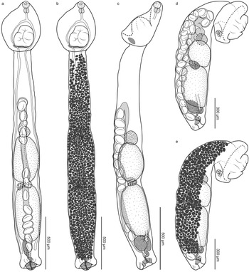
Crassiphiala bulboglossa Van Haitsma, 1925 (figs 2 and 3)
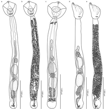
Crassiphiala bulboglossa. (a) hologenophore 1, ventral view with vitellarium omitted; (b) hologenophore 2, ventral view with vitellarium shown; (c) voucher 1, relaxed, lateral view; (d) voucher 2, contracted, lateral view with vitellarium omitted; (e) voucher 3, contracted, lateral view with vitellarium shown.

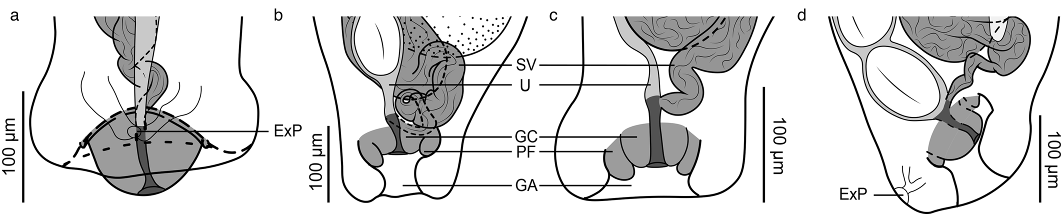
Posterior end of opisthosoma of Crassiphiala bulboglossa with vitellarium omitted. (a) hologenophore 1, ventral view, genital cone everted; (b) hologenophore 2, ventral view; (c) voucher 1, lateral view; (d) hologenophore 3, lateral view. Abbreviations: ExP, excretory pore; GA, genital atrium; GC, genital cone; PF, prepucial fold; SV, seminal vesicle; U, uterus.

Taxonomic summary
Type host: Megaceryle alcyon (Linnaeus) (Coraciiformes: Alcedinidae).
Site of infection: small intestine.
Type locality: Douglas Lake Michigan, USA.
Collection locality in this study: Grand Forks Co., North Dakota, USA (47°58′56.3″N 97°15′36.3″W).
Infection rate: numerous C. bulboglossa were found in single examined M. alcyon from North Dakota.
Type material: slides deposited in the National Museum of Natural History (NMNH), Washington DC, under accession number USNPC 071491.00.
New specimens deposited: 38 mature specimens deposited in the HWML. Vouchers: HWML 216888, labelled ex. Megaceryle alcyon, small intestine, Grand Forks Co., North Dakota, USA, June 07, 2018, coll. T. Achatz. Hologenophores (five slides): HWML 216889–216893, label identical to the vouchers.
Hologenophore DNA sequences: cox1: OP688075 (HWML 216889), OP688076 (HWML 216890), OP688077 (HWML 216891), OP688079 (HWML 216892), OP688081 (HWML 216893).
Previously published genetic lineage name: Crassiphiala sp. lineage 2 of Achatz et al. (Reference Achatz, Pulis, Fecchio, Schlosser and Tkach2019b)
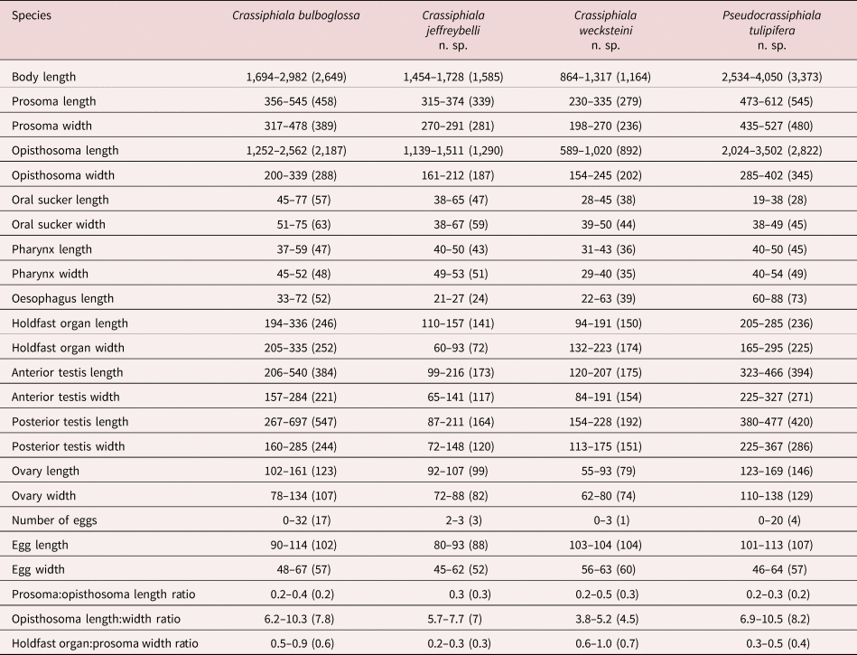
Description. Based on 38 adult specimens. Measurement ranges of series given in text and table 2. Body 1,694–2,982 long, consists of distinct prosoma and opisthosoma; prosoma oval, with shallow concavity, 356–545 long, widest at level of holdfast organ, 317–478; opisthosoma elongated, cylindrical, 1,252–2,562 × 200–339; opisthosoma length:width ratio 6.2–10.3. Prosoma:opisthosoma length ratio 0.2–0.4. Tegument unarmed. Oral sucker subterminal, 45–77 × 51–75. Pseudosuckers absent. Holdfast organ oval, with longitudinal aperture, armed with fine spines, proximal portion glandular, 194–336 × 205–335; holdfast organ:prosoma width ratio 0.5–0.9. Proteolytic gland consisting of diffuse gland cells. Prepharynx absent. Pharynx subspherical, 37–59 × 45–52. Oesophagus 33–72 long. Caecal bifurcation in anterior 40% of prosoma length. Caeca slender, extend to near level of seminal vesicle.
Morphometric characters of diplostomids described in the present study.

Ranges provided followed by mean in parentheses.
Testes two, tandem, rounded, entire, anterior testis 206–540 × 157–284, posterior testis 267–697 × 160–285. Seminal vesicle post-testicular, proximal portion pouch-like, followed by winding distal portion that joins distal part of metraterm to form short hermaphroditic duct. Hermaphroditic duct opens at apex of small, muscular genital cone. Genital cone surrounded by small prepucial fold, positioned within genital atrium. Prepucial fold small or not observable when genital cone everted (fig. 3a vs. fig. 3b–d). Genital atrium with terminal opening.
Ovary pretesticular, subspherical, 102–161 × 78–134. Oötype and Mehlis’ gland intertesticular (not illustrated). Vitelline follicles limited to opisthosoma, distributed from near level of prosoma–opisthosoma junction to near posterior end of opisthosoma. Vitelline reservoir intertesticular. Uterus ventral to gonads, extends anteriorly beyond level of ovary before turning and extending posteriorly. Uterus in our specimens containing up to 32 eggs. Eggs 90–114 × 48–67.
Excretory pore subterminal, ventral.
Remarks
Historically, descriptions of many diplostomoideans were based on laterally oriented specimens: for example see the numerous illustrations in the monograph by Dubois (Reference Dubois1968). The same is true in the case of the original description and illustrations of C. bulboglossa (Van Haitsma, Reference Van Haitsma1925). Our newly collected adult specimens of C. bulboglossa allowed us to study these digeneans in both ventrodorsal and lateral orientations. Although the description by Van Haitsma (Reference Van Haitsma1925) lacked many essential measurements, the description and illustrations closely resemble our contracted, laterally positioned specimens (fig. 2d, e). Our newly collected digeneans demonstrated substantial morphological variation in appearance. Some of this variation may be the result of different ages of the diplostomids or the crowding effect, since the host studied was infected with many hundreds of these digeneans. The extent of body contraction and eversion of the genital cone are at least partly responsible for the observed morphological variation (figs 2 and 3). The partial cox1 sequences of C. bulboglossa obtained from specimens of different sizes and states of contraction were identical (table 1).
Crassiphiala jeffreybelli n. sp. Achatz, Von Holten, Fecchio et Tkach (figs 4 and 5)
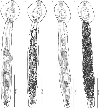
Crassiphiala jeffreybelli n. sp. (a) holotype, ventral view with vitellarium omitted; (b) holotype, ventral view with vitellarium shown; (c) paratype, ventral view with vitellarium omitted; (d) paratype, ventral view with vitellarium shown.

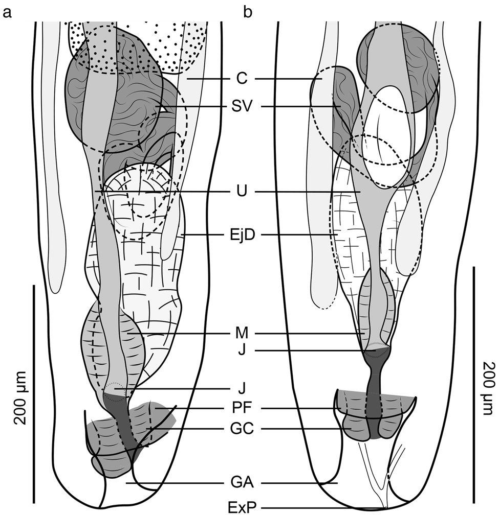
Posterior end of opisthosoma of Crassiphiala jeffreybelli n. sp. with vitellarium omitted. (a) holotype, ventral view; (b) paratype, ventral view. Abbreviations: C, ceca; EjD, ejaculatory duct; ExP, excretory pore; GA, genital atrium; GC, genital cone; J, joining site of male and female ducts; M, metraterm; PF, prepucial fold; SV, seminal vesicle; U, uterus.

Taxonomic summary
Type host: Megaceryle torquata (Linnaeus) (Coraciiformes: Alcedinidae).
Other host: Chloroceryle americana (Gmelin) (Coraciiformes: Alcedinidae).
Site of infection: small intestine.
Type locality: Pantanal, Fazenda Retiro Novo, Municipality of Poconé, Mato Grosso State, Brazil (16°21′53″S 56°17′31″W).
Infection rate: one of four of Megaceryle torquata and one of three of Chloroceryle americana were infected by six and one specimens of C. jeffreybelli n. sp., respectively.
Type-material: the type series consists of six mature specimens deposited in the MPEG and HWML. Holotype: MPEG 000335, labelled ex. Megaceryle torquata, small intestine, Pantanal, Fazenda Retiro Novo, Municipality of Poconé, Mato Grosso State, Brazil, June 07 2017, coll. A. Fecchio. Paratypes: MPEG 000336–000339 (lot of four slides). Hologenophore: HWML 216896, label identical to the holotype.
Hologenophore DNA sequences: cox1: OP688085 (HWML 216896).
Zoobank registration: urn:lsid:zoobank.org:act:153FC1A5-7E02-498D-8E71-F1BD95A24473.
Etymology: the species is named after Dr Jeffrey A. Bell for his contributions to our knowledge of parasites of South American birds and for being a terrific colleague and team member during field trips.
Description. Based on six adult specimens. Measurements of holotype in text; measurements of entire series given in table 2. Body 1,589 long, consists of distinct prosoma and opisthosoma; prosoma oval, flattened, posterior extremity with slight ventral concavity, 345 long, widest at level of holdfast organ, 291; opisthosoma elongated, cylindrical, 1,244 × 161; opisthosoma length:width ratio 7.7. Prosoma:opisthosoma length ratio 0.3. Tegument unarmed. Oral sucker subterminal, 38 × 61. Pseudosuckers absent. Holdfast organ oval, with longitudinal opening, proximal portion glandular, 143 × 60; holdfast organ:prosoma width ratio 0.2. Proteolytic gland consisting of diffuse gland cells. Prepharynx absent. Pharynx subspherical, 41 × 52. Oesophagus 21 long. Caecal bifurcation in anterior 40% of prosoma length. Caeca slender, extend to level of seminal vesicle.
Testes two, tandem, generally rounded, entire, anterior testis 142 × 102, posterior testis 141 × 104. Seminal vesicle post-testicular, proximal portion pouch-like, followed by winding distal portion connected to ejaculatory duct; ejaculatory duct strongly dilated, pouch-like, elongated, glandular, joining distal part of metraterm to form short hermaphroditic duct. Hermaphroditic duct opens at apex of small, muscular genital cone. Genital cone with prepucial fold, positioned within genital atrium; genital atrium with terminal opening.
Ovary pretesticular, subspherical, 98 × 77. Oötype and Mehlis’ gland intertesticular (not illustrated). Vitelline follicles limited to opisthosoma, distributed from near level of prosoma to near posterior end of opisthosoma. Vitelline reservoir intertesticular. Uterus ventral to gonads, extends anteriorly beyond level of ovary before turning and extending posteriorly. Terminal part of uterus and proximal part of hermaphroditic duct muscular. Uterus contains three eggs in holotype (up to three eggs in paratypes). Eggs 80–93 × 45–62.
Excretory pore subterminal on ventral side.
Remarks
Crassiphiala jeffreybelli n. sp. belongs to Crassiphiala based on several morphological features, including a distinctly bipartite body, vitellarium confined to the opisthosoma, presence of a prepucial fold associated with the genital cone, unarmed tegument and the absence of pseudosuckers, ventral sucker and genital atrium sucker.
Unlike other Crassiphiala spp., except for C. ceryliformis, the ejaculatory duct is dilated and pouch-like. Crassiphiala jeffreybelli n. sp. and C. ceryliformis are similar in size (body length 1,454–1,728 in the new species vs. 1,440–1,648 in C. ceryliformis), but most organs/structures of C. ceryliformis are much smaller (e.g. oral sucker 38–65 × 38–67 in C. jeffreybelli n. sp. vs. 17.5–25 × 24–25 in C. ceryliformis; pharynx 40–50 × 49–53 in C. jeffreybelli n. sp. vs. pharynx 21 × 18–23 in C. ceryliformis).
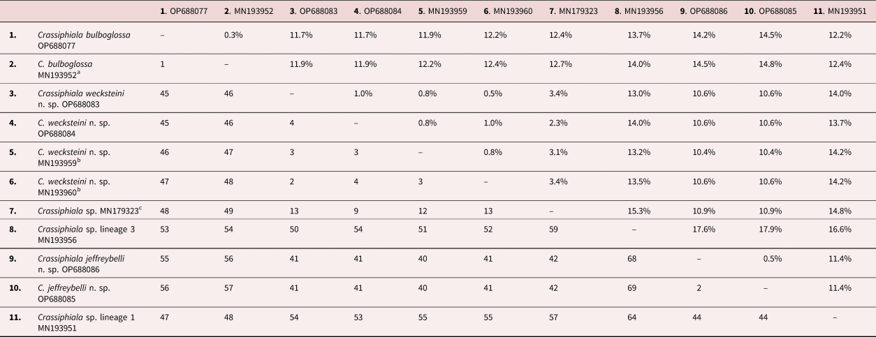
The eggs of C. ceryliformis (40–80) are smaller than in C. jeffreybelli n. sp. (80–93), with the size ranges barely overlapping. The posterior testis of C. ceryliformis is bilobed, while the posterior testis of C. jeffreybelli n. sp. is not lobed. In addition, C. ceryliformis is only known from India, whereas the new species described herein was collected in Brazil. Crassiphiala jeffreybelli n. sp. differs from its congeners (except for Crassiphiala sp. lineage 1) by 0.9–1.1% in the partial sequences of 28S (table 3) and 10.4–17.9% in partial sequences of cox1 (table 4). No DNA sequences of C. ceryliformis are available. Crassiphiala jeffreybelli n. sp. and Crassiphiala sp. lineage 1 of Achatz et al. (Reference Achatz, Pulis, Fecchio, Schlosser and Tkach2019b) have identical partial sequences of 28S (table 3), but differ by 11.4% in partial sequences of cox1 (table 4). Unfortunately, the adult specimens of Crassiphiala sp. lineage 1 of Achatz et al. (Reference Achatz, Pulis, Fecchio, Schlosser and Tkach2019b) from M. alcyon in Minnesota, USA are in too poor condition to describe or use for morphological differentiation.
Pairwise comparisons of partial 28S rDNA gene sequences among Crassiphiala spp.

Percentage differences are given above the diagonal, and number of variable nucleotide positions are given below the diagonal. Results based on a 1,108 bp long alignment.
a Previously published as Crassiphiala sp. lineage 2 of Achatz et al. (Reference Achatz, Pulis, Fecchio, Schlosser and Tkach2019b).
Pairwise comparisons of partial cox1 mitochondrial DNA gene sequences among Crassiphiala spp.

Percentage differences are given above the diagonal, and number of variable nucleotide positions are given below the diagonal. Results based on a 386 bp long alignment.
a Previously published as Crassiphiala sp. lineage 2 of Achatz et al. (Reference Achatz, Pulis, Fecchio, Schlosser and Tkach2019b).
b Previously published as Crassiphiala sp. lineage 5 of Achatz et al. (Reference Achatz, Pulis, Fecchio, Schlosser and Tkach2019b).
c Previously published as Crassiphialinae gen. sp. of López-Hernández et al. (Reference López-Hernández, Locke, Alves de Assis, Drago, Melo, Rabelo and Pinto2019).
Crassiphiala wecksteini n. sp. Achatz, Von Holten, Fecchio et Tkach (figs 6 and 7)
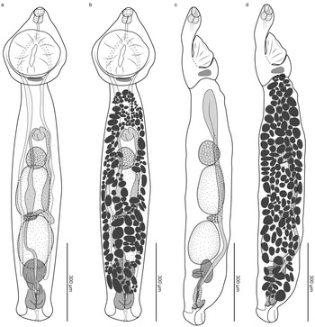
Crassiphiala wecksteini n. sp. (a) holotype, ventral view with vitellarium omitted; (b) holotype, ventral view with vitellarium shown; (c) paratype, lateral view with vitellarium omitted; (d) paratype, lateral view with vitellarium shown.

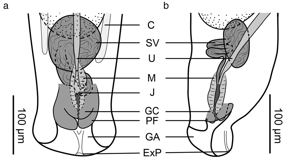
Posterior end of opisthosoma of Crassiphiala wecksteini n. sp. with vitellarium omitted. (a) holotype, ventral view; (b) paratype, lateral view. Abbreviations: C, caeca; ExP, excretory pore; GA, genital atrium; GC, genital cone; J, joining site of male and female ducts; M, metraterm; PF, prepucial fold; SV, seminal vesicle; U, uterus.

Taxonomic summary
Type host: Megaceryle torquata (Linnaeus) (Coraciiformes: Alcedinidae).
Site of infection: small intestine.
Type locality: Pantanal, Fazenda Retiro Novo, Municipality of Poconé, Mato Grosso State, Brazil (16°21′53″S 56°17′31″W).
Infection rate: one of four M. torquata was infected with eight C. wecksteini n. sp.
Type-material: the type series consists of eight mature specimens deposited in the MPEG and HWML. Holotype: MPEG 000349, labelled ex. Megaceryle torquata, small intestine, Pantanal, Fazenda Retiro Novo, Municipality of Poconé, Mato Grosso State, Brazil, June 07 2017, coll. A. Fecchio. Paratypes: MPEG 000350–000354 (five slides), HWML 216894 (one slide), labels identical to the holotype. Hologenophore: HWML 216895, label identical to the holotype.
Other specimens: HWML 216014.
Hologenophore DNA sequences: 28S: OP687979 (HWML 216895), cox1: OP688083 (HWML 216895).
Previously published genetic lineage name: Crassiphiala sp. lineage 5 of Achatz et al. (Reference Achatz, Pulis, Fecchio, Schlosser and Tkach2019b).
Zoobank registration: urn:lsid:zoobank.org:act:689706E1-C42B-4021-90AA-E282BF2CED42.
Etymology: the species is named after Dr Jason D. Weckstein in recognition of his numerous contributions to knowledge of South American birds and their parasites.
Description. Based on eight adult specimens. Measurements of holotype in text; measurements of entire series given in table 2. Body 1,317 long, consists of distinct prosoma and opisthosoma; prosoma oval, generally flattened, posterior portion with slight concavity, 355 long, widest at level of holdfast organ, 270; opisthosoma elongated, cylindrical, 982 × 220; opisthosoma length:width ratio 4.5. Prosoma:opisthosoma length ratio 0.4. Tegument unarmed. Oral sucker subterminal, 45 × 44. Pseudosuckers absent. Holdfast organ oval, with longitudinal aperture, armed with fine spines, proximal portion glandular, 191 × 222; holdfast organ:prosoma width ratio 0.8. Proteolytic gland consisting of diffuse gland cells. Prepharynx absent. Pharynx subspherical, 38 × 40. Oesophagus 63 long. Caecal bifurcation in anterior half of prosoma length. Caeca slender, extend to level of seminal vesicle.
Testes two, tandem, rounded, entire; anterior testis 177 × 133, posterior testis 215 × 135. Seminal vesicle post-testicular, proximal portion pouch-like, followed by winding distal portion that joins distal part of metraterm to form short hermaphroditic duct. Hermaphroditic duct opens at apex of muscular genital cone. Genital cone surrounded by prepucial fold, positioned within genital atrium; genital atrium with terminal opening.
Ovary pretesticular, subspherical, 86 × 80. Oötype and Mehlis’ gland intertesticular (not illustrated). Vitelline follicles limited to opisthosoma, distributed from near level of prosoma to near posterior end of opisthosoma. Vitelline reservoir intertesticular. Uterus ventral to gonads, extends anteriorly beyond level of ovary before turning and extending posteriorly. Terminal part of uterus and proximal part of hermaphroditic duct surrounded by distinct layer of muscles. Uterus contains no eggs in holotype (up to three in paratype). Eggs 103–104 × 56–63.
Excretory pore subterminal on ventral side.
Remarks
The new species belongs to Crassiphiala based on morphological features, including a distinctly bipartite body, vitellarium confined to the opisthosoma, presence of a prepucial fold associated with the genital cone, unarmed tegument and the absence of pseudosuckers, ventral sucker and genital atrium sucker. The molecular phylogeny also positioned this species in a strongly supported clade (100% BI; 99% ML) with C. bulboglossa (fig. 1).
Crassiphiala wecksteini n. sp. can be easily distinguished from C. jeffreybelli n. sp. and C. ceryliformis by the absence of a dilated, pouch-like ejaculatory duct. In contrast, C. wecksteini n. sp. and C. bulboglossa are extremely similar morphologically. The majority of measurements of C. wecksteini n. sp. are smaller than those of C. bulboglossa (table 2) and the opisthosoma length:width ratio is somewhat smaller in C. wecksteini n. sp. (3.8–5.2; average 4.5) compared with C. bulboglossa (6.2–10.3; average 7.8). The most obvious difference is that the prepucial fold surrounding the genital cone in C. wecksteini n. sp. (fig. 7) is more pronounced than in C. bulboglossa (fig. 3). The metraterm is well-developed in C. wecksteini n. sp., but not in C. bulboglossa. Based on molecular studies, C. wecksteini n. sp. has only been reported from the Neotropics, whereas C. bulboglossa is only known from the Nearctic, although there is a single report of C. bulboglossa from an unknown kingfisher in Brazil (Dubois, Reference Dubois1970). It is possible, however, that these digeneans may have broader distributions than currently recognized and may even occur in sympatry. Megaceryle torquata (the host of C. wecksteini n. sp.) is distributed between the southern USA and Tierra del Fuego, while M. alcyon (the host of C. bulboglossa) is distributed between the northern part of North America and northern edge of South America (Brush, Reference Brush and Poole2020; Kelly et al., Reference Kelly, Bridge, Hamas and Poole2020).
The partial 28S sequences of C. wecksteini n. sp. differ by at least 0.2% from its congeners (table 3), and the partial cox1 sequences of this new species differ by at least 11.7% from C. bulboglossa and by at least 10.4% from C. jeffreybelli (table 4).
Family Diplostomidae Poirier, 1886
Genus Pseudocrassiphiala n. gen. Achatz, Von Holten, Fecchio et Tkach
Diagnosis. Body distinctly bipartite; prosoma strongly concave, cup-shaped, much shorter than cylindrical opisthosoma. Tegument of prosoma and anterior part of opisthosoma armed with fine spines. Oral sucker present; ventral sucker and pseudosuckers absent. Holdfast organ subspherical, with longitudinal aperture, most of holdfast organ positioned inside prosoma concavity. Pharynx present; caeca reach level of or posterior to level of seminal vesicle. Testes two, tandem. Seminal vesicle post-testicular, winding, with pouch-like expansion at proximal end. Ejaculatory duct short, joins distal part of metraterm to form a short hermaphroditic duct. Hermaphroditic duct opens at apex of short genital cone. Genital cone with ventrolateral prepucial fold, opens into genital atrium. Genital atrium with terminal opening. Ovary pretesticular; oötype intertesticular. Vitellarium distributed throughout opisthosoma; vitelline reservoir intertesticular. Excretory pore subterminal on ventral side. In kingfishers. Neotropics.
Type species: Pseudocrassiphiala tulipifera n. sp. Achatz, Von Holten, Fecchio et Tkach.
Zoobank registration: urn:lsid:zoobank.org:act:F8E6CFEB-7F32-4499-9677-6AC4045E36CF.
Etymology: the name of the new genus refers to its morphological similarity to Crassiphiala.
Remarks
While molecular comparisons revealed the presence of two members in the new genus (fig. 1; 0.4% and 7.5–7.8% divergence between lineages in 28S and cox1 sequences, respectively), we only had quality adult specimens of the type-species. The differentiation below relies only on the type-species and may require amendment once morphological data from the second member become available.
Pseudocrassiphiala n. gen. belongs to the Diplostomidae based on the presence of a sucker-like holdfast organ and absence of a paraprostate. The new genus can be distinguished from most other diplostomids, with the exceptions of Cercocotyla and Crassiphiala, based on the absence of pseudosuckers and ventral sucker. It is worth noting that members of these genera were included in our 28S phylogenetic analyses (fig. 1). Members of Cercocotyla possess a genital atrium sucker which is absent in the type-species of Pseudocrassiphiala n. gen.
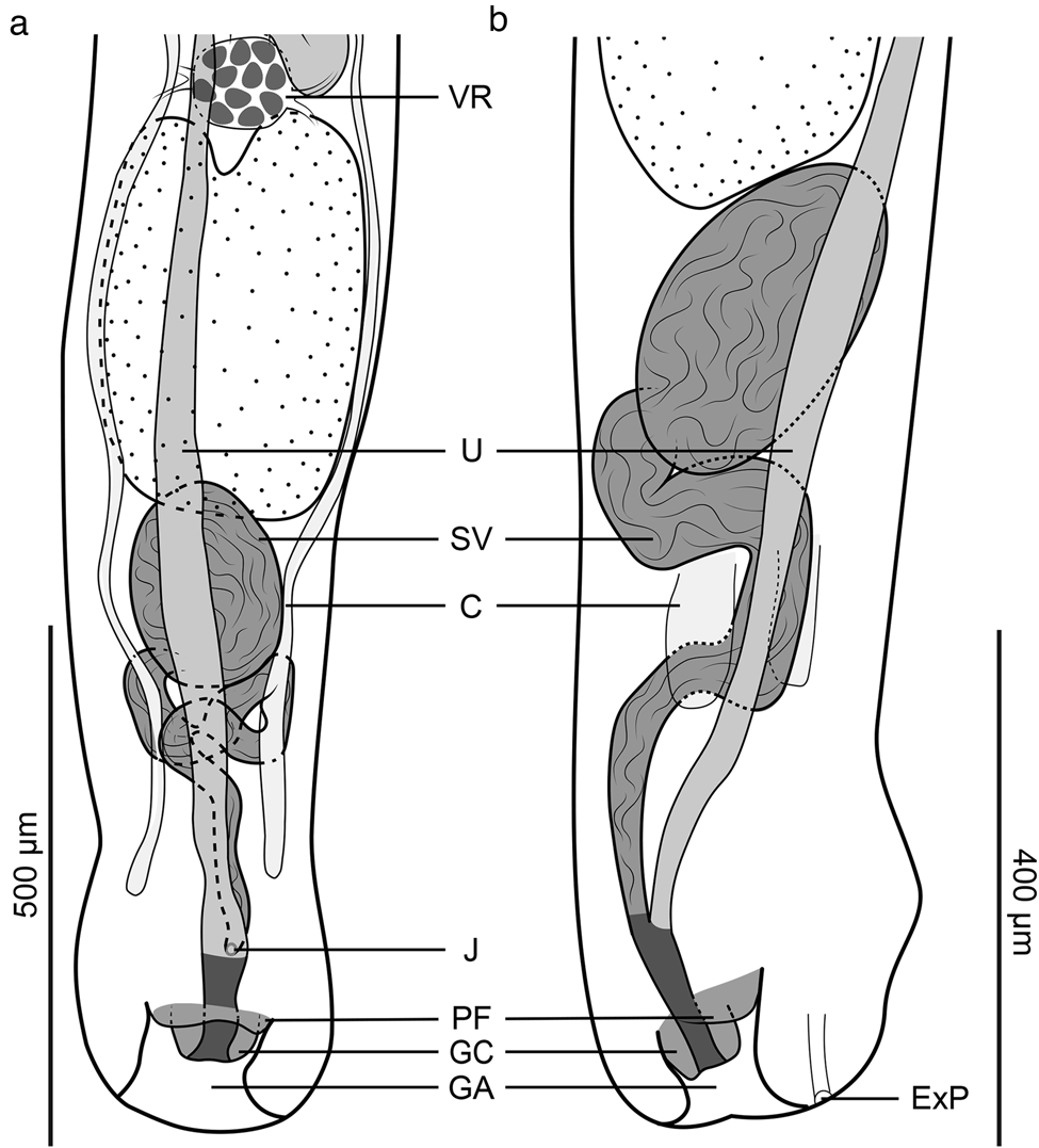
The new genus and Crassiphiala are morphologically similar and their reliable differentiation requires quality adult specimens. Based on light microscopy, in Pseudocrassiphiala n. gen. the tegument of the prosoma and anterior part of opisthosoma are armed with fine spines, while in Crassiphiala the tegument is unarmed. The type-species of Pseudocrassiphiala n. gen. has a cup-like prosoma with a deep concavity that opens anteriorly (fig. 8), while the prosoma of Crassiphiala is generally flattened or has only a shallow concavity that opens ventrally (figs 2, 4 and 6). In the type-species of Pseudocrassiphiala n. gen., most of the holdfast organ is positioned within the deep prosoma concavity, while most of the holdfast organ in Crassiphiala spp. is not within prosoma concavity.
Pseudocrassiphiala tulipifera n. sp. (a) holotype, ventral view with vitellarium omitted; (b) holotype, ventral view with vitellarium shown; (c) paratype 1, ventral view with vitellarium omitted; (d) paratype 2, lateral view with vitellarium omitted; (e) paratype 2, lateral view with vitellarium shown.

Pseudocrassiphiala tulipifera n. sp. Achatz, Von Holten, Fecchio et Tkach (figs 8 and 9)
Posterior end of opisthosoma of Pseudocrassiphiala tulipifera n. sp. with vitellarium omitted. (a) holotype, ventral view; (b) paratype 2, lateral view. Abbreviations: C, caeca; ExP, excretory pore; GA, genital atrium; GC, genital cone; J, joining site of male and female ducts; PF, prepucial fold; SV, seminal vesicle; U, uterus; VR, vitelline reservoir.

Taxonomic summary
Type host: Megaceryle torquata (Linnaeus) (Coraciiformes: Alcedinidae).
Other host: Chloroceryle americana (Gmelin) (Coraciiformes: Alcedinidae).
Site of infection: small intestine.
Type locality: Pantanal, Fazenda Retiro Novo, Municipality of Poconé, Mato Grosso State, Brazil (16°21′53″S 56°17′31″W).
Other locality: Lábrea municipality, Amazonas State, Brazil.
Infection rate: one in four M. torquata was infected with 12 P. tulipifera n. sp. in Pantanal and a single examined specimen of M. torquata from Lábrea was infected with two P. tulipifera n. sp.
Type-material: the type series consists of 12 mature specimens deposited in the MPEG and HWML. Holotype: MPEG 000340, labelled ex. Megaceryle torquata, small intestine, Pantanal, Fazenda Retiro Novo, Municipality of Poconé, Mato Grosso State, Brazil, June 07 2017, coll. A. Fecchio. Paratypes: MPEG 000341–000348 (lot of eight slides), HWML 216897 (lot of two slides), labels identical to the holotype. Hologenophore: HWML 216898, label identical to the holotype.
Other specimens: HWML 216013.
Hologenophore DNA sequences: 28S: MN200258, cox1: MN193957.
Previously published genetic lineage name: Crassiphiala sp. lineage 4 of Achatz et al. (Reference Achatz, Pulis, Fecchio, Schlosser and Tkach2019b).
Zoobank registration: urn:lsid:zoobank.org:act:0CECC018-8A0A-4B5F-88E0-88951A62986A.
Etymology: the specific epithet refers to the tulip-shaped prosoma in the new species.
Description. Based on 12 adult specimens. Measurements of holotype in text; measurements of entire series given in table 2. Body 3,945 long, consists of distinct prosoma and opisthosoma; prosoma deeply concave, cup-like, 547 long, widest at level of holdfast organ, 489; opisthosoma elongated, cylindrical, 3,398 × 324; opisthosoma length:width ratio 10.5. Prosoma:opisthosoma length ratio 0.2. Tegument of prosoma and anterior part of opisthosoma armed with fine spines. Oral sucker subterminal, 32 × 48. Pseudosuckers absent. Holdfast organ subspherical, with longitudinal aperture, most of holdfast organ positioned within prosoma concavity, proximal portion glandular, 235 × 185; holdfast organ:prosoma width ratio 0.4. Proteolytic gland dorsal to holdfast organ, consists of diffuse gland cells. Prepharynx absent. Pharynx subspherical, 47 × 52. Oesophagus 76 long. Caecal bifurcation in anterior-most third of prosoma length. Caeca slender, reach level of, or posterior to, level of seminal vesicle.
Testes two, tandem, rounded, entire, anterior testis 403 × 260, posterior testis with shallow invagination on anterior margin, 440 × 275. Seminal vesicle post-testicular, proximal portion expansive, pouch-like, followed by winding distal portion that joins distal part of metraterm to form short hermaphroditic duct. Hermaphroditic duct opens at apex of small genital cone. Genital cone with ventrolateral prepucial fold, positioned within genital atrium; genital atrium with terminal opening.
Ovary pretesticular, subspherical, 160 × 136. Oötype and Mehlis’ gland intertesticular (not illustrated). Vitelline follicles limited to opisthosoma, distributed from near level of prosoma to near posterior end of opisthosoma. Vitelline reservoir intertesticular. Uterus ventral to gonads, extends anteriorly to beyond level of ovary before turning and extending posteriorly. Uterus in holotype does not contain eggs (up to 20 in paratype). Eggs 101–113 × 46–64.
Excretory pore subterminal, ventral.
Discussion
Our phylogenetic analyses (fig. 1) demonstrated that members of Subuvulifer, Pseudocrassiphiala, Crassiphiala, Posthodiplostomoides and Uvulifer form a strongly supported monophyletic group. Although the interrelationships among genera have not been resolved, all genus-level clades were strongly supported and revealed the presence of at least two unknown species-level lineages of Crassiphiala and one additional species of Pseudocrassiphiala. Unfortunately, specimens representing these three additional species were either adults in poor condition or metacercariae and thus not suitable for descriptions.
Van Haitsma (Reference Van Haitsma1925) erected Crassiphiala for diplostomids collected from the intestine of M. alcyon in Michigan, USA. Until recently, the genus was viewed as monotypic and limited in its distribution to the Nearctic (Preble & Harwood, Reference Preble and Harwood1944; Dubois & Rausch, Reference Dubois and Rausch1948 Hoffman, Reference Hoffman1956; Dubois, Reference Dubois1968; Boyd & Fry, Reference Boyd and Fry1971; Scott, Reference Scott1984; Niewiadomska, Reference Niewiadomska, Gibson, Jones and Bray2002; Muzzall et al., Reference Muzzall, Cook and Sweet2011), except for a single report by Dubois (Reference Dubois1970) who identified C. bulboglossa among specimens from an unknown species of kingfisher in Brazil collected by A. Lutz. Based on DNA sequences, Achatz et al. (Reference Achatz, Pulis, Fecchio, Schlosser and Tkach2019b) demonstrated the presence of at least five species-level lineages of Crassiphiala throughout the New World (three in North America and two in South America). The present data reveals the presence of at least two additional closely related species-level lineages in the New World. We have provided morphological descriptions for four of these seven species-level lineages, which include representatives of a new genus (Pseudocrassiphiala n. gen.) as well as Crassiphiala (tables 1 and 2). It is worth noting that Crassiphialinae gen. sp. collected from Biomphalaria straminea (Dunker) in Belo Horizonte, State of Minais Gerais, Brazil by López-Hernández et al. (Reference López-Hernández, Locke, Alves de Assis, Drago, Melo, Rabelo and Pinto2019), is potentially conspecific with C. wecksteini n. sp. based on the low level of genetic divergence. The two forms differ by only 2.3–3.4% in partial sequences of cox1 (table 4). However, previous studies (Achatz et al., Reference Achatz, Chermak and Martens2021b and references therein) have demonstrated that interspecific variation between diplostomid species may be as low as 3.4% in this fragment of cox1. Intraspecific variability of cox1 sequences of Crassiphiala spp. in our study showed minimal variation (0.5% in C. jeffreybelli, up to 0.3% in C. bulboglossa, and up to 1% in C. wecksteini). The two partial cox1 sequences of P. tulipifera differed by only 1.3%, despite originating from distant (about 1,400 km) geographic locations in Brazil.
In the original description of C. bulboglossa, Van Haitsma (Reference Van Haitsma1925) inaccurately referred to an expanded portion of the seminal vesicle as an ejaculatory pouch. An ejaculatory pouch in diplostomids is a muscular/glandular structure that surrounds at least part of the ejaculatory duct (Achatz et al., Reference Achatz, Martens, Kudlai, Junker, Boe and Tkach2022b). This structure is absent in Crassiphiala spp., but present in members of other diplostomid genera, including Uvulifer (Niewiadomska, Reference Niewiadomska, Gibson, Jones and Bray2002; Achatz et al., Reference Achatz, Curran, Patitucci, Fecchio and Tkach2019a, Reference Achatz, Martens, Kudlai, Junker, Boe and Tkach2022b).
Vidyarthi (Reference Vidyarthi1938) described C. ceryliformis based on specimens collected from the intestine of the pied kingfisher Ceryle rudis (Linnaeus) in India. Crassiphiala ceryliformis was described as lacking a ventral sucker, but having an ejaculatory pouch (Vidyarthi, Reference Vidyarthi1938). Bhalerao (Reference Bhalerao1942) transferred this species into Uvulifer based on the smaller holdfast organ compared with C. bulboglossa and stated that the relative holdfast organ size held more taxonomic importance than the presence/absence of the ventral sucker. It is clear that Uvulifer ceryliformis (Vidyarthi, 1938) is more morphologically similar to C. jeffreybelli n. sp. than to any member of Uvulifer. Based on the illustration by Vidyarthi (Reference Vidyarthi1938), the ejaculatory pouch of U. ceryliformis is probably a dilated ejaculatory duct, similar to the condition in C. jeffreybelli n. sp. Both U. ceryliformis and C. jeffreybelli n. sp. lack a ventral sucker and have a holdfast organ that does not occupy much of the prosoma width. Based on morphological comparisons, we return U. ceryliformis to Crassiphiala as Crassiphiala ceryliformis (Vidyarthi, 1938).
The fauna of diplostomids parasitic in New World kingfishers is likely much richer than previously known. Until 2018, only a single species of Crassiphiala and five species of Uvulifer were known from kingfishers in the New World. The present study and recent publications (López-Jiménez et al., Reference López-Jiménez, Pérez-Ponce de León and García-Varela2018; Achatz et al., Reference Achatz, Curran, Patitucci, Fecchio and Tkach2019a, Reference Achatz, Pulis, Fecchio, Schlosser and Tkachb) have revealed four additional species/species-level lineages of Crassiphiala and seven additional species/species-level lineages of Uvulifer in the New World. The diversity of these diplostomids from kingfishers in the New World is further expanded by the members of Pseudocrassiphiala n. gen. (two species/species-level lineages), Sphincterodiplostomum Dubois, 1936 (one species) and Posthodiplostomum Dubois, 1936 (one species) (Achatz et al., Reference Achatz, Bell, Melo, Fecchio and Tkach2021a, Reference Achatz, Chermak and Martensb). Based on the current knowledge, it is certain that at least four species of Uvulifer as well as additional Crassiphiala (two species) and Pseudocrassiphiala n. gen. (one species) require description when suitable specimens are available. Future parasitological surveys should attempt to collect quality adult specimens of these diplostomids for morphological study.
We have provided the first DNA sequence data from a member of Subuvulifer (S. glandulaxiculus) and U. semicircumcisus. Subuvulifer is a small genus with only three nominal species: Subuvulifer halcyonae (Gogate, 1940), Subuvulifer sabahensis (Fischthal et Kuntz, 1973) and S. glandulaxiculus. Members of this genus are only known to parasitize kingfishers and have only been reported in Southeastern Asia and India. It would not be surprising if Subuvulifer also contains several currently undescribed species. More research is required to further explore the diversity and relationships of diplostomids, and other digeneans, parasitic in kingfishers.
Acknowledgements
We are grateful to Dr Jason D. Weckstein (Drexel University, Philadelphia, Pennsylvania, USA) for his help with collecting the specimens from Lábrea and Dr João B. Pinho (Universidade Federal de Mato Grosso, Cuiabá, Mato Grosso, Brazil) for his invaluable help with obtaining collecting permits and organizing field trips in Pantanal. We are indebted to Dr Rob Moyle and Dr Carl Oliveros (University of Kansas, Lawrence, Kansas, USA) for organizing expeditions in the Philippines and providing birds for parasitological examination.
Financial support
This study was supported by the National Science Foundation (VVT, grant number DEB-1120734; University of North Dakota School of Medicine & Health Sciences, REU Site award number 1852459), National Institutes of Health, USA (VVT, grant number R15AI092622), University of North Dakota (TJA, Joe K. Neel Memorial Award, Esther Wadsworth Hall Wheeler Award, Dissertation Research Award and Pre-PostDoc Research and Grant Writing Experience), American Society of Parasitologists (TJA, Willis A. Reid, Jr Student Research Grant), University System of Georgia Stem Initiative IV (Middle Georgia State University) and National Institute of General Medical Sciences of the National Institutes of Health (University of North Dakota School of Medicine & Health Sciences, Institutional Development Award (IDeA) grant number P20GM103442).
Conflicts of interest
None.
Ethical standards
The authors assert that all procedures contributing to this work comply with the ethical standards of the relevant national and institutional guides on the care and use of animals.