INTRODUCTION
There is a significant disparity between recent neuroimaging work that detects measurable changes in brain structure and white matter integrity many years following mild traumatic brain injury (TBI) (Kraus, Susmaras, Caughlin, Walker, Sweeney, & Little, Reference Kraus, Susmaras, Caughlin, Walker, Sweeney and Little2007; Lo, Shifteh, Gold, Bello, & Lipton, Reference Lo, Shifteh, Gold, Bello and Lipton2009; Niogi et al., Reference Niogi, Mukherjee, Ghajar, Johnson, Kolster and Sarkar2008; Rutgers, Toulgoat, Cazejust, Fillard, Lasjaunias, & Ducreux, Reference Rutgers, Toulgoat, Cazejust, Fillard, Lasjaunias and Ducreux2008; Wozniak et al., Reference Wozniak, Krach, Ward, Mueller, Muetzel and Schnoebelen2007) and documentation of persisting memory deficits that may exist in well motivated, nonlitigating, nondepressed, ostensibly “recovered” individuals (Belanger & Vanderploeg, Reference Belanger and Vanderploeg2005; Gentilini et al., Reference Gentilini, Nichelli, Schoenhuber, Bortolotti, Tonelli and Falasca1985; Iverson, Lovell, & Smith, Reference Iverson, Lovell and Smith2000; Ponsford et al., Reference Ponsford, Willmott, Rothwell, Cameron, Kelly and Nelms2000). This discrepancy has led some to question the ecological validity and sensitivity of neuropsychological assessment to detect persisting cognitive changes in patients with a history of mild TBI (Satz et al., Reference Satz, Alfano, Light, Morgenstern, Zaucha and Asarnow1999; Silver, Reference Silver2000). We set out to address this disparity by comparing neuroimaging measures with verbal memory performance in a sample of nonlitigating, nondepressed, chronic, mild TBI patients. Rather than rely upon composite measures of learning and memory, we focused on trial-by-trial performance on a measure of verbal memory.
Acute mild TBI is commonly associated with symptoms including visual disturbance, sensitivity to noise/light, nausea/vomiting, and headache (Ropper & Brown, Reference Ropper and Brown2005) as well as alterations in cognition and behavior with specific impairments in memory (Belanger & Vanderploeg, Reference Belanger and Vanderploeg2005), attention (Kwok, Lee, Leung, & Poon, Reference Kwok, Lee, Leung and Poon2008; Rao et al., Reference Rao, Mukundan, Jamuna, Das, Shastry and Hegde1997), working memory (McAllister, Flashman, McDonald, & Saykin, Reference McAllister, Flashman, McDonald and Saykin2006), processing speed (Willmott, Ponsford, Hocking, & Schönberger, Reference Willmott, Ponsford, Hocking and Schönberger2009), executive functioning (Wozniak et al., Reference Wozniak, Krach, Ward, Mueller, Muetzel and Schnoebelen2007), and mood (Jorge, Acion, Starkstein, & Magnotta, Reference Jorge, Acion, Starkstein and Magnotta2007). Neuropsychological studies in acute mild TBI have demonstrated that the majority of individuals recover and that cognitive and behavioral difficulties remit over a period of weeks to months (Macciocchi, Barth, Alves, Rimel, & Jane, Reference Macciocchi, Barth, Alves, Rimel and Jane1996; Maddocks & Saling, Reference Maddocks and Saling1996; Teasdale & Engberg, Reference Teasdale and Engberg1997). While postconcussive memory complaints are frequently reported in chronic mild TBI (Sigurdardottir, Andelic, Roe, Jerstad, & Schanke, Reference Sigurdardottir, Andelic, Roe, Jerstad and Schanke2009), several studies in chronic mild TBI patients have included participants with psychiatric disturbance or those involved in litigation which raises questions about effort and motivation.
Memory studies in acute mild TBI have traditionally assessed memory using measures of total immediate learning and delayed recall (Chamelian & Feinstein, Reference Chamelian and Feinstein2006; Ettenhofer & Abeles, Reference Ettenhofer and Abeles2009; Mooney, Speed, & Sheppard, Reference Mooney, Speed and Sheppard2005). These studies, having found no objective evidence of memory complaints, attributed chronic complaints to psychological or motivational factors rather than the pathophysiology of the injury itself (Ettenhofer & Abeles, Reference Ettenhofer and Abeles2009; Larrabee, Reference Larrabee1997; Mooney & Speed, Reference Mooney and Speed2001; Mooney et al., Reference Mooney, Speed and Sheppard2005). Others have argued such methods may miss more subtle functional deficits in learning and memory experienced in chronic mild TBI (Bigler, Reference Bigler2008; Gioia & Isquith, Reference Gioia and Isquith2004; Yeates & Taylor, Reference Yeates and Taylor2005).
As a cognitive domain, memory is a highly integrated series of functions dependent on a distributed neural network. In formal neuropsychological assessments, learning and memory are characterized by juxtaposing total recall across multiple presentations with total recall following a delay. In an information-processing framework, encoding encompasses the end result of fundamental steps of both attending to and acquiring the information at first trial to the final trial. New information is deemed encoded once it is able to be recalled following both immediate presentation and following a delay (Bauer, Grande, & Valenstein, Reference Bauer, Grande and Valenstein2003). Within this specific framework, there are three crucial steps for memory formation including acquisition, encoding, and retrieval. In this model, acquisition refers to ability to recall information following a single presentation whereas encoding encompasses all information retrieved across all presentations. In the case where materials are presented only once, both acquisition and encoding reflect the same process.
Characterizing the specific nature of the memory complaint (e.g., acquisition, encoding, retrieval) is often left to a neuropsychologist. It is not uncommon to hear reports of memory deficits offered by mild TBI patients and their collaterals. Further inquiry often elicits statements such as, “he can remember if I tell him two times” coupled with the admission that under such circumstances, the patient then demonstrates an intact ability to learn and recall information. Traditional neuropsychological assessments designed to evaluate memory (i.e., mesial temporal lobe/dienchephalic dependent functions) use repeated presentation of material to then make comparisons of the gross ability to encode information versus what information was later recalled. Given the nature of the memory complaint described by many mild TBI patients, the traditional method of neuropsychological interpretation may miss subtle, but functionally significant deficiencies in acquisition of material presented a single time. Indeed, when confronted with well-motivated, nonlitigating, nonsomatizing, nondepressed and ostensibly recovered clinical patients, the subjective day-to-day memory complaint of some with mild TBI proves perplexing.
In the current study, we explore the possibility that chronic mild TBI patients may demonstrate a subtle initial learning deficit that can be explained by changes in white matter integrity using diffusion tensor imaging (DTI). Diffuse axonal injury is the primary injury in mild TBI regardless of etiology and as such, can be used to quantify pathology providing a neuroanatomic basis for alterations in memory performance (Kraus et al., Reference Kraus, Susmaras, Caughlin, Walker, Sweeney and Little2007). The overarching hypothesis is that patients with mild TBI exhibit decreased initial learning ability relative to healthy controls. The secondary hypothesis is that this deficit resolves with repeated presentations of material. Furthermore, in light of the speculation that cognitive complaints are at least in part attributable to psychological factors such as somatization in patients with chronic mild TBI (Chamelian & Feinstein, Reference Chamelian and Feinstein2006; Suhr & Gunstad, Reference Suhr and Gunstad2002), we examined the relationship between the cognitive variables of interest, injury variables, and various mood measures.
METHODS
Participants
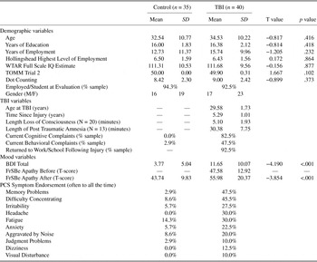
Forty participants with a history of mild closed head TBI (23 females, 17 males) at least 6 months from injury were recruited from the University of Illinois Medical Center via advertisements in the community seeking individuals who had ever sustained a closed head injury, concussion, brain injury, or traumatic brain injury. None were recruited from active clinical practice. Thirty-five healthy controls (19 females, 16 males) were also recruited from the community to match the TBI on age, years of education, years of employment, and estimated premorbid intelligence (see Table 1). Highest level of occupational achievement was determined using the Hollingshead Four Factor Index of Socioeconomic Status Occupational Scale with values ranging from 1 (e.g., menial labor) to 9 (e.g., executive) (Hollingshead, Reference Hollingshead1975). All participants provided written informed consent and experimental procedures complied with the code of ethics of the World Medical Association, the University of Illinois Institutional Review Board, and Declaration of Helsinki.
Participant demographics, mood and behavioral data, and frequency of reported post-concussive symptoms complaints

Participants (control and TBI) were excluded if they had a history of psychiatric disorder before the TBI, substance abuse/dependency, current or past litigation, failure on a formal measure of effort, or any other neurologic or medical condition that could result in cognitive changes (e.g., hypertension, severe chronic pain). Participants were not receiving any psychiatric medication or medications used for cognitive enhancement at the time of the study. The criteria used for defining mild TBI follow the guidelines set forth by the American Congress of Rehabilitation Medicine (1993) including endorsement of at least one of the following: any period of loss of consciousness; any loss of memory for events immediately before or after the accident; any alteration in mental state at the time of the accident (e.g., feeling dazed, disoriented, or confused); and focal neurological deficit (American Congress of Rehabilitation Medicine, 1993; Cassidy et al., Reference Cassidy, Carroll, Peloso, Borg, von Holst and Holm2004). For this study, participants were categorized as moderate and subsequently excluded if the duration of loss of consciousness (LOC) was greater than 30 min, post-traumatic amnesia (PTA) was greater than 24 h, there was positive radiologic finding of contusion or bleed, or evidence of skull fracture suggesting significant trauma to the head. Beyond self-report, witness reports, discharge notes from the emergency department, and previous medical records were used to confirm severity. These criteria help ensure that the only patients remaining were, in fact, mild severity (LOC less than 30 min; PTA less than 24 h, and/or the Glasgow Coma Scale greater than 13) (American Congress on Rehabilitation Medicine, 1993; Cassidy et al., Reference Cassidy, Carroll, Peloso, Borg, von Holst and Holm2004; Levin, Reference Levin1992; Tagliaferri, Compagnone, Korsic, Servadei, Kraus, Reference Tagliaferri, Compagnone, Korsic, Servadei and Kraus2006). For the individuals who had information on duration of loss of consciousness and/or posttraumatic amnesia confirmed by witness reports, the average reported LOC was 5.1 min (n = 20; range, 0–30 min) and average reported PTA was 27.9 min (n = 13; range, 0–60 min). For patients without specific information regarding LOC (n = 20) or PTA (n = 27), we relied upon estimates of self-reported and witness reported of duration of LOC or PTA and discharge note diagnosis from emergency departments when available for the purpose of study inclusion only.
The mechanism of injury varied and included motor vehicle accidents (n = 10), pedestrian MVA (n = 3), assault (n = 3), sports related (n = 11), and falls or blows to the head (n = 13). No TBI patient evidenced any frank structural lesion suggestive of focal injury on neuroimaging. Fourteen patients reported experiencing more than one TBI (range, 2–7). Analyses were conducted without these 14 participants with no effect on statistical significance on learning and memory or DTI analyses. As such, all participants were included in subsequent analyses. Demographic data and injury related variables are presented in Table 1.
MATERIALS AND PROCEDURE
Neuropsychological Assessment
As detailed in prior work, participants completed an extensive neuropsychological test battery that was assembled to assess executive function, attention, and memory (Kraus et al., Reference Kraus, Susmaras, Caughlin, Walker, Sweeney and Little2007). The California Verbal Learning Test-Second Edition (CVLT-II) was used to assess memory. Our motivation for using this tool, rather than a customized learning task, is that the CVLT-II is a widely available clinical tool. The CVLT-II consists of two different lists of words (List A and List B). Each list is comprised of sixteen words from four related categories presented in a pseudo-random manner. List A is administered five times followed immediately by the sole presentation of List B. Participants receive a point for each accurately recalled item.
Participants were also administered the Beck Depression Inventory-Second Edition (BDI-II) to assess for mood disturbance. While our TBI participants endorsed a significantly higher number of depressive symptoms than controls (see Table 1), the TBI group mean of 11.08 (SD = 10.07) is within the “minimal” depression criterion category. Concentration difficulties, sleep disturbance, and fatigue are commonly reported following mild TBI (Lundin, de Boussard, Edman, & Borg, Reference Lundin, de Boussard, Edman and Borg2006; Orff, Ayalon, & Drummond, Reference Orff, Ayalon and Drummond2009) and examination of specific BDI items found a high frequency of these items endorsed in the mild TBI group versus endorsement of sadness or loss of pleasure. Further review, however, also determined the presence of seven individuals in the TBI group with BDI-II scores within the moderate range (above 20). Analyses of memory and DTI variables were conducted with and without these seven participants and demonstrated no effect on the pattern of results. As such, all participants were included in reported analyses.
The Post-Concussion Syndrome Checklist (PCSC) (Gouvier, Cubic, Jones, Phillip, & Cutlip, Reference Gouvier, Cubic, Jones, Phillip and Cutlip1992) was used to rate subjective frequency of various post-concussive symptoms (PCS). Almost half (47.5%) of the TBI participants reported experiencing memory difficulties and 45.0% reported attention/concentration problems with high frequency. During clinical interview, participants were also asked if they experienced cognitive (e.g., memory, attention) or behavioral (e.g, irritability, fatigue) difficulties. The Frontal Systems Behavior Rating Scale-Self Version (FrSBe), a self-report behavior rating scale, was used to assess for the presence of postinjury behavioral syndromes of apathy, disinhibition and executive dysfunction (Reid-Arndt, Nehl, & Hinkebein, Reference Reid-Arndt, Nehl and Hinkebein2007). The TBI and control participants differed only on reported current level of apathy (Table 1) with TBI participants endorsing higher rates of apathy. Finally, TBI and control participants also completed two measures of effort (i.e., Test of Memory Malingering, Dot Counting) and all participants achieved scores in the valid range on the respective measure (Table 1).
DTI Data Acquisition
Imaging studies were conducted using a 3.0-Tesla whole body scanner (General Electric Medical Systems, Waukesha, WI) using a customized DTI pulse sequence with a quadrature head coil. The DTI sequence is based on a single-shot EPI with the capability of compensating eddy currents induced by the diffusion gradients via dynamically modifying the imaging gradient waveforms (Poonawalla & Zhou, Reference Poonawalla and Zhou2004). The sequence used 27 diffusion gradient directions, b-values of 0, 750 s/mm2, and voxel sizes of 1.5 × 1.5 × 5 mm3. A 3D high resolution anatomical scan was also acquired to allow coregistration with the DTI data and normalization to the Montreal Neurological Institute template (MNI) with a spatial resolution of 0.85 × 0.64 × 1.5 mm3 (Kraus et al., Reference Kraus, Susmaras, Caughlin, Walker, Sweeney and Little2007, for additional details).
DTI Data Analysis
The 28 diffusion directions were used to calculate the fractional anisotropy (FA) as the primary indicator of white matter integrity. The images were reconstructed and FA calculated using DTI Studio (Wakana et al., Reference Wakana, Jiang, Nagae-Poetscher, van Zijl and Mori2004). The 28 diffusion weighted images were examined for image quality and head movement. Head movement was required to be less than 2 mm. Voxels with very low signal (indicating nonbrain voxels) were masked out of the analysis before calculation of pixel-wise FA (background noise = 125). The FA map was then converted to ANALYZE. Statistical Parametric Mapping (SPM2, Wellcome Department of Imaging Neuroscience, London, UK) Was used to co-register the DTI with corresponding T1 images and then convert the DTI to normalized space (Montreal Neurologic Institute T1 template). None of the mild TBI in this investigation had significant atrophy or pathology making normalization to the MNI template accurate.
Region of Interest Analysis
All region of interest (ROI) analyses were carried out on data from each individual participant. The ROIs were drawn on a group averaged (including both controls and mild TBI) normalized FA map referencing not only the grayscale FA map but also a color-coded directionality map. This color-coded map allows visualization of intersecting fiber bundles and provides information as to where specific tracts begin and end. The masks were then overlaid on the FA maps from the remaining participants and visually checked for accuracy. The specific ROIs included the following: anterior and posterior corona radiata, corticospinal tracts (including parts of the corticopontine tract and superior thalamic radiation), external capsule, cingulum, forceps minor, forceps major, inferior fronto-occipital fasciculus, superior longitudinal fasciculus, uncinate fasciculus, sagittal stratum, and body, genu, and splenium of the corpus callosum. Right and left were analyzed separately where appropriate and combined when no differences reached significance. Detailed descriptions of these regions of interest can be found in (Kraus et al., Reference Kraus, Susmaras, Caughlin, Walker, Sweeney and Little2007).
These masks were then applied to the FA map from each individual participant. One concern when using ROI masks drawn on representative participants is that either gray matter or cerebral spinal fluid would be included in the calculation of mean FA. To ensure that FA was only calculated from white matter tissue, a threshold of FA = 0.20 was applied before extraction of FA for each ROI.
Statistical Analysis
For the CVLT-II, we examined group comparisons of the first trial of List A and the single List B learning trial using independent sample t tests We then conducted a mixed-design repeated-measure analysis of variance with Trial as the within-subject factor (recall on Trials 1, 2, 3, 4, 5) and Group (control, TBI) as the between-subject factor. We also conducted a mixed-design repeated-measures analysis of variance (ANOVA) of single trial learning as the within-subject factor (recall on Trial 1 and List B) and Group as the between-subject factor. In addition to raw rates of recall across trials, data from each individual were fitted to a power function (Equation 1). The power function, which is commonly applied in the behavioral learning literature (Anderson, Reference Anderson1982; Logan, Reference Logan1998), was applied to data from each participant to allow extraction of both the y-intercept (represented by y in Equation 1) and slope (represented by b in Equation 1):

For the DTI data, tests of independent means were conducted between groups for the body, genu, splenium, and total corpus callosum. For the 11 remaining ROIs for which measurements could be taken for each hemisphere, we conducted repeated measures ANOVAs with the left/right ROI as the within-subject measures by Group (control vs TBI) as the between-subject comparison.
To determine the amount of unique variance accounted for by DTI variables in performance on single trial learning, ROIs that demonstrated group differences were entered into a stepwise regression analyses with CVLT Trial 1 as the dependent variable.
RESULTS
CVLT-II
To test the primary hypothesis that mild TBI show reductions in single trial learning (acquisition), an independent samples t test was used to compare TBI and control recall on Trial 1 of List A. The mild TBI group achieved lowers scores than the control group on the first trial of the CVLT-II (Figure 1A), t(73) = 2.341; p = .020; η2 = 0.070.
California Verbal Learning Test-Second Edition (CVLT-II) learning analyses. A: CVLT-II raw recall findings across trials 1 through 5 for controls and patients with mild traumatic brain injury (TBI). Statistically significant difference between groups was only observed on the first learning trial. Lines represent best fit power function for each group. B: Average exponential rate of learning for controls and mild TBI patients. Error bars represent 1 ± SEM.

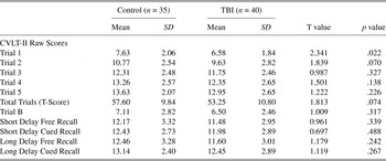
Although there was a trend for reduced performance in TBI, a repeated-measures ANOVA demonstrated that the groups did not differ in performance across the five total immediate learning trials, F(1,73) = 3.288; p = .074; η2 = 0.043. Additionally, there was no Group × Trial interaction. Unlike List A Trial 1, the groups did not differ on List B, t(73) = 1.009; p = .317; η2 = 0.014 (Table 2) and repeated-measures revealed no Group × List interaction.
Raw scores of California Verbal Learning Test-Second Edition (CVLT-II) performance

Further analyses of each CVLT-II trial demonstrated that the mild TBI participants were significantly different from controls on the first trial, but not on any subsequent trial (Trials 2–5) of List A. However, the analysis of slope (or rate) from the individually fit power functions demonstrated a significant difference between the groups with reduced rate in the mild TBI group, t(73) = 2.514; p = .014; η2 = 0.080 (see Figure 1B). As would be expected from the Trial 1 effect, there was also a difference in the y-intercept, t(73) = 2.118; p = .038; η2 = 0.058 between groups. In terms of overall list learning, the groups did not differ on the total five-trial verbal learning composite score, t(73) = 1.813; p = .074; η2 = 0.043, short-delay free recall, t(73) = 0.961; p = .343; η2 = 0.013, or long delay free recall, t(73) = 1.179; p = .242; η2 = 0.019. The groups performed similarly on both the cued short-delay t(73) = 0.697; p = .488; η2 = 0.007 and long-delay, t(73) = 1.119; p = .267; η2 = 0.017.
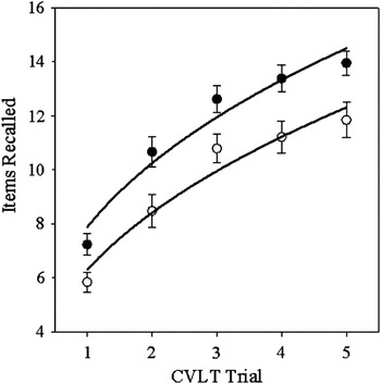
Analyses were also conducted comparing CVLT-II performance between TBI participants who reported memory complaints on the PCSC versus those who did not. As detailed in Figure 2, the TBI participants with reported memory complaints achieved lower scores on all trials of the CVLT-II than TBI without complaints (all comparisons, p < .05). TBI patients with subjective memory complaints also achieved lower scores on List B and the delayed memory score (p < .05).
California Verbal Learning Test-Second Edition (CVLT-II) learning by traumatic brain injury (TBI) with and without memory complaints. CVLT-II raw recall findings across trials 1 through 5 for TBI patients with (filled circles) and without (open circles) memory complaints as measured by the PCSC.

Relationship between Verbal Learning and Mood
Pearson product-moment correlations found no relationship between initial verbal learning with total BDI-2 depression score (r = −0.059; p = .715), frequency of PCS anxiety (r = −0.046; p = .907) and FrSBe ratings of apathy after injury (r = −0.133; p = .420). There was also no relationship detected between the composite total learning score or any delayed memory variables with depression, anxiety, or apathy (all p’s > .05).
Neuroimaging-DTI Analysis
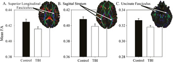
Consistent with earlier findings (Kraus et al., Reference Kraus, Susmaras, Caughlin, Walker, Sweeney and Little2007) and as depicted in Figure 3A–C, the mild TBI group had significantly lower FA compared with controls in the superior longitudinal fasciculus, F(1,73) = 4.608; p = .035; η2 = 0.059, sagittal stratum, F(1,73) = 5.695; p = .020; η2 = 0.072, and uncinate fasciculus, F(1,73) = 10.600; p = .002; η2 = 0.127. The TBI group did not differ from controls in any other ROI.
Fractional anisotropy (FA) by group comparisons. Mean FA for the left/right superior longitudinal fasciculus (A), left/right sagittal stratum (B), and left/right uncinate fasciculus (C) for controls and mild TBI participants.

Relationship Between DTI and Single Trial Learning
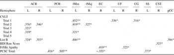
Linear regression analyses were conducted to determine the amount of unique variance that FA in these ROIs could account for in the single-trial learning measure. These analyses demonstrated that only FA of the left uncinate fasciculus, t(39) = 2.549; p = .016 and left superior longitudinal fasciculus t(39) = 2.059; p = .047 accounted for a significant amount of variance (14% and 9%) in the first learning trial of the CVLT-II. However, neither ROI was a significant predictor of total learning, learning rate, or short or long delay memory performance (all p’s > .05). Table 3 details the Pearson-product moment correlations between the remaining ROIs, single-trial learning measures, sustained attention measures, and mood and behavior variables.
Significant bivariate correlation in regions of interest with cognitive and behavioral variables

** p = 0.01 level
* p = 0.05 level
Note. ACR = anterior corona radiata; PCR = posterior corona radiata; fMin = forceps minor; fMaj = forceps major; EC = external capsule; UF = uncinate fasciculus; CG = cingulum; SS = the sagittal stratum including the optic radiations; CST = corticospinal tracts, which included parts of the corticopontine tract and parts of the superior thalamic radiation; gCC = genu corpus callosum.
DISCUSSION
The individual components of memory (i.e., attend, acquire, encode, consolidate, and recall information) are likely not given much consideration by an individual who reports “memory” problems; an individual simply experiences a failure to remember. If an individual is unable to initially acquire information following one trial, the memory complaint is of the same verisimilitude as that experienced by an individual with an impaired ability to encode information over many trials. In daily interactions such as conversations, lectures, and work-place instructions, individuals are often presented with information one time, rather than being given the benefit of repeated “trials.” For this reason and others, studies have questioned the ecological validity of composite learning and memory domain scores in elucidating the nature of memory complaints following TBI (Silver, Reference Silver2000; Silverberg & Millis, Reference Silverberg and Millis2009; Wood, Reference Wood2009; Yeates & Taylor, Reference Yeates and Taylor2005)
Consistent with our hypothesis, participants with a history of chronic mild TBI did differ significantly from the control participants on a measure of initial verbal learning (acquisition). Furthermore, there was no significant group difference on the total composite learning score or delayed memory variables. Taken together, despite reported memory difficulties by 47% of the mild TBI participants, these findings suggest no generalized deficit in the broadly conceptualized memory system per se. The TBI participants were able to encode information over the five trials; what they did encode, they were able to recall. Given the objective finding of differences on Trial 1 and slower rate of overall learning, interpreting only the total immediate or delayed memory scores on the CVLT-II may not be ecologically valid. The mild TBI patients demonstrate an inefficiency when presented with information for the first time which could easily be exacerbated by the qualities of day-to-day interaction versus constitute a generalized encoding, consolidation, and/or retrieval-based “memory” deficit. These findings have translational value in that they support clinical recommendations such as rehearsal of information to facilitate encoding and recall.
While historically considered a “benign” injury with complete recovery expected within 3 months (Carroll et al., Reference Carroll, Cassidy, Peloso, Borg, Von Holst and Holm2004; Lange, Iverson, & Franzen, Reference Lange, Iverson and Franzen2009; Levin et al., Reference Levin, Mattis, Ruff, Eisenberg, Marshjall and Tabaddor1987), it is believed that a minority of individuals experience postconcussive symptoms beyond this period (Pagulayan, Temkin, Machamer, & Dikmen, Reference Pagulayan, Temkin, Machamer and Dikmen2006; Rothweiler, Temkin, & Dikmen, Reference Rothweiler, Temkin and Dikmen1998; Sterr, Herron, Hayward, & Montaldi, Reference Sterr, Herron, Hayward and Montaldi2006; Vanderploeg, Berlanger, & Curtiss, Reference Vanderploeg, Belanger and Curtiss2009; Vanderploeg, Curtiss, & Belanger, Reference Vanderploeg, Curtiss and Belanger2005; Wood, Reference Wood2004). That mild TBI is the casual factor underlying these complaints is controversial, especially given the inconsistent objective evidence supporting the complaints (e.g., evidence of neuropsychological impairment, observable lesions on MRI). Furthermore, in part because postconcussive symptoms are not specific to traumatic brain injury (Lees-Haley, Fox, & Courtney, Reference Lees-Haley, Fox and Courtney2001; Smith-Seemiller, Fow, Kant, & Franzen, Reference Smith-Seemiller, Fow, Kant and Franzen2003), some clinicians have suggested that the majority of complaints are complicated by or solely attributable to psychological or motivational factors rather than involve, at least in part, the pathophysiology of the injury itself (Bay & Bergman, Reference Bay and Bergman2006; Ettenhofer & Abeles, Reference Ettenhofer and Abeles2009; Jacobson, Reference Jacobson1995; Karzmark, Hall, & Englander, Reference Karzmark, Hall and Englander1995; Larrabee, Reference Larrabee1997; Mooney & Speed, Reference Mooney and Speed2001; Mooney et al., Reference Mooney, Speed and Sheppard2005; Williams, Lees-Haley, & Brown, Reference Williams, Lees-Haley and Brown1993). In fact, many studies demonstrate that psychiatric comorbidities influence PCS reporting and reports of memory complaints (Chamelian & Feinstein, Reference Chamelian and Feinstein2006; Suhr & Gunstad, Reference Suhr and Gunstad2002; Vanderploeg et al., Reference Vanderploeg, Belanger and Curtiss2009). However, our data suggests Trial 1 differences exist independently of primary mood disturbance, apathy, or anxiety. Moreover, CVLT-II Trial 1 did not correlate significantly with any TBI grading parameters (e.g., duration of posttraumatic amnesia, duration of loss of consciousness, posttraumatic seizures, posttraumatic headache) as it has in other studies (MacKenzie et al., Reference MacKenzie, Siddiqi, Babb, Bagley, Mannon and Sinson2002). It is important to note that this lack of relationship may reflect the accuracy of self-report rather than a lack of true relationship. Most critically, the finding of diminished recall for Trial 1 was observed in well motivated (i.e., as assessed by effort measures), nonlitigating, nondepressed, and gainfully employed individuals many years after sustaining a mild TBI.
Of interest is the lack of significant difference between groups on the second word list (List B). Initially, we speculated that increased task familiarity would explain improved performance for both groups. However, this was not the case. In fact, as detailed in Table 2, the TBI participants performed at a comparable level on both Trial 1 of List A and List B where control participants declined slightly. One alternative explanation for the lack of improvement on List B may be attributed to proactive interference (PI) effects which are common in semantic clusters (Delis, Kramer, Kaplan, & Obers, Reference Delis, Kramer, Kaplan and Obers2000). Based on the performance of the normative sample of the CVLT-II and other patient populations (Ivory, Knight, Longmore, & Caradoc-Davie, Reference Ivory, Knight, Longmore and Caradoc-Davie1999), PI effects are expected and demonstrated when the number of words recalled on the single trial of List B is lower than what was recalled on the first trial of List A (Delis et al., Reference Delis, Kramer, Kaplan and Obers2000). In our sample, the lack of a significant difference between groups on List B may illustrate the expected modest PI effects demonstrated by the control participants and the lack of PI effects in the TBI participants. While the repeated measures analysis did not demonstrate a significant group by trial interaction effect, this may have been due to lack of power, but raises a question of whether there is a reduction in PI in mild TBI.
The question is then raised as to the underlying mechanism for slower acquisition on Trial 1. In mild TBI, acceleration–deceleration forces and related diffuse axonal injury is generally found to be the only significant pathology (Bazarian, Zhong, Blyth, Zhu, Kavcic, & Peterson, Reference Bazarian, Zhong, Blyth, Zhu, Kavcic and Peterson2007; Inglese et al., Reference Inglese, Makani, Johnson, Cohen, Silver and Gonen2005; Medina et al., Reference Medina, DeToledo-Morrell, Urresta, Gabrieli, Moseley and Fleischman2006). Given the nature of diffuse axonal injury and its potential impact on distributed neurobehavioral networks, injury along these pathways could result in wide-spread cognitive and behavioral dysfunction. In examining ROIs which demonstrated differences in FA relative to controls, the left uncinate fasciculus accounted for a significant amount of variance in Trial 1. This specific tract has been implicated previously in studies of memory (Niogi et al., Reference Niogi, Mukherjee, Ghajar, Johnson, Kolster and Sarkar2008). The uncinate fasciculus connects temporal and prefrontal areas so it is not surprising to find its involvement in learning and memory. The relationship between memory and FA of the uncinate fasciculus has been demonstrated in patients with TBI (Niogi et al., Reference Niogi, Mukherjee, Ghajar, Johnson, Kolster and Sarkar2008) with poor memory performance being correlated with reduced FA. While our data did not demonstrate a significant difference between groups on overall memory performance or a relationship between memory performance and the uncinate fasciculus, our data did demonstrate a significant relationship between Trial 1 and the left uncinate fasciculus.
Similarly, the relationship between Trial 1 performance and FA in the superior longitudinal fasciculus is also to be expected as this tract is thought to be composed of three component parts and has been purported to play a role in visual awareness, maintenance of attention, initiation of complex motor behavior, phonemic and articulatory aspects of language, and lexical decision making (Gold, Powell, Xuan, Jiang, & Hardey, Reference Gold, Powell, Xuan, Jiang and Hardy2007; Schmahmann, Smith, Eichler, & Filley, Reference Schmahmann, Smith, Eichler and Filley2008). Damage to this tract has also been reported previously in TBI (Bendlin et al., Reference Bendlin, Ries, Lazar, Alexander, Dempsey and Rowley2008; Cho et al., Reference Cho, Yang, Shon, Kim, Kim and Choi2008; Kraus et al., Reference Kraus, Susmaras, Caughlin, Walker, Sweeney and Little2007). In our study, FA of the left superior longitudinal fasciculus was a significant predictor of Trial 1 performance. To our knowledge, this is the first study to demonstrate the involvement of this tract in verbal learning in TBI. Integrity of this tract has also been reported to have a role in other verbally mediated tasks such as verbal repetition (Breier, Hasan, Zhang, Men, & Papanicolaou, Reference Breier, Hasan, Zhang, Men and Papanicolaou2008), which may explain its involvement in the CVLT-II.
The slower rate of learning across trials in TBI warrants further investigation. Again, we would suspect that successful recall of items is influenced by how well one consistently uses a recall strategy (Chan, Kwoka, Chiub, Lamb, Pangb, & Chow, 2000; Gongvatana, Woods, Taylor, Vigil, Grant, & Group, Reference Gongvatana, Woods, Taylor, Vigil, Grant and Group2007; Luek, Reference Luek1976; Ribeiro, Guerreiro, & De Mendonça, Reference Ribeiro, Guerreiro and De Mendonça2007). It is possible that the TBI group was slow to recognize and then use a successful strategy, or they adopted a less efficient strategy (e.g., serial recall) across trials. Examining the strategic and arguably, higher-order (i.e., executive/frontal lobe), aspects of learning could be informative in appreciating the complexities of the initial verbal learning inefficiency (Wood, Reference Wood2009). Our groups did not differ on formal neuropsychological measures of executive function. However, the administered executive tasks are partially externally facilitated (e.g., changing visual sorting contingencies, visual planning to match a model) and do not require the same internally derived strategy formation used in verbal list learning. However, without an analysis of the strategies used within trial and across trials on the CVLT-II, this contention is speculative. Future work will test the validity of this hypothesis.
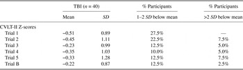
Another concern in many TBI studies is the inclusion of participants with a history of mild TBI without witness confirmation of LOC or PTA. Given the reliance on retrospective report, it is possible that some of these individuals did not sustain a TBI. Indeed, post hoc analyses examining differences between control participants and the mild TBI subgroups (witness corroborated vs only subjective report of TBI) on the data presented herein found that significant differences on Trial 1 were only observed between controls and TBI patients with witness corroborated TBI. A study with restrictions to inclusion for only witnessed or objectively verified TBI parameters would be compelling. However, the inclusion of self-reported participants in this study only serve to increase the likelihood of supporting the null hypothesis rather than biasing in favor of finding group differences. Similarly, only approximately half of our mild TBI participants reported memory difficulties suggesting that the data presented may actually underestimate the magnitude of the effect. Consistent with this, when converted to standardized scores, these patients fall into the low end of normal performance (Table 4). Indeed, the 19 TBI participants, who reported experiencing memory difficulties, performed more poorly on all CVLT-II learning variables and likely represent the more severe end of the mild TBI continuum.
California Verbal Learning Test-Second Edition (CVLT-II) standard scores

Finally, 14 TBI participants reported a history of multiple mild TBI. While analyses conducted without these individuals did not change the significance of the findings, their inclusion raises the possibility that findings could be driven, in part, by changes attributable to multiple mild injuries as has been suggested by others (Weber, Reference Weber2007).
To our knowledge, this is the first study to examine verbal learning with a focus on single-trial learning and white matter integrity in a nonlitigating, nondepressed, employed population with mild TBI. Our data suggest that chronic mild TBI patients demonstrate deficits in the acquisition of information which are supported by evidence of chronic damage to white matter microstructure. However, we collected no data regarding the functional significance of any cognitive complaints and as such it is unclear if initial trial learning difficulties relate to functional deficits. Future studies comparing initial learning and more specific outcome variables (e.g., difficulties at work/school) would prove especially informative in this regard.
ACKNOWLEDGMENTS
This work is supported in part by NIH grant K23 MH068787 from the National Institute of Mental Health (MFK), the Marshall Goldberg Traumatic Brain Injury Fund (DML), and a grant from the Department of Defense/Congressionally Directed Medical Research Program grant PT 075675. The contents of this study are solely the responsibility of the authors and do not necessarily represent the official views of the University of Illinois at Chicago, Department of Defense, or the National Institute of Mental Health. We thank Dr. Xiaohong Joe Zhou for providing the pulse sequence used to collect the DTI data.