Ventricular septal defects are the most common congenital heart defects with perimembranous ventricular septal defects accounting for ~70–80%. The current standard of treatment is open-heart surgery which carries the risk of heart block, residual shunt, complications from cardiopulmonary bypass, and wound infection.Reference Meijboom, Szatmari and Utens 1 – Reference Yuh, Vricella, Yang and Doty 3 Transcatheter closure of perimembranous ventricular septal defects using devices such as Amplatzer perimembranous ventricular septal defects occluder (AGA Medical Corp, Plymouth, Minnesota, United States of America) and Amplatzer membranous ventricular septal defect occluder, also known as the “eccentric device” (AGA Medical Corp., Golden Valley), in clinical trials in the United States of America have been attempted in older infants and children. These trials have raised concerns about procedural complications including complete heart block, arrhythmia, and new valve regurgitation.Reference Fu, Bass and Amin 4 – Reference Sullivan 6 Similar concerns have been raised by reports outside the United States of America.Reference Pinto, Dalvi and Sharma 7 – Reference Zuo, Xie and Yi 10 Masura et al in a multicentre follow-up study using the eccentric device showed that patients developed anterior hemi-block, right bundle branch block, and complete heart block. At present, there is no FDA-approved device for transcatheter closure of perimembranous ventricular septal defects. Small studies and case reports have shown the use of various catheter-based devices in an off-label management; however, there are no large studies to show their efficacy.Reference Pinto, Dalvi and Sharma 7 , Reference Lee, Song and Choi 11 – Reference Yang, Sheng and Cao 15 To our knowledge, no study or case report has described the use of the Amplatzer Vascular Plug-II (AVP-II, St. Jude Medical, Minneapolis, Minnesota, United States of America) for transcatheter closure of perimembranous ventricular septal defects.
In this report, we describe our experience using the Amplatzer Vascular Plug-II for transcatheter closure of perimembranous ventricular septal defects. We sought to establish the safety and efficacy of this device in our patient cohort.
Methods
We conducted a 4-year retrospective chart review of patients with perimembranous ventricular septal defects and associated ventricular septal aneurysm who were greater than 6 months of age and underwent attempted transcatheter closure using the Amplatzer Vascular Plug-II from August, 2010 to August, 2014 at Valley Children’s Hospital. This study was approved by our local Institutional Review Board. Patients were considered for transcatheter therapy upon referral from their primary-care cardiologist. As this report is a retrospective review, there were no a priori inclusion criteria. Patients with evidence of aortic valve prolapse or aortic valve hinging at the superior margin of the defect or lacked a ventricular septal aneurysm were not considered for transcatheter therapy. Our study cohort included 16 patients. We recorded the patient’s age, weight, the defect size, left ventricle dimension, left atrium-to-aorta ratio, clinical symptoms, and residual shunt. We also assessed the tricuspid, mitral, and aortic valve for regurgitation before and after the procedure.
Before the procedure the morphology of the perimembranous ventricular septal defect and the estimation of left-sided volume load – left atrial and left ventricular dimensions – were determined by transthoracic echocardiogram. At the time of catheterisation the size of the defect was re-confirmed by transoesophageal echocardiogram while the patient was under general anaesthesia. Both the left- and right-sided margins of the ventricular defect and the aneurysm were measured. The ventricular septal aneurysm typically formed a cone-shaped structure with the base at the left ventricular end of the defect and with the apex at the right ventricular end. We recognised that the apex of the cone in many instances was fenestrated. The largest fenestration was recorded as the primary defect size on the right ventricular end. All attempts were made to fill the defect and ventricular septal aneurysm without obstructing the left and/or right ventricular outflow tract or potentially distorting the aortic valve while covering the primary defect and any associated fenestration.
Procedure
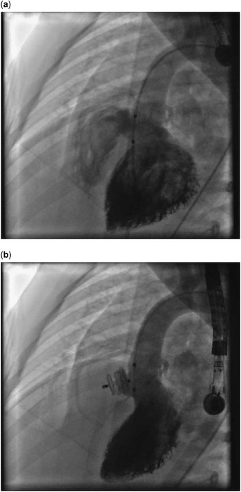
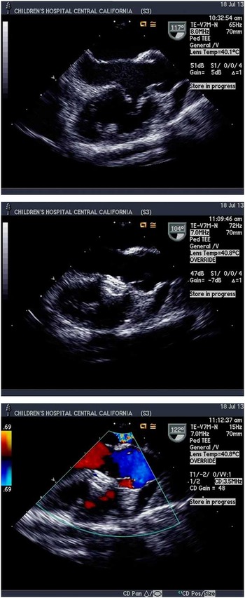
The procedures were performed under general anaesthesia with transoesophageal echocardiography guidance. Heparin and antibiotics were administered intravenously during the procedure. Access was through the right femoral vein and artery; the left femoral vessels were used when we were unable to access the right side. An angiogram within the left ventricle was performed to visualise the size, location, and shape of the defect (Fig 1a). The Amplatzer Vascular Plug-II device size was chosen by the operator taking into consideration the most consistent measurements obtained by both angiogram and transoesophageal echocardiography of the ventricular septal aneurysm size and the largest diameter of the defect at the base. Care was taken to ensure that the occlusion device had sufficient diameter to occlude the defect without obstructing other anatomical structures or cause impingement to the aortic valve. The ventricular septal defect was approached from the left ventricular side using a Judkins right coronary catheter (Cook Medical, Bloomington, Indiana, United States of America). An exchange length Glide Wire (Terumo Corporation, Somerset, New Jersey, United States of America) was directed across the defect, snared in the main pulmonary artery, and brought out through the femoral vein thus creating an arteriovenous circuit. A long 5- or 6-Fr Flexor sheath (Cook Medical) was advanced from the femoral vein over the guide wire to the descending aorta. An appropriate-sized Amplatzer Vascular Plug-II was back-loaded and advanced through the long sheath. Under transoesophageal echocardiography and fluoroscopic guidance, the distal disk was re-configured in the left ventricle and brought against the ventricular septum in the left ventricular outflow tract or within the ventricular septal aneurysm. Care was taken not to distort the aortic valve or entrap the mitral apparatus. The central disk was re-configured in the ventricular septal aneurysm and the proximal disk on the right ventricle side of the ventricular septal aneurysm. A left ventricular angiogram was performed to confirm proper device configuration before release. Following device release, haemodynamic assessment was repeated including a left ventricular angiogram (Fig 1b). In addition, a follow-up transoesophageal echocardiography was performed to assess device placement (Fig 2), degree of residual shunt, any changes in valve insufficiency, and any evidence of outflow tract obstruction.
( a ) Typical left ventricle (LV) angiogram showing the perimembranous ventricular septal defect (pmVSD) with a left-to-right shunt. ( b ) The Amplatzer Vascular Plug-II (AVP-II) device implanted within the ventricular septal aneurysm (VSA), closing the pmVSD. There is a trivial residual shunt.

Top: Pre-device transoesophageal echocardiogram showing the ventricular septal aneurysm (windsock appearance) and deficient aortic rim. Middle: transoesophageal echocardiography (TEE) showing the device placement in the ventricular septal aneurysm (VSA) and away from the aortic valve. Bottom: After device placement with colour Doppler showing no residual shunt and no flow disturbance across the left ventricular outflow tract.

Follow-up
The patients were observed overnight after device implantation for any evidence of device failure, outflow tract obstruction, development of haemolysis, or rhythm changes. Electrocardiogram and echocardiography were obtained before discharge. Our institutional standard is to re-evaluate patients who have undergone device closure of their ventricular septal defect at 1 week, 1 month, 6 months, 12 months, and then annually for 5 years with assessment of clinical status. Physical examination, electrocardiogram, and typically an echocardiogram are performed to assess residual shunt and development of valve insufficiency.
Statistics
Continuous variables are presented as median and range. Comparisons between baseline values and values after treatment were tested for significance using the two-tailed paired t-test. The null hypothesis was rejected with a p-value of less than 0.05.
Results
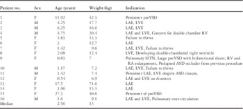
All 16 patients underwent successful Amplatzer Vascular Plug-II device closure of their perimembranous ventricular septal defects. All defects had an associated ventricular septal aneurysm forming a windsock appearance. Deficient aortic rim did not preclude candidacy for device implantation. The majority of patients, in fact, had no aortic rim. Indications for closure were based on the findings of the referring cardiologist and include: left-sided enlargement, Qp:Qs>1.5, failure to thrive, concern for worsening perimembranous ventricular septal defect, with developing left-sided enlargement, no signs of closure, or enlargement of the perimembranous ventricular septal defect, developing double-chamber right ventricle, and/or volume overload despite closure of other defects (Table 1). Patient 9 had Down syndrome along with reactive pulmonary hypertension, patent ductus arteriosus, and atrial septal defect. The patient underwent intervention due to prolapse of a previously placed atrial septal defect occluder device with a small residual shunt and persistent pulmonary over-circulation from the perimembranous ventricular septal defect. The atrial septal defect occluder was replaced with a larger occluder and the perimembranous ventricular septal defects were closed percutaneously. Patient 3 had a trivial patent ductus arteriosus that was coil-embolised during the same procedure.
Patient characteristics and indications for intervention.

ASD=atrial septal defect; HTN=hypertension; LAE=left atrium enlargement; LVE=left ventricular enlargement; pmVSD=perimembranous ventricular septal defect; RA=right atrium; RV=right ventricle
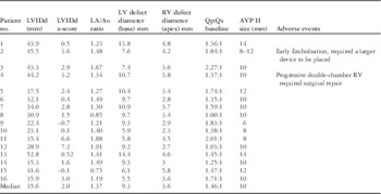
The patients’ demographic data are summarised in Table 1. The median age was 2.56 years (range: 0.5–27.3). Their median weight was 13.0 kg (range: 6.9–71.6). From the transoesophageal echocardiography and angiogram, the left ventricular median defect size was 9.3 mm (range: 5.9–14.4). The right ventricular defect had a primary median defect size of 3.6 mm (range: 2.3–5.8). The median Qp:Qs at baseline was 1.46:1 (range: 1–2.27:1) (Table 2). The median Qp:Qs following device implantation was 1.0:1 (range: 1–1.36:1). The Amplatzer Vascular Plug-II device size used ranged between 6 and 14 mm with the most common size being 10 mm. Devices used tended to be 1–2 mm greater than the left ventricle defect size or equal to the left ventricle defect size at the base.
Pre-echocardiographic and procedural data.

LA/Ao=left atrium-to-aorta ratio; LV=left ventricle; LVIDd=left ventricle internal diameter, diastole; RV=right ventricle
The LVIDd, LVIDd z-score, LA/Ao ratio were obtained via transthoracic echocardiogram before catheterisation. The LV and RV defect diameters and Qp:Qs ratio were obtained during catheterisation and before the device placement
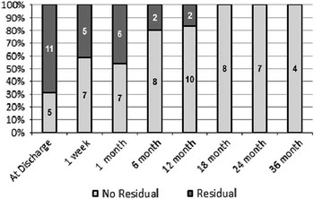
At discharge, five patients had complete closure of their ventricular septal defect. Eleven patients had traces of mild residual shunts – six with residual shunts around the device and five with residual shunts through the centre of the device – which improved at each follow-up. The median follow-up period was 12 months (range: 0–36). By 12 months, 83% (10 of 12) of the patients had complete closure of their perimembranous ventricular septal defects (Fig 3). The other two patients continued to have a residual shunt around the device with complete closure at the 24-month follow-up. We considered any flow detected by transthoracic echocardiogram around or through the device as residual shunt. There has been no haemodynamic instability in any of the patients. All residual shunts were felt to be haemodynamically insignificant by the patients’ primary-care cardiologists. No surgical intervention has been required so far for the management of residual shunt or device failure.
Number of patients with residual shunt at each follow-up.

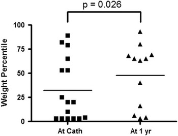
Of the three patients diagnosed with failure to thrive, patient 5, 7, and 10, there were minimal differences in their weight percentile at the 12-month follow-up; however, as a group, the entire cohort had a significant increase in weight at the 1-year follow up (median 20th percentile at baseline, 64th percentile at 1 year; p=0.026; Fig 4).
Graph depicting the increase in the weight percentile from the time of the procedure to the 1-year follow-up.

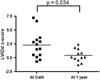
From the pre- and post-transcatheter echocardiogram, there was a difference in the left ventricle internal diameter diastole z-score and the left atrium-to-aorta root ratio, showing improvement of the left ventricle dimension. Before device implantation, the median left ventricle internal diameter diastole was 35.6 mm (range: 22.4–52.8) with a z-score of 2.0 (range: −0.7 to 7.2). The left atrium-to-aorta root ratio was 1.37 (range: 0.85–1.91). At the 1-year follow-up, the left ventricle internal diameter diastole was 33.0 mm (range: 25.7–50.6) with a z-score of 0.1 (range: −1.3 to 2.4). The left atrium-to-aorta root ratio was 1.22 (range: 0.96–1.69). The difference in left ventricle internal diameter diastole z-score measurements was significant at p=0.034 (Fig 5).
Graph depicting the improvement in left ventricle internal diameter, diastole (LVIDd) z-score from the time of the procedure to the 1-year follow-up.

During follow-up, we also monitored all the heart valves for any change in regurgitation. We considered any backflow detected by transthoracic echocardiogram, whether it was physiological, trivial, trace, or mild as regurgitation. None of the patients developed valve insufficiency that required medication or intervention. In total, 14 patients had trace tricuspid regurgitation before the procedure with 2 resolving at the 1-month and the 6-month follow-up, respectively. Moreover, two patients developed trace tricuspid regurgitation at 1 week after the procedure, which remained stable at the 12- and 24-months follow-ups. All other patients’ tricuspid regurgitation remained stable; two patients had aortic valve insufficiency before catheterisation, with one resolving at discharge. The second patient’s aortic insufficiency resolved at discharge, but at the 1-week follow-up trace insufficiency was found and has remained stable thereafter; five patients developed new trace-to-trivial aortic insufficiency, four noted at 1 week and one at 24 months after device implantation. Of the five patients, two had their aortic insufficiencies resolved at 1 and 12 months after intervention. The other two remained stable at the 12- and 18-month follow-ups. The patient with aortic insufficiency at 24 months had it resolved at 36 months after intervention. In addition, five patients had mitral regurgitation before the procedure with two resolving at discharge and at 12 months after the procedure, while the other three remained stable Furthermore, eight patients developed trace mitral regurgitation, three at discharge, four at the 1-month follow-up, and one at the 12 month-follow-up, and four self-resolved at the 6-, 12- and 18-month follow-ups. The other four remained stable at discharge and at the 1-, 12-, and 24-month follow-ups. Of the five patients with previous mitral regurgitation, one resolved at discharge, two at 12 and 18 months after the intervention. The other two remained stable with no haemodynamic instability.
Among our study cohort there were no cases of arrhythmia, heart block, haemolysis, or death after the procedure or during follow-up. Only one procedure was complicated by early device embolisation. The patient was noted to have a new-onset murmur while recovering in the post-anaesthesia care unit. A transthoracic echocardiogram and chest x-ray showed dislodgement of the device. The patient was taken back to the cardiac catheterisation laboratory where the device was retrieved from the pulmonary artery. A larger device was deployed with successful occlusion of the ventricular septal defect without further complication. Two patients had a mild right ventricular outflow tract obstruction with concern for developing double-chambered right ventricle. Although patient 9 had complete resolution of the double-chambered right ventricle, patient 4 developed progression of obstruction and required surgical repair at 26 months following catheter intervention. The patient’s double-chambered right ventricle was surgically repaired with right ventricular outflow tract myectomy. The surgeon electively removed the Amplatzer Vascular Plug-II device and patched the ventricular septal defect in a traditional manner as the occluder device was adherent to a part of the muscle and redundant tricuspid tissue.
Discussion
Ventricular septal defect is a common congenital heart defect with perimembranous ventricular septal defect being the most common subtype. The current standard of treatment is open-heart surgery. In general, this is carried out in the first 6 months of life in patients with evidence of pulmonary over-circulation, left heart enlargement, and congestive heart failure symptoms. Common adverse effects of surgical repair are well recognised and include wound infections, heart block, arrhythmias, and prolonged hospital stay and recovery period.Reference Meijboom, Szatmari and Utens 1 – Reference Yuh, Vricella, Yang and Doty 3 Transcatheter interventions using the Amplatzer perimembranous ventricular septal defect occluder devices have been attempted but have shown to be a challenging procedure. There are small studies and case reports showing them to be effective in the closure of perimembranous ventricular septal defects.Reference Fu, Bass and Amin 4 , Reference Tzikas, Ibrahim and Velasco-Sanchez 13 However, studies have also shown complete heart block, which range from 3.5 to 22%,Reference Sullivan 6 , Reference Yang, Kong and Sheng 9 as one of the concerns following device implantation, which in part has precluded FDA approval here in the United States of America.
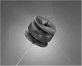
The Amplatzer Vascular Plug-II is an FDA-approved device used for arterial and venous embolisation in the peripheral vasculature. Within congenital heart disease this device has found a niche in the management of pulmonary arteriovenous malformations and select patent ductus arteriosus. We hypothesised that the Amplatzer Vascular Plug-II may have potential advantages in the transcatheter closure of perimembranous ventricular septal defects. The device is designed with three parts to its body: the distal, the central, and the proximal disk (Fig 6). Leveraging this unique design, we conceptualised the distal disk to lay in the left ventricular outflow tract covering the left ventricle side of the defect. The central disk fills the ventricular septal aneurysm, whereas the proximal disk lies on the right ventricle side of the ventricular septal aneurysm, theoretically preventing the device from embolising into the left-sided circulation. The device is made of self-expandable Nitinol mesh that helps facilitate thrombus formation and eventual endothelialisation. In addition, its softness helps the closure of the ventricular septal defect in a relatively atraumatic manner, and thus potentially decreasing the risk of heart conduction complications. This is different from the Amplatzer perimembranous ventricular septal defect occluder that “stents” the defect, putting pressure along the rim of the ventricular septal defect where the conduction system runs and where potentially heart blocks occur.
Amplatzer Vascular Plug-II device.

Our cohort tended to be comprised of older patients (median age of 2.5 years) who for various reasons were able to avoid surgical repair as an infant. Patients needed to have a prominent ventricular septal aneurysm forming a windsock appearance for proper placement of the device leading to some selection bias.
Of note, patient 9 on previous catheterisation had pulmonary vascular reactivity to inhaled nitric oxide and oxygen in the setting of a large atrial septal defect, moderate-sized ventricular septal defect, patent ductus arteriosus, and baseline pulmonary hypertension. There was no left ventricle enlargement due to “pop-off” through the atrial septal defect. The patient had transcatheter atrial septal defect and patent ductus arteriosus repaired first as the ventricular septal defect was thought to be more restrictive at that time. Not until after the atrial septal defect repair did the significance of the ventricular septal defect shunt become apparent. With the prolapse of the atrial septal defect occluder device, the decision was made to address the perimembranous ventricular septal defect at the same time as the atrial septal defect device replacement. At the 1.5-year follow-up, there was no evidence of ongoing pulmonary hypertension with tricuspid regurgitation velocity of 2.4 m/s and normal septal configuration. The patient is reported to be thriving.
After closure, patients had a significant improvement of their left heart dimension as seen on the echocardiogram. Patients’ growth improved with appropriate catch-up weight gain at the 1-year follow-up. The increase in the weight percentile suggests that the haemodynamic significance of these defects may have been under-appreciated. Moreover, they experienced an increase in daily activities, exercise tolerance, and decrease in respiratory symptoms as reported by patients and/or parents.
To our knowledge no published studies have reported on the use of the Amplatzer Vascular Plug-II in closing perimembranous ventricular septal defects. Although there were new valve regurgitations seen on the follow-up echocardiogram, no haemodynamic instability was appreciated at intermediate follow-up. In fact, more than half of the patients had resolution of their new valve regurgitation without any treatment. None of the patients needed initiation of medication or surgical interventions.
Among our patients, two of them experienced significant adverse events. The single device embolisation event occurred early in our experience. In retrospect, the original device was clearly under-sized for the defect. The second patient required surgical repair of progressive right ventricular outflow tract obstruction in the context of a double-chambered right ventricle. At the time of original catheterisation, the peak right ventricular outflow tract gradient before device implantation was 30 mmHg. The operator had hoped the gradient would remain stable or regress. All the other patients remained clinically stable throughout follow-up.
Study limitations
The number of patients in our study was small. Our patient selection was biased as they tended to be older and referral for intervention was based on the preferences of primary-care cardiologists. Of note, to perform the procedures, patients needed to have a prominent ventricular septal aneurysm, again leading to selection bias. Thus, not all potential complications may have been observed. As a retrospective study, clear inclusion criteria were not defined. Finally, although all the procedures were successful, we do not have long term follow-up data.
Conclusion
Application of the Amplatzer Vascular Plug-II for closure of selected patients with perimembranous ventricular septal defects appears to be a safe and effective treatment option. Patients need to have a ventricular septal aneurysm as part of the ventricular septal defect to allow for proper implantation of the device. Larger studies looking at the safety and efficacy of the Amplatzer Vascular Plug-II in the closure of perimembranous ventricular septal defects, potentially with a prospective design randomiszing surgical and transcatheter groups using the Amplatzer Vascular Plug-II, and comparing the long-term outcomes are warranted.
Acknowledgements
Authors wish to thank Ms. Ashley Bily for her assistance in research coordination and preparation of this manuscript.
Financial Support
This research received no specific grant from any funding agency, commercial, or not-for-profit sectors.
Ethical Standards
This was a retrospective chart review study and did not require ethics approval. The study was approved by our local Institutional Review Board.