Introduction
The beetle family Staphylinidae, or rove beetles, is the largest animal family, currently comprising over 55,000 nominal species (Grebennikov and Newton, Reference Grebennikov and Newton2009). Scydmaeninae, or ant-like stone beetles, is a large and diverse evolutionary lineage within staphylinids, with nearly 4,900 species (Grebennikov and Newton, Reference Grebennikov and Newton2009). These minute beetles have constituted a separate family of Staphylinoidea for nearly 200 years, and only recently Grebennikov and Newton (Reference Grebennikov and Newton2009), based on a comprehensive phylogenetic analysis, included scydmaenines in the megadiverse rove beetles.
Scydmaeninae is currently divided into four supertribes, which are highly unequal in terms of species richness. The extinct Cretaceous Hapsomelitae is ill-defined and comprises only several enigmatic species known from Burmese amber (Poinar and Brown, Reference Poinar and Brown2004; Chatzimanolis et al., Reference Chatzimanolis, Engel, Newton and Grimaldi2010); their phylogeny, relationships and systematic position remain unclear. Mastigitae is also a small group of less than a hundred species classified into nine genera, and internal relationships of this group were recently studied, with not entirely conclusive results (Jałoszyński, Reference Jałoszyński2012a). Cephenniitae includes three tribes with 26 genera and less than 500 species; the phylogeny of this lineage and its well-supported monophyly was a subject of recent studies (Jałoszyński, Reference Jałoszyński2012b, Reference Jałoszyński2014). Scydmaenitae, after recent exclusion of tribes now forming the Cephenniitae, comprises five tribes with 65 genera and ca. 4400 species; this is the largest supertribe, but also the least studied and most problematic in terms of evolutionary relationships between genera. Many genera of Scydmaenitae, especially those of the largest tribe Glandulariini (=Cyrtoscydmini), require revision. The monophyly of Scydmaenitae was recently challenged, as the tribe Scydmaenini was placed as a sister group of Mastigitae (O’Keefe, Reference O’Keefe2005; Jałoszyński, Reference Jałoszyński2012b, Reference Jałoszyński2014).
Mastigitae is an intriguing group because of its distribution, morphological structures and biology. It includes the largest known Scydmaeninae, reaching the body length of 10 mm, which is a gigantic size for a subfamily in which the majority of species do not exceed 2 mm and the smallest forms do not reach 0.5 mm. In addition, a diurnal and exposed mode of life is common among some tribes of Mastigitae, while nearly all other members of Scydmaeninae are cryptic beetles living in leaf litter, soil, rotten wood, and other moist substrates, or within ant and termite colonies (O’Keefe, Reference O’Keefe2005). Genera of Mastigitae share the following characters: antennal scape strongly elongate, much longer than pedicel; antennae distinctly geniculate between scape and pedicel (distal part of antenna bent ventrally); labial palpomere III pointed; maxillary palps longer than head with palpomere IV either larger than III or smaller, but then strongly asymmetrical and never dome-shaped; and eyes located in the anterior part of the head (O’Keefe et al., Reference O’Keefe, Pike and Poinar1997; O’Keefe, Reference O’Keefe2005; Jałoszyński, Reference Jałoszyński2012a). This supertribe is currently divided into three tribes. Leptomastacini includes small (2–3 mm), flattened and often lightly pigmented beetles associated with leaf litter or deeper soil layers, occurring mostly in northern and north-eastern Mediterranean subregion and in the Balkans. Clidicini comprises beetles of various lengths, from 1.57 to 10 mm, with three genera showing disjunctive distributions: Clidicus Laporte occurs in SE Asia and N Australia, Leptochromus Motschulsky in Central and South America, and Papusus Casey in SW United States and Mexico (Jałoszyński, Reference Jałoszyński2012a). The placement of Papusus in Clidicini remains problematic (Jałoszyński, Reference Jałoszyński2012a). This tribe is also most diverse in terms of the biology: small, light brown, short-legged and flattened species of Papusus live under stones on arid deserts; moderately large, light brown, long-legged and strongly convex species of Leptochromus are associated with leaf litter of humid tropical forests; and large, often darkly pigmented, long-legged and convex species of Clidicus show a similar habitat preferences as Leptochromus, but one species is known to have a diurnal and gregarious activity and bicolorous pigmentation (head and femora dark brown, pronotum and elytra reddish) (O’Keefe and Monteith, Reference O’Keefe and Monteith2000). Diurnal forms also predominate in Mastigini, which comprises wingless and large (4–10 mm) beetles with long legs and antennae, living often in dense populations and vividly running on the ground, bushes and even climbing trees in southern Europe and South Africa, showing a large and puzzling distributional gap in between. Interestingly, two genera of Mastigini, Mastigus Latreille and Stenomastigus Leleup, occur only in South Africa, whereas Palaeostigus Newton is known from S Europe and RSA. European species are uniformly black, while among South African Mastigini yellowish, reddish and often bicolorous forms with dark head and pronotum and light elytra are common.
Fossils of Scydmaeninae are known from Baltic, Burmese, Lebanese, Mexican, Sicilian, Canadian, Spanish, and Dominican amber (O’Keefe et al., Reference O’Keefe, Pike and Poinar1997; Newton and Franz, Reference Newton and Franz1998; Chatzimanolis et al., Reference Chatzimanolis, Engel, Newton and Grimaldi2010; Jałoszyński, Reference Jałoszyński2012a; Kirejtshuk and Azar, Reference Kirejtshuk and Azar2013; Peris et al., Reference Peris, Chatzimanolis and Delclòs2014). Descriptions of older authors are usually brief and inaccurate, and type specimens of some taxa described from German or eastern European amber collections were destroyed or lost during World War II. Systematic placement of some fossil genera is therefore unclear and impossible to verify. Only the most recent descriptions are accurate and amply illustrated, but some important characters remain undescribed because they were not visible in fossil specimens. The oldest known named taxa of Scydmaeninae were first described from the Late Cretaceous (earliest Cenomanian, ca. 99 Ma) Burmese amber (Poinar and Brown, Reference Poinar and Brown2004; Chatzimanolis et al., Reference Chatzimanolis, Engel, Newton and Grimaldi2010). Jałoszyński (Reference Jałoszyński2012a) briefly discussed their systematic placement and problems related with unclear diagnoses. Within Mastigitae, the focal taxon of the present study, unambiguously placed taxa were described by O’Keefe et al. (Reference O’Keefe, Pike and Poinar1997), O’Keefe (Reference O’Keefe2002) and Jałoszyński (Reference Jałoszyński2012a). The oldest is Palaeoleptochromus schaufussi O’Keefe, 1997 from the Upper Cretaceous (Campanian) Canadian amber, dated 79 Ma. Two remaining taxa are much younger: Leptochromus palaeomexicanus O’Keefe, Reference O’Keefe2002 is known from Chiapas Miocene amber (15–20 Ma) and most recently discovered Euroleptochromus sabathi Jałoszyński, Reference Jałoszyński2012a from the lower to middle Eocene (37–54.5 Ma) Lithuanian amber. These three species were placed in Clidicini, and this placement was strongly supported by results of phylogenetic analyses comprising morphological characters of extant and extinct taxa (O’Keefe, Reference O’Keefe2002; Jałoszyński, Reference Jałoszyński2012a).
Other fossils placed by older authors in various groups within the present-day Mastigitae are problematic. Palaeomastigus helmi Schaufuss from Eocene Baltic amber was treated as incertae sedis within Scydmaeninae by Newton and Franz (Reference Newton and Franz1998) but as a representative of Mastigitae by O’Keefe et al. (Reference O’Keefe, Pike and Poinar1997). As discussed by Jałoszyński (Reference Jałoszyński2012a), the latter view seems correct, but it was not possible to unambiguously place Palaeomastigus in any tribe of Mastigitae. Clidicus balticus Schaufuss from Baltic amber was described inadequately and again, it is not possible to firmly support the placement of this species in Clidicus or even within Clidicini. Schaufuss (Reference Schaufuss1891) described also another intriguing species from the Baltic amber, Aenictosoma doenitzi, which he placed in Cerambycidae, but Vitali (Reference Vitali2006) transferred this genus to Scydmaeninae, Clidicini. The type specimens of all these problematic taxa are lost, and it may never be possible to clarify their systematic placement.
The disjunctive distribution of extant Mastigitae and fossil genera found in distant localities (Palaeoleptochromus in Canada and Euroleptochromus in Northern Europe, while the phylogenetically closest relatives are known from Central America) strongly suggest that extinctions played a significant role in shaping the currently observed biogeography of this supertribe. One of the puzzling mysteries is the evolution of Mastigini, a tribe with all extant species showing a strong asymmetry in male genital structures, with one aedeagal paramere long and the other one much shorter or even obliterated (Jałoszyński et al., Reference Jałoszyński, Matsumura and Beutel2015). Not only all remaining Mastigitae have symmetrical aedeagi, but also the sister group of this supertribe, the Scydmaenini, has symmetrical genitalia (except asymmetrical endophalli). Therefore, the Mastigini must have evolved from a symmetrical ancestor. In this paper a new Eocene genus of Mastigini is described, which shows an interesting mosaic of plesiomorphic and derived character states, among others an apparently symmetrical aedeagus. Characters of the new genus are included in a phylogenetic analysis and their interpretation sheds light onto the evolution of this interesting tribe of still poorly studied ant-like stone beetles.
Materials and methods
Specimen handling, imaging and measurements
Habitus images of amber inclusions were taken with a KY-F75U digital camera (JVC, Yokohama, Japan) mounted on a Leica M205C stereoscopic microscope (Leica Microsystems, Wetzlar, Germany); image stacks were processed using Auto-Montage Pro (Synoptics Ltd., Cambridge, UK). Details of morphological structures were figured by freehand drawing, with exact proportions and general shapes traced from photographs.
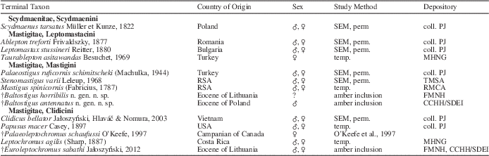
For the purpose of phylogenetic analysis, morphological structures of several extant and extinct Scydmaeninae genera were examined (Table 1). Specimens studied are deposited in the following collections: cPJ, Paweł Jałoszyński collection, Wrocław, Poland; MHNG, Muséum d’histoire naturelle de la Ville de Genéve, Switzerland; RMCA, Musee Royal de l’Afrique Centrale, Tervuren, Belgium; TMSA, Ditsong National Museum of National History, Pretoria, RSA (most of them were examined previously; see Jałoszyński, Reference Jałoszyński2012a). Morphological structures were studied either by the scanning electron microscopy or by light microscopy. For light microscopy, KOH-macerated, disarticulated and Canada balsam-embedded specimens were prepared as permanent slide preparations, while some unique vouchers were examined as intact specimens in temporary glycerol slides. Scanning electron microscopy was performed on fully disarticulated and gold-coated specimens using a LEO 435 VP scanning electron microscope.
Terminal taxa and data of specimens used in the phylogenetic analysis.

Study methods are as follows: perm., disarticulated specimen in permanent slide preparations; SEM, gold-coated disarticulated specimen examined with scanning electron microscopy; temp., intact specimen in temporary slide preparation; if characters were extracted from literature, an appropriate citation is given instead of the study method.
The measurements are as follows: length of antennae is a sum of antennomere lengths measured separately; body length is the sum of the lengths of the head, pronotum and elytra, measured separately; length of elytra was measured along the suture; width of elytra is the maximum combined width; width of head was measured across eyes; length of head was measured from the occipital constriction to anterior margin of clypeus; length of pronotum was measured along midline; width of pronotum is at the widest place.
The map presented in Figure 5 is based on an image obtained from the Demis World Map Sever open source (http://webmap.iwmi.org/DataSrc.htm).
Phylogenetic analysis
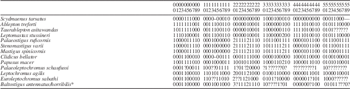
The analysis was largely based on previously published data matrix (Jałoszyński, Reference Jałoszyński2012a) with minor modifications and additional genera not included previously. The in-group taxa are comprised of adult representatives of all known genera currently placed in Mastigitae. The out-group taxa included Scydmaenus, a representative of Scydmaenini, previously found to be a sister group of Mastigitae (O’Keefe, Reference O’Keefe2005; Jałoszyński, Reference Jałoszyński2012b, Reference Jałoszyński2014). Phylogenetic analysis was based on 60 (numbered from 0) non-additive and unordered adult morphological characters; inapplicable character states were assigned a gap value (“–“) and treated equivalent to missing data (“?”). The data matrix was assembled in Nexus Data Editor for Windows v. 0.5.0 (Page, Reference Page2001); parsimony analysis was conducted in TNT (Goloboff et al., Reference Goloboff, Farris and Nixon2008) under equal weights and using the implicit enumeration strategy; the analysis was rooted with Scydmaenus. Standard bootstrap analysis (1000 replicates) was also conducted in TNT, and character mapping was made in WinClada v. 1.00.08 (Nixon, Reference Nixon1999). Trees were displayed in WinClada and annotated in COREL DRAW 9. The data matrix is presented in Table 2; the character list and states are given below:
Morphological data matrix for phylogenetic analysis of the supertribe Mastigitae.
* Character states of both species are combined.
‘–’ indicates unapplicable characters; ‘?’ indicates missing data.
0 General body vestiture: strongly erect (0); only slightly suberect or recumbent (1).
1 Modified, broadened and flattened setae on thorax: absent (0); present (1).
2 Modified, broadened and flattened setae on antennae: absent (0); present (1).
3 Anterior margin of frons: expanded anteriorly in middle (0); not expanded anteriorly (1).
4 Median part of frons: demarcated laterally by grooves or impressions and forming distinct subtriangular convexity (0); confluent with surrounding areas (1).
5 Frontoclypeal groove: present, entire (0); strongly reduced (marked only in middle) or obliterated (1).
6 Vertex + frons: strongly transverse (0); weakly transverse or subquadrate (1).
7 Median longitudinal groove on vertex: absent (0); present (1).
8 Posteromedian impression on vertex: absent (0); present (1).
9 Posterior margin of vertex: without pair of long and thick lateral setae and without large chaetopores (0); with a pair of distinctly enlarged lateral setae inserted in large chaetopores (1).
10 Postgenal setae: absent (0); present (1).
11 Postgenal process: absent (0); present (1).
12 Eyes: large, composed of at least several dozens of ommatidia (0); rudimentary, composed of one to several ommatidia (1).
13 Labrum: with anterior margin straight (0); with anteromedian emargination (1).
14 Anteromedian emargination of labrum: bordered at each side by small tooth (0); without tooth at each side (1).
15 Setal process on maxillary palpomere II: absent (0); present (1).
16 Maxillary palpomere II: in distal half strongly broadening (0); in distal half nearly parallel-sided (1).
17 Distal margin of maxillary palpomere III: perpendicular to the long axis of palpomere or only slightly oblique (0); strongly oblique (1).
18 Maxillary palpomere IV: broader than III (0); narrower than III (1).
19 Apex of maxillary palpomere IV: rounded (0); subtriangular (1).
20 Maxillary palpomeres III+IV: confluent or nearly confluent (i.e., without constriction between them), strongly broadened (more than twice as broad as apex of palpomere II) (0); confluent or nearly confluent, weakly broadened (less than twice as broad as apex of palpomere II) (1); distinctly demarcated by constriction and only slightly broader than apex of palpomere II (2); distinctly demarcated by constriction and much broader than apex of palpomere II (3).
21 Anterior margin of mentum: straight or rounded (0); deeply and broadly emarginate (1).
22 Insertions of labial palps: broadly separated (0); approximate (1).
23 Labial palpomere II: unremarkable (0); strongly enlarged (1).
24 Antennal insertions: approximate (0); broadly separated (1).
25 Length of antennal scape: much shorter than head (0); about as long as head or slightly longer (1); much longer than head (2).
26 Width of scape: as broad as pedicel or only slightly broader (0); distinctly broader than pedicel (1).
27 Length of pedicel: short, up to twice as long as broad and comparable in length with subsequent flagellomeres (0); strikingly elongated, three or more times as long as broad and much longer than subsequent flagellomeres (1).
28 Two rows of very long bristles on scape and pedicel: absent (0); present (1).
29 Flagellomeres (except XI): all or nearly all elongate (0); mostly broader than long or some about as long as broad (1).
30 Posterior pronotal ‘collar’: distinct, demarcated from disc by transverse groove or row of pits (0); indistinct or absent (1).
31 Basisternal part of prosternum: about as long as procoxal sockets or only slightly longer (0); at least twice as long as procoxal sockets (1).
32 Procoxal sockets: broadly open (0); closed by hypomeral lobes (1).
33 Coxal region of prosternum: distinctly delimited from basisternal region (0); confluent with basisternal region (1).
34 Mesoventral anterior ridge: with subtriangular median projection (0); without median projection (1).
35 Mesoventrite: with pair of lateral impressions (0); without lateral impressions (1).
36 Mesoventral intercoxal process: narrow, carinate (0); broad and flat (1).
37 Posterior part of mesoventral intercoxal process: subrectangular (0); subtrapezoidal (1); subtriangular (2).
38 Elytral punctures: deep and large, at least partly arranged in regular longitudinal rows (0); superficial and fine, not arranged in rows or rows barely discernible (1).
39 Metaventrite: in posterior part narrowing and with rounded sides (0); in posterior part broadening or subquadrate and with nearly straight lateral margins (1).
40 Mesocoxal cavities: posteriorly confluent with median portion of metaventrite (0); demarcated by carinae (1).
41 Postmesocoxal setose pits on metaventrite: absent (0); present (1).
42 Anterolateral setal pits on metaventrite: absent (0); present (1).
43 Lateral admetacoxal margins of metaventrite: not carinate (0); distinctly carinate (1).
44 Median projection or lobe of admetacoxal margin of metaventrite: absent (0); present (1).
45 Metepimera: nearly parallel-sided (0); distinctly and usually strongly broadening posteriorly (1).
46 Metendosternite: Y- or V-shaped, with insertions of furcal arms approximate (0); with insertions of furcal arms broadly separated (1).
47 Distance between mesal margins of metacoxae: distinctly (usually much) shorter than 1/3 of maximum metaventral width (0); equal to or distinctly wider than 1/3 of maximum metaventral width (1).
48 Mesocoxal lateroventral bristles: absent (0); present (1).
49 Protrochanteral bristles: absent (0); present (1).
50 Profemoral bristles: absent (0); present (1).
51 Femora: strongly and nearly abruptly clavate (0); weakly and gradually clavate (1).
52 Suture between metaventrite and the first visible abdominal sternite: distinct (0); nearly obliterated, abdominal sternite fused with posterior margin of metaventrite (1).
53 Length of the first visible (i.e., III) abdominal sternite: similar to sternite IV or only slightly longer, shorter than sternites IV+V (0); about twice as long as sternite IV, usually as long or longer than sternites IV+V (1).
54 Posterior margin of abdominal sternite VIII in males: distinctly emarginate (0); arcuate (1).
55 Median lobe of aedeagus: strongly sclerotized and darkly pigmented (0); weakly sclerotized and nearly transparent (1).
56 Flagellum: short and straight or nearly straight, never looped (0); very long, forming several loops (1).
57 Permanently everted membranous apical part of copulatory piece: absent (0); present (1).
58 Parameres: symmetrical or nearly symmetrical (0); strongly asymmetrical, one paramere much shorter than the other one (1).
59 Apices of parameres: strongly and rapidly bent mesally (0); not bent mesally or only slightly curved (1).
Systematic paleontology
Holotypes are deposited in The Field Museum of Natural History, Chicago, USA (FMNH) and the Collection Christel and Hans Werner Hoffeins, Hamburg, Germany, with final depository at the Senckenberg Deutsches Entomologisches Institut Müncheberg, Germany (CCHH/SDEI). The nomenclature and morphological terms are used after Jałoszyński (Reference Jałoszyński2012a).
Family Staphylinidae Latreille, Reference Latreille1802
Subfamily Scydmaeninae Leach, Reference Leach1815
Supertribe Mastigitae Fleming, Reference Fleming1821
Tribe Mastigini Fleming, Reference Fleming1821
Genus Baltostigus new genus
Type species
Baltostigus antennatus new species, here designated.
Diagnosis
Differs from all other genera of Mastigini in strongly enlarged and axe-shaped maxillary palpomere IV, which is more than twice as broad as palpomere III; angulate humeral calli; fully developed hind wings; deep elytral punctures partly arranged in longitudinal rows; and symmetrical parameres.
Etymology
The genus name is composed of the prefix Balto- derived from the Baltic Sea and the stem -stigus derived from Mastigus, the type genus of Mastigini. Sex masculine.
Occurrence
Eocene of northern Europe.
Remarks
The new genus is classified within Mastigitae on the basis of the strongly elongate antennal scape with ventrally located notch, so that the pedicel with flagellum can bend ventrally, and the maxillary palp, which is longer than the head. Baltostigus n. gen. is placed within Mastigini based on two longitudinal rows of robust spines on scape and pedicel, the pedicel distinctly enlarged in relation to flagellomeres and the maxillary palpomere IV broader than III. Within Mastigini, Baltostigus is unique in having distinct, angulate humeral calli (indistinct and rounded in all other genera), a structure associated with fully developed hind wings (all other genera are wingless); deep and large punctures on the elytra (superficial and fine punctures in all other genera); and the maxillary palpomere IV strongly flattened, axe-shaped and much broader than long (weakly flattened or subcylindrical and strongly elongate in all other genera). The male of Baltostigus antennatus n. spec. has a symmetrical parameres, whereas the aedeagi of all other genera of Mastigini are strongly asymmetrical, with one paramere much shorter than the other one (Palaeostigus and Mastigus) or even rudimentary (Stenomastigus). When the apex of median lobe or the copulatory piece is visible through the genital opening of any extant species of Mastigini, the apical portion of only one paramere can be seen. In Baltostigus, apices of two parameres project to an equal extent from the genital opening.
Two available specimens of Baltostigus differ in several characters and two alternative hypotheses were considered: (1) they represent different species, and (2) they represent a male and female of the same species. The first hypothesis is supported and the other one excluded by observations based on characters known in all extant taxa. Males and females of Mastigini differ (often remarkably) in the shape of elytra, which in females are broader and often of a different shape. The two amber inclusions studied do not differ in the shape of elytra. Females of extant Mastigini have the antennae shorter than males (in relation to the body length); in the studied fossil material, the specimen unambiguously identified as a male (on the basis of partly visible genital characters) has the antennae much shorter in relation to the body length than the other specimen, whose sex remains unknown, and whose antennae are much longer than the body. Also the scape in the male specimen is only 1.33 times as long as the head, while in the other specimen the scape is 1.75 times as long as the head. These observations strongly suggest that the specimen whose sex is unknown cannot represent a female of the same species as the male.
Baltostigus antennatus new species
Photographs of Baltostigus n. gen.: (1, 2) Holotype of Baltostigus antennatus n. sp. (specimen CCHH/SDEI 835-1) in lateral (1) and dorsal (2) views; (3, 4) holotype of Baltostigus horribilis n. sp. (specimen FMNH INS3101601) in laterodorsal (3) and lateroventral (4) views; all scale bars are 1 mm.
Morphological details of male holotype of Baltostigus antennatus n. sp. (specimen CCHH/SDEI 835-1) with arrows indicating characters and character states: (1) Entire specimen in lateral view, scale bar represents 1 mm; (2) head and right antenna in lateral view, scale bar represents 0.25 mm; (3) abdominal apex in ventrolateral view showing parameres (pm), apex of median lobe (aml), tergite IX (tIX), and sternite IX (stIX), scale bar represents 0.25 mm; (4) abdominal apex in ventral view, showing parameres (pm), apex of median lobe (aml), tergites IX (tIX) and X (tX), sternites VIII (stVIII) and IX (stIX), scale bar represents 0.25 mm.
Holotype
Male; CCHH/SDEI 835-1, Eocene Baltic amber, Poland; precise collecting data unknown.
Diagnosis
Body length <3.50 mm; antenna shorter than body; scape 1.33 times as long as head and only 1.2 times as long as pronotum; antennomere IV <1.5 times as long as III.
Etymology
The name antennatus was chosen to underline the strikingly modified antennae.
Occurrence
Known only from the lower to middle Eocene (37–54.5 Ma) amber of Poland.
Description
Body (Figs. 1.1, 1.2, 2.1) elongate and uniformly brown, including vestiture; body length 3.40 mm.
Head (Fig. 2.2) longer than broad, broadest at eyes, length 0.67 mm, width 0.60 mm; occipital constriction deep and distinct, tempora rounded and convergent posteriorly, about as long as eye diameter; eyes very large, slightly oval and strongly convex, finely faceted, vertex flattened; frons in lateral view forms a distinctly obtuse angle with the plane of vertex, its posterior margin delimited by antennal sockets and deeply bisinuate with narrow median longitudinal ridge between antennae, median part of frons slightly convex and covered with indistinct, sparse punctures and sparse, short, erect setae; maxillary palp with minute and weakly elongate palpomere I, strongly elongate and slender palpomere II, subtriangular and flattened palpomere III broadest at apex, and axe-shaped palpomere IV much broader than long, with weakly rounded distal margin and base slightly narrower than palpomere III. Labial palp with enlarged palpomere II and small, subconical and strongly elongate palpomere III much narrower than II. Antennae (Fig. 2.1, 2.2) distinctly shorter than body, length 2.97 mm, lengths of antennomeres: I, 0.90 mm; II, 0.42 mm; III, 0.12 mm; IV, 0.17 mm; V, 0.20 mm; VI, 0.22 mm; VII, 0.17 mm; VIII, 0.20 mm; IX, 0.20 mm; X, 0.17 mm; XI, 0.17 mm; scape strongly elongate and broadening distally, 1.33 times as long as head, 1.2 times as long as pronotum and slightly shorter than half length of elytra, with two lateroventral rows of robust bristles (six in internal row and five in external row); pedicel 0.45 times as long as scape, also with two lateroventral rows of long bristles each composed of five or six bristles (the number unclear because some bristles seem to be broken off but insertion sites are not visible in the specimen); all flagellomeres strongly elongate, with sparse, long and strongly erect setae.
Pronotum (Figs. 1.1, 1.2., 2.1) elongate and broadest near anterior third, length 0.75 mm, width 0.65 mm; anterior and posterior margins arcuate, without dorsal grooves or pits. Pronotal disc covered with distinct, unevenly distributed and moderately dense punctures, those along midline distinctly sparser. Prothoracic hypomera not demarcated laterally; details of prosternum not visible.
Elytra (Figs. 1.1, 1.2., 2.1) much more convex than pronotum, with the highest and broadest place located distinctly behind middle, length 1.97 mm, width 1.25. Each elytron with distinct angulate humerus and three slightly irregular impressed rows of large punctures well visible in anterior third, punctures on remaining elytral surface seem nearly randomly distributed; elytral apex subtriangular. Vestiture distinct, composed of sparse, short and suberect setae directed posteriorly.
Hind wings not visible.
Details of mesoventrite not visible, except prominent mesocoxal projections and flat (non-carinate) mesoventral intercoxal process of unclear shape.
Metaventrite only partly visible, subrectangular, with broad and slightly concave metaventral intercoxal process and broad metepimera.
Abdomen about as long as metaventrite; abdominal sternites not visible except terminal ones, sternite VIII with rounded posterior margin.
Legs (Figs. 1.1, 1.2., 2.1) long and slender; coxae subconical; trochanters subtriangular (protrochanter unmodified), trochantero-femoral junction strongly oblique; femora indistinctly clavate, covered with dense, short and nearly recumbent setae; tibiae straight and slender, with nearly parallel margins, protibia shorter than profemur, meso- and metatibia about as long as corresponding femora, all tibiae densely covered with short and nearly recumbent vestiture with several long and erect setae scattered along entire length; each tarsus distinctly longer than half length of corresponding tibia, tarsomeres distinctly flattening from I to V; tarsomere I strongly elongate and only slightly broadened toward apex, tarsomere II shorter than I but similar in shape, tarsomeres III and IV each much shorter than II and subtriangular, slightly broadened distally; tarsomere V only slightly longer than broad, broader than IV, with curved and pointed claws.
Aedeagus (Fig. 2.3, 2.4) slightly projecting from genital opening, tips of slightly curved and symmetrical parameres and apex of median lobe visible.
Baltostigus horribilis new species
Morphological details of male holotype of Baltostigus horribilis n. sp. (specimen FMNH INS3101601) with arrows indicating characters and character states: (1) entire specimen in laterodorsal view, scale bar represents 1 mm; (2) entire specimen in ventrolateral view; (3) head in anteroventral view with antennae in ventral view, scale bar represents 0.25 mm.
Holotype
Sex unknown; FMNH INS3101601, Lithuanian Eocene Baltic amber; precise collecting data unknown.
Diagnosis
Body length >3.70 mm; antenna longer than body; scape 1.75 times as long as head and nearly 1.6 times as long as pronotum; antennomere IV >2 times as long as III.
Etymology
Latin adjective horribilis (horrible) refers to the terrifying appearance rendered by the enlarged and spinose scape and pedicel.
Occurrence
Known only from the lower to middle Eocene (37–54.5 Ma) amber of northern Lithuania.
Description
Body (Figs. 1.3, 1.4, 3.1, 3.2) elongate and uniformly black, including vestiture; body length 3.77 mm.
Head (Fig. 3.3) longer than broad, broadest at eyes, length 0.70 mm, width 0.62 mm; occipital constriction deep and distinct, tempora rounded and convergent posteriorly, about as long as eye diameter; eyes very large, nearly circular and strongly convex, finely faceted, vertex flattened; frons in lateral view forms a slightly obtuse angle with the plane of vertex, its posterior margin delimited by antennal sockets and deeply bisinuate with narrow median longitudinal ridge between antennae, median part of frons slightly convex and covered with indistinct, sparse punctures and sparse, short erect setae, anterior margin of frons separated from clypeus by transverse impression; shape of clypeus not visible, its dorsal surface with several very long and curved suberect setae directed anteriorly; labrum with anteromedian emargination at each side bordered by small tooth and with several long dorsal setae directed anteriorly; apex of each mandible with long and sharp apical tooth and similar in shape but slightly shorter dorsal subapical tooth; maxillary palp with minute and weakly elongate palpomere I, strongly elongate and slender palpomere II, subtriangular and flattened palpomere III broadest at apex, and axe-shaped palpomere IV distinctly broader than long, with rounded distal margin and base slightly narrower than palpomere III. Labial palps appear large and robust, but details are unclear. Antennae (Fig. 3.3) distinctly longer than body, length 4.12 mm, lengths of antennomeres: I, 1.22 mm; II, 0.62 mm; III, 0.17 mm; IV, 0.37 mm; V, 0.27 mm; VI, 0.27 mm; VII, 0.25 mm; VIII, 0.25 mm; IX, 0.25 mm; X, 0.22 mm; XI, 0.20 mm; scape extremely elongate and broadening distally, 1.75 times as long as head, nearly 1.6 times as long as pronotum and slightly longer than half length of elytra, with two lateroventral rows of multiple robust bristles (left scape, internal row composed of 11 bristles, external row with 6 bristles; right scape, internal row with 7 bristles, external row with 6 bristles); pedicel 0.53 times as long as scape, also with two lateroventral rows of long bristles composed of 5 bristles each (one bristle broken off in each row of right pedicel); all flagellomeres strongly elongate, with sparse, long and strongly erect setae.
Pronotum (Fig. 3.1) elongate and broadest near anterior third, length 0.77 mm, width not possible to measure; anterior and posterior margins arcuate, without dorsal grooves or pits, with nearly vertical elongate impression on lateral surface dividing prothorax into anterior (precoxal and coxal) and posterior (postcoxal) parts. Pronotal disc covered with distinct, unevenly distributed and moderately dense punctures. Prothoracic hypomera not demarcated laterally; prosternum elongate, prosternal process subtriangular, not separating procoxae.
Elytra (Figs. 1.3, 3.1) much more convex than pronotum, with the highest and broadest place located distinctly behind middle, length 2.30 mm, width not possible to measure. Each elytron with distinct angulate humerus; shallow adsutural stria well visible in anterior half; and sharp, narrow and deep marginal stria separating narrow marginal carina running from humerus to apex; elytral apex rounded. Elytra covered with dense, unevenly distributed and distinct punctures separated by spaces 0.5–1.5 times as long as diameters of punctures, punctures are of various diameters and depths and are distributed almost randomly, except for three slightly irregular striae extending from humerus along lateral elytral margin. Vestiture distinct, composed of sparse, short and suberect setae directed posteriorly, and sparsely distributed long and erect setae well visible in anterior half of elytra.
One hind wing is visible, extending from under elytra, slightly shorter than twice as long as elytra.
Mesoventrite (Fig. 3.2) barely visible, except for broad mesocoxal projection; mesoventral intercoxal process flat and distinctly separating mesocoxae.
Metaventrite (Fig. 3.2) large, broader than long, convex at middle and slightly impressed in posteromedian part, with distinctly concave posterior margin between metacoxae; posterior adcoxal margin deeply concave; coxal suture indistinctly marked; ventrite covered with moderately dense, short and nearly recumbent setae. Metepimeron well visible in ventral and lateral view, nearly parallel-sided.
Abdomen (Fig. 3.2) longer than metaventrite; six abdominal sternites visible.
Legs (Figs. 1.3, 1.4, 3.1, 3.2) very long and slender; coxae subconical; trochanters subtriangular, trochantero-femoral junction strongly oblique; femora indistinctly clavate, in lateral view broadest near distal third, covered with dense, short and nearly recumbent basic vestiture with several long and strongly erect setae in distal part; tibiae straight and slender, with nearly parallel margins, protibiae shorter than femora, meso- and metatibiae longer than femora, all tibiae densely covered with short and nearly recumbent vestiture with several long and erect setae scattered along entire length; each tarsus slightly longer than half length of corresponding tibia, tarsomeres distinctly flattening from I to V; tarsomere I strongly elongate and only slightly broadened toward apex, tarsomere II shorter than I but similar in shape, tarsomeres III and IV each much shorter than II and subtriangular, broadened distally; tarsomere V about as long as broad, broader than IV, with curved and pointed claws.
Results
The new genus Baltostigus was found to have several unique characters, not known in extant Mastigini: fully-developed hind wings and prominent humeral calli; deep elytral punctures partly arranged in longitudinal rows; and symmetrical parameres. The remaining character states clearly place it within the tribe Mastigini.
In the phylogenetic analysis, two equally parsimonious trees were obtained (Fig. 4), tree length=107 steps, consistence index, CI=0.59; retention index, RI=0.75. In both trees, Baltostigus was placed as a sister group to Stenomastigus + (Palaeostigus + Mastigus), and this lineage is supported by the bootstrap value 79. The tribe Mastigini, including Baltostigus, is characterized by five unique apomorphies: approximate insertions of labial palps (character 22(1)); strongly elongated pedicel, distinctly longer than flagellomeres (27(1)); two rows of bristles present on scape and pedicel (28(1)); mesoventral intercoxal process relatively broad and nearly flat, not carinate (36(1)); and metaventrite with nearly straight lateral margins, not rounded at sides (39(1)).
Results of the parsimony analysis of the phylogenetic relationships within all genera of extant and extinct Mastigitae; (1, 2) Two equally parsimonious trees (tree length=107; consistency index=0.59; retention index=0.75) representing two alternative phylogenetic hypotheses. The trees were rooted on Scydmaenus. Standard bootstrap support values are shown below branches in the bottom tree (values <50 are omitted). Unambiguously optimized character changes are plotted along the internodes. Black circles indicate unique character changes; white circles indicate parallelisms or reversals; character numbers are above circles; character states below circles.
Within Mastigitae, only Leptomastacini (bootstrap 99), Mastigini and a part of Clidicini were placed as monophyletic clades; unstable position of Papusus and the placement of Clidicus in two alternative and equally parsimonious topologies (Fig. 4.1, 4.2) render Clidicini polyphyletic. The ‘Leptochromus lineage’ composed of Palaeoleptochromus + (Leptochromus + Euroleptochromus) is placed as monophyletic and supported by the bootstrap value 77, but Clidicus is sister to the ‘Leptochromus lineage’ + Mastigini.
Discussion
Baltostigus is the first extinct genus of Scydmaeninae unambiguously assigned to Mastigini. The phylogeny of Mastigitae is far from being resolved and the new results do not support previous hypotheses of the monophyly of Clidicini, and Mastigini as a sister group of a clade composed of all remaining Mastigitae (Jałoszyński, Reference Jałoszyński2012a). Moreover, the placement of Papusus, previously suggested as unclear, in the present analysis was also problematic. These issues will require further study, but the monophyly of Mastigini seems already well established and supported by several unique apomorphies, both in the present and previous phylogenetic analysis (Jałoszyński, Reference Jałoszyński2012a). Baltostigus was here placed as a sister group to a clade composed of all remaining, extant genera of Mastigini. Combination of apparently plesiomorphic and derived characters suggests that Baltostigus represents a lineage that branched off the ancestral stem and while some ‘primitive’ structures remained conserved, some others differentiated to an almost monstrous extent.
All species of Mastigitae except the extant Mastigini, and also Scydmaenini (the most plausible sister group of Mastigitae) have a symmetrical aedeagus. The strong asymmetry of the aedeagus in Palaeostigus, Mastigus, and especially Stenomastigus, with one paramere long and the other short or even obliterated, is therefore a derived condition. Circumstances that might have promoted a profound elongation and further asymmetrization of male genitalia in Mastigini were studied by Jałoszyński et al. (Reference Jałoszyński, Matsumura and Beutel2015), and processes related to a reproductive competition were suggested to play a significant role in the evolution of the aedeagus and associated structures. Baltostigus has an apparently symmetrical aedeagus, or at least the parameres (and the asymmetry of parameres is crucial for mating strategies in extant Mastigini). This is clearly an ancestral condition.
Another interesting and clearly ancestral character state is the presence of fully developed hind wings (visible in B. horribilis), accompanied by prominent and angulate humeral calli (visible in both newly described species). As the size of humeral calli usually correlates with the presence (large calli) or absence (small or missing calli) of hind wings, B. antennatus may also be a winged species. Most extant species of Scydmaeninae, with some exceptions, especially within Cephenniitae, have long and functional wings, but only in Mastigitae the lack of wings is a typical and predominating condition. In all extant Mastigini not only the wings and humeral calli are entirely absent, but also the elytral sutures are fused (Leleup, Reference Leleup1968; Jałoszyński, Reference Jałoszyński2012a). Most members of Scydmaeninae are tiny cryptic beetles that live in substrates rich in decaying plant matter, where their prey (mites, springtails, or other small organisms) is abundant (O’Keefe, Reference O’Keefe2005; Jałoszyński, Reference Jałoszyński2012d, e; Jałoszyński and Olszanowski, Reference Jałoszyński and Olszanowski2013, Reference Jałoszyński and Olszanowski2015). They are characterized by low population densities and the ability to fly may be an important mean of dispersal, finding new habitats and mating partners. In contrast, species of Mastigini usually live in large groups, their bodies are several times as long as those typical of the majority of scydmaenines, they have very long legs and move quickly on the surface of ground and plants in a broad daylight (Jałoszyński et al., Reference Jałoszyński, Matsumura and Beutel2015). Encounters and interactions with potential prey and mating partners under such circumstances are frequent, and this mode of life very likely compensates for the loss of hind wings. However, it may not entirely compensate for the loss of flight-associated dispersal capabilities, as Mastigini seem to be prone to extinctions driven by habitat fragmentation. Dense local populations observed by the author in South Africa in 2012 often occupy small territories surrounded by large barriers (e.g., dry savannas) that seem impenetrable for flightless beetles whose larvae are soft-bodied and sensitive to desiccation. The habitat fragmentation may, therefore, be at least partly responsible for the present disjunctive distribution of Mastigini; the continuous areas showed in Figure 5 are in fact composed of many isolated ‘islands’ where various species and subspecies occur. The relatively small Baltostigus shows long legs, typical of extant fast-running Mastigini, but its fully-developed wings suggest better dispersal abilities.
Distribution of extant species of Mastigini.
Another interesting character of Baltostigus are deep elytral punctures partly arranged in longitudinal striae. In all extant Mastigini the elytra bear fine and superficial punctures, mostly randomly distributed, but in some species forming indistinct and shallow longitudinal rows (Leleup, Reference Leleup1968; Bordoni and Castellini, Reference Bordoni and Castellini1973; Jałoszyński, Reference Jałoszyński2012c, Reference Jałoszyński2012d). In the remaining tribes of Mastigitae deeply punctate longitudinal striae are common (e.g., Castellini, Reference Castellini1996; O’Keefe, Reference O’Keefe2002; O’Keefe and Monteith, Reference O’Keefe and Monteith2000). They are absent in the sister group (Scydmaenini) of Mastigitae and in all remaining Scydmaeninae. It is therefore possible that this is a derived synapomorphy of Mastigitae, reversed in extant Mastigini.
The extremely elongate, monstrous antennal scape with multiple very long bristles seems to be a derived condition. The scape is enlarged in all Mastigitae, and only in Mastigini it is provided with rows of bristles, but in no extant genus this antennomere is so huge as that in Baltostigus. However, it cannot be excluded that the scape has initially become extremely large to subsequently reduce in size. This hypothesis is based on observations of extant Palaeostigus and Stenomastigus (Jałoszyński, unpublished observations, 2012, 2014). Large and spiny scape and pedicel, combined with the movable joint between them, appear as a sort of a catching or trapping device, similar to that of a ground beetle Loricera Latreille, which catches springtails using a ‘cage’ formed by similarly spiny proximal antennomeres (Bauer, Reference Bauer1982). However, under laboratory conditions, tested species of Mastigini did not use the spiny antennae to capture prey. If these peculiar structures indeed evolved to play a role in catching jumping or otherwise fast-moving prey, they likely might have reached the size seen in Baltostigus. However, if subsequently Mastigini shifted their feeding habits toward slowly moving organisms (and extant species can feed on caterpillars or scavenge dead arthropods), the scape and pedicel might have lost their catching function and secondarily reduced in size. Only further discoveries of fossil Mastigini may shed light on this interesting problem.
It seems that the currently known genera of Mastigini represent only a fraction of the true diversity of this lineage, and extinct taxa may indeed be crucial for understanding the evolution of this supertribe. Already three extinct genera of Mastigitae are known: Baltostigus, Euroleptochromus, and Palaeoleptochromus. The latter genus of Clidicini from the Upper Cretaceous (Campanian) Canadian amber is strikingly similar to extant Leptochromus, which demonstrates that this group has differentiated before 79 Ma and has little changed since then.
Acknowledgments
I express my sincere thanks to Albert Allen, who generously donated the amber specimen to the FMNH, and to Christel and Hans Werner Hoffeins who sent me another interesting specimen for study. Anonymous reviewers are thanked for their valuable comments.