Case description
We report a case of a 20-month-old girl who was born with the diagnosis of perimembranous and complex muscular ventricular septal defects and prematurity. She was initially treated with anti-congestive medications and high calorie formula, subsequently undergoing pulmonary artery banding due to the failure of conservative managements. Her repeat echocardiogram before planned surgical treatment revealed the presence of a large complex mid-muscular ventricular septal defect with two different shunt trajectories across the defect. The perimembranous ventricular septal defect was partially covered by redundant tricuspid valve tissue and was determined to be pressure restrictive per echocardiographic measurements (Fig 1a and b).
Echocardiographic images of mid-muscular and perimembranous ventricular septal defects. (a) Muscular (mVSD) and perimembranous (pVSD) ventricular septal defects and muscle bands. (b) Complex muscular ventricular septal defect with two colour jets across the defect (LV = left ventricular; RV = Right ventricular).

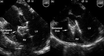
She underwent a repeat median sternotomy for perventricular device closure of the muscular ventricular septal defect on a beating heart. Under transoesophageal echocardiographic guidance, a 0.021 × 1 inch Argon access needle (AGA Medical Corp., Plymouth, Minnesota, United States of America) was introduced into the right ventricular cavity through a 5-0 polypropylene purse-string suture. A 0.021 inch × 40 centimetre Argon J-wire was passed through the needle and manipulated into the left ventricular cavity through the defect. A 6-French Terumo Pinnacle Sheath (Terumo Medical, Elkton, Maryland, United States of America) was carefully advanced into the left ventricular cavity. Initially, a 16-millimetre self-expandable Amplatzer Muscular VSD Occluder (AGA Medical Corporation, Golden Valley, Minnesota, United States of America) was introduced and deployed successfully on the left ventricular side; however, deployment of the right ventricular disc was unsuccessful. The device was removed and downsized to 14 millimetres. The same problem was experienced on the right ventricular side, with the entrapment of the disc preventing it from full deployment (Fig 2a). Nonetheless, the transoesophageal echocardiogram revealed a small residual shunt in the lower aspect of the device with a pressure-restrictive perimembranous ventricular septal defect. The initial decision was to leave the device in place, given haemodynamically insignificant shunting across the residual muscular and restrictive perimembranous septal defects, and to re-evaluate the shunts after pulmonary artery debanding.
(a) Entrapped right ventricular (RV) disc in the muscle bands with full deployment of the left ventricular (LV) disc. (b) Full deployment of the discs after resection of the muscle bundles.

Therefore, the patient was placed on cardiopulmonary bypass via single aortic and bicaval cannulation using warm perfusion with a beating heart. The band was taken down, and pulmonary artery reconstruction was performed in an end-to-end manner. The transoesophageal echocardiogram revealed moderate shunting at the lower aspect of the device and at the perimembranous septal defect, which appeared more significant than before debanding. Given the above findings and poorly seated device in the right ventricular side, the decision was made to go back to cardiopulmonary bypass for closure of the defects.
The patient was placed on full cardiopulmonary bypass and under moderate hypothermia and cardioplegic arrest, and the ventricular septal defects were exposed and explored through a right atrial approach. The perimembranous ventricular septal defect was moderate in size and was closed using an autologous pericardial patch tanned in glutaraldehyde solution. Inspection of the device revealed entrapment of the disc in the moderator band and trabecular septum. This was released by dividing the bands, which led to full expansion of the disc. The patient was separated from cardiopulmonary bypass with good biventricular function and in normal sinus rhythm. Intra-operative transoesophageal echocardiogram revealed complete closure of the muscular and perimembranous ventricular septal defects, with complete expansion of both disc plates (Fig 2b). The patient did well and was discharged home 5 days after the surgery on diuretic therapy and a baby aspirin. At 2 months after her surgery, she had reached normal somatic growth and was off of her medications with a well-seated device.
Discussion
The first successful perventricular device closure of a ventricular septal defect on a beating heart was reported in an animal model and subsequently carried over into clinical practice.Reference Amin, Berry, Foker, Rocchini and Bass1–Reference Bacha, Cao, Starr, Waight, Ebeid and Hijazi3 Some of the advantages of this procedure include avoidance of cardiopulmonary bypass or shorter bypass time and cardioplegic arrest, avoidance of ventriculotomy or muscle bundle resection, immediate confirmation of adequate closure, and avoidance of vascular access complications or injuries. However, there are some anatomical variants that may prohibit and complicate muscular ventricular septal defect device closure.
The procedure was performed in the operating room suite because of it being a re-operation with possible re-entry injury, debanding of the pulmonary artery, possible closure of perimembranous ventricular septal defect, and any procedural mishaps, which would require cardiopulmonary bypass. Complete dissection of the heart and vessels was accomplished, and the aortic and caval purse strings were placed in case of arrhythmia or injury to major structures requiring emergent cardiopulmonary support. Transoesophageal echocardiographic guidance was used to locate a site on the right ventricular free wall for insertion of the needle and guidance of the wire and sheath to avoid false passage through the septum or perforation of the left ventricular free wall, mitral or aortic valve.
There are some potential complications that can occur with perventricular ventricular septal defect device closure, with two major ones being cardiac perforation and device embolisation.Reference Diab, Cao and Hijazi4 Another potential challenge that may result in unsuccessful closure is the inability to deploy the right ventricular disc as the result of apical location of the muscular septal defect or heavy trabeculations of the right ventricular sinus.Reference Diab, Hijazi, Cao and Bacha5–Reference Crossland, Wilkinson, Cochrane, d'Udekem, Brizard and Lane9 In this particular case, the initial ventricular septal defect device had to be downsized because of entrapment of the disc in the right ventricular muscle bundles. This tactic is sometimes necessary in order to avoid crossing muscle bands for full expansion of the disc. However, in this instance it was not successful, resulting in incomplete expansion of the disc and incomplete closure of the muscular ventricular septal defect on the right ventricular side. There was also significant shunting across the perimembranous ventricular septal defect after pulmonary artery debanding, which collectively required surgical intervention and use of cardiopulmonary bypass. The right ventricular disc was entrapped in the moderator band and trabecular muscle bundles, which were divided to free the disc. The perimembranous ventricular septal defect size was underestimated because of the presence of tricuspid aneurysmal tissue and the pulmonary artery band requiring full exploration of the defect and successful closure. Intra-operative post-repair transoesophageal echocardiogram showed complete closure of the perimembranous and muscular ventricular septal defects and complete deployment of both discs.
Conclusion
The hybrid approach to perventricular closure of the dominant muscular ventricular septal defect proved to be useful in this case by placing the closure device across the complex muscular septal defects, which would have been very difficult to identify on an empty non-beating heart. It also decreased the time needed for cardiopulmonary support and ischaemia during perimembranous ventricular septal defect closure, resulting in good ventricular function and early post-operative recovery. In this particular case, the presence of heavy trabeculations prohibited successful deployment of the right ventricular disc requiring division of the muscle bands entrapping the disc plate. The presence of the pulmonary artery band with associated high right ventricular pressure also underestimated the degree of residual shunt across the muscular septal defect and the size of the perimembranous ventricular septal defect, requiring open surgical exploration and closure of the defects.