Ebstein malformation of tricuspid valve is a relatively uncommon form of congenital tricuspid valve dysplasia and right ventricular cardiomyopathy that accounts for less than 1% of cases with CHD. Association with right- and left-sided cardiomyopathies including noncompaction left ventricle has been reported previously, but co-occurrence with other forms of left-sided cardiomyopathies is less commonly seen. Reference Attenhofer Jost, Connolly, Dearani, Edwards and Danielson1–Reference van Engelen, Postma and Van De Meerakker3 Here we present a young patient with concomitant left and right-sided cardiomyopathy due to Ebstein and hypertrophic cardiomyopathies.
Case
A 33-year-old male presented with exertional dyspnoea, fatigue and mild lower extremity oedema since 3 months. He mentioned no previous illnesses, drug use or family history of cardiovascular disease. Physical examination revealed III/VI systolic murmur in the left sternal border. He had +1 pitting peripheral oedema, no ascites and no clinical cyanosis. The lungs were clear to auscultation.
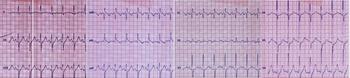
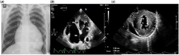
Twelve lead ECG showed normal sinus rhythm, left axis deviation and incomplete right bundle branch block pattern (Fig 1). Chest X-ray was remarkable for cardiomegaly (Fig 2a).
Twelve lead ECG depicting left axis deviation and incomplete right bundle branch block.

Chest X-ray with cardiomegaly ( a ), Transthoracic 4 chamber ( b ), and short axis ( c ) views showing Ebstein malformation of tricuspid valve of tricuspid valve with septal leaflet displacement (arrow) and left ventricular hypertrophy.

Transthoracic echocardiography revealed normal left ventricular size with preserved systolic function (EF = 55% but reduced GLS: −11.4, GCS: −21.6), severe left ventricular hypertrophy mostly in mid ventricle (maximum size at mid interventricular septum: 2.3 cm and posterior wall: 1.6 cm), no outflow tract or intra cavitary obstruction at rest and mild diastolic dysfunction(E/E' = 12). There was considerable apical displacement of septal and inferior leaflets of tricuspid valve (displacement index = 1.6 cm/m2). Anterior leaflet was severely tethered, elongated and attached to the right ventricular free wall with multiple chordae resulting in severe tricuspid regurgitation compatible with Ebstein malformation of tricuspid valve of tricuspid valve. The aortic arch was right sided (Fig 2b and c).
The patient underwent cardiac MRI depicting asymmetric LVH with a pattern consistent with hypertrophic cardiomyopathy. Maximum wall thickness was 21 mm in basal inferoseptal walls and remarkable increased LV mass index (138 g/m2) with a low-grade scar index of 7%. There was no LV outflow tract obstruction. The patient also had right-sided aortic arch with aberrant retro-esophageal left subclavian artery. There was typical Ebstein malformation of tricuspid valve of tricuspid valve with 18.5 mm/m2 septal leaflet displacement and tethered, elongated anterior tricuspid leaflet (video 1 and video 2).
The patient did not show clinical and laboratory findings in favour of metabolic cardiomyopathies such as LAMP2 cardiomyopathy, glycogen storage diseases, or Fabry disease. As there was significant RV dysfunction, the patient was scheduled for TV repair which he has so far deferred due to COVID-19 pandemic.
Discussion
Ebstein malformation of tricuspid valve is a rare congenital form of tricuspid valve dysplasia associated with right ventricular cardiomyopathy. Affected individuals exhibit both functional and structural right ventricular abnormalities. There are variable degrees of septal and inferior leaflet displacement resulting from abnormal adherence to the underling myocardium and failure of normal delamination process leading to severe tricuspid regurgitation and global right ventricular dysfunction. Genetic alterations have been implicated in this anomaly including mutations in MYH7 and NKX2.5 genes. Reference Attenhofer Jost, Connolly, Dearani, Edwards and Danielson1–Reference Maron, Maron and Olivotto4 The MYH7 gene encodes the beta (β)-myosin heavy chain protein that is found in sarcomeres. Reference van Engelen, Postma and Van De Meerakker3,Reference Maron, Maron and Olivotto4 Hypertrophic cardiomyopathy is the most common genetic cardiovascular disease and the most common cause of sudden cardiac death in young adults. Mutations in MYH7 gene are also the most common mutations found in patients with hypertrophic cardiomyopathy that is characterised by genetic left ventricular hypertrophy of various patterns and a potential for syncope, sudden cardiac death, and progression to heart failure. Reference Maron, Maron and Olivotto4,Reference Saedi, Oraii and Hajsheikholeslami5 Despite the role of MYH7 gene mutation in both hypertrophic cardiomyopathy and Ebstein malformation of tricuspid valve, simultaneous clinical presentation of both disorders is not commonly seen, and this report might provide insights into the genetic pathogenesis of both cardiac anomalies. Few previously reported cases in the literature involve older patients. Reference de Agustín, de Isla and Zamorano6,Reference Lee, Fu and Fang7 Treatment for Ebstein malformation of tricuspid valve revolves around surgical repair or replacement of tricuspid valve, whereas family screening, drug therapy mostly with beta blockers, and measures to prevent sudden cardiac death are the mainstays of management in patients with hypertrophic cardiomyopathy.
Supplementary material
To view supplementary material for this article, please visit https://doi.org/10.1017/S1047951121003644
Acknowledgements
The authors would like to thank the RHC imaging department staff for their kind assistance.
Financial support
This research received no specific grant from any funding agency, commercial, or not-for-profit sectors.
Conflicts of interest
None.
Ethical standards
Institutional ethics committee approved of the manuscript.