Introduction
Echinococcus remains a significant public health problem worldwide and, in several regions, the aetiological agents of cystic hydatid disease/echinococcosis are extending their range (Eckert et al., Reference Eckert, Schantz, Gasser, Torgerson, Bessonov, Movsessian, Thakur, Grimm, Nikogossian, Eckert, Gemmell, Meslin and Pawlowski2001). During the past 40 years, observations from laboratory and field studies have revealed considerable phenotypic and genetic variability between isolates of Echinococcus granulosus from different species of intermediate host (Thompson & McManus, Reference Thompson, McManus, Eckert, Gemmell, Meslin and Pawlowski2001). Increasingly, this phenotypic variability has been found to correlate with genetic differences, and from this the concept of a series of host-adapted strains of E. granulosus was developed. Molecular epidemiological studies have also revealed that the genetic differences between what were perceived to be host-adapted strains of E. granulosus are conserved and occur consistently in isolates derived from different species of intermediate hosts throughout the world (Thompson & McManus, Reference Thompson, McManus, Eckert, Gemmell, Meslin and Pawlowski2001, Reference Thompson and McManus2002; McManus & Thompson, Reference McManus and Thompson2003). This has been well demonstrated for the sheep, horse, cattle, pig and camel strains in Europe, Iran and Africa.
It has been proposed that the morphological, biological and genetic differences that separate strains of Echinococcus are sufficient to justify species rank in at least two forms – the horse (E. equinus) and cattle (E. ortleppi) strains – thus increasing the number of recognized species of Echinococcus from four to six (Thompson & McManus, Reference Thompson and McManus2002; Lavikainen et al., Reference Lavikainen, Lehtinen, Meri, Hirvelä-Koski and Meri2003; Obwaller et al., Reference Obwaller, Schneider, Walochnik, Gollackner, Deutz, Janitschke, Aspöck and Auer2004). Although the strongest arguments can be made for recognition of species status for E. equinus and E. ortleppi, the camel, pig and cervid forms also warrant taxonomic status. However, it is considered that more isolates of these forms require molecular characterization from different geographical areas. Unlike the horse and cattle forms, which have a high degree of intermediate host specificity, the camel strain/genotype is not host specific for camels, and pigs may harbour more than one closely related genotype.
Cystic echinococcosis due to E. granulosus occurs in most countries of South America and is an important public health and economic problem in the southern part of South America, particularly Chile, Argentina, Uruguay, Brazil and parts of Peru (Schantz et al., Reference Schantz, Chai, Craig, Eckert, Jenkins, Macpherson, Thakur, Thompson and Lymbery1995). In these areas, the sheep ‘strain’, E. granulosus, is the predominant form that is perpetuated in a typical dog–sheep cycle. In contrast, cystic echinococcosis has not been recognized as a public health problem in Mexico where a pig–dog cycle appears to be the most important cycle for maintaining the parasite (Cruz-Reyes & Martinez-Lopez, Reference Cruz-Reyes and Martinez-Lopez1990; Schantz et al., Reference Schantz, Chai, Craig, Eckert, Jenkins, Macpherson, Thakur, Thompson and Lymbery1995). An abattoir survey in 1992 reported a prevalence of 0.27% in approximately 40,000 pigs slaughtered in Los Reyes, La Paz (Vargas-Rivera et al., 1995). Although there have been isolated reports of E. oligarthrus in a wild cat and E. ortleppi in a human (Salinas-López et al., Reference Salinas-López, Jiménez-Guzmán and Cruz-Reyes1996; Maravilla et al., 2004) there is no evidence that either species is being maintained in Mexico. In Europe, although the pig strain has been identified in a number of patients in eastern Europe, overall, epidemiological data suggest that the pig strain of E. granulosus is poorly infective to humans (Thompson & McManus, Reference Thompson, McManus, Eckert, Gemmell, Meslin and Pawlowski2001), which may explain the paucity of reported cases of human cystic echinococcosis in Mexico (Martinez-Lopez et al., Reference Martinez-Lopez, Jaramillo and Cruz-Reyes1990; Schantz et al., Reference Schantz, Chai, Craig, Eckert, Jenkins, Macpherson, Thakur, Thompson and Lymbery1995; Maravilla et al., 2004).
In this paper we describe the molecular and morphological characterization of E. granulosus from pigs in Mexico.
Materials and methods
Parasite isolates
Isolates of larval E. granulosus were obtained from the livers of seven pigs slaughtered in Zacatecas state, north-central Mexico, all of which were used for morphological characterization and three for molecular analysis. Protoscoleces were preserved in 10% formalin for morphology and 90% ethanol for molecular characterization. In addition, DNA from a number of reference isolates in the reference collection of the WHO Collaborating Centre for the Molecular Epidemiology of Parasitic Infections were used for polymerase chain reaction (PCR)–restriction fragment length polymorphism (RFLP) and random amplified polymorphic DNA (RAPD) analysis.
Morphology
Individual protoscoleces were mounted in polyvinyl lactophenol (RA Lamb) with sufficient coverslip pressure to cause the hooks to lie flat. The hook components measured were as in Hobbs et al. (Reference Hobbs, Lymbery and Thompson1990) and were made on three large and three small hooks per rostellum from each of ten protoscoleces for each isolate. Measurements were made using an Olympus BX50 microscope with a 100 × objective and an Optimas image analyser.
rDNA internal transcribed spacer 1 PCR-RFLP analysis
DNA was extracted as described previously (Thomson et al., 2006). PCR amplification was performed in 50 μl volumes containing DNA (not quantified), 50 mM KCl, 10 mM Tris–HCl, 3 mM MgCl2, 200 mM of each deoxynucleoside triphosphate (dNTP), 12.5 pmoles of each primer BD1 and 4S (Bowles & McManus, Reference Bowles and McManus1993) and 2 U Tth plus (Fisher-Biotech, Perth, Western Australia). Thermocycler conditions were: initial denaturation of 95°C for 2 min, 55°C annealing for 1 min, 72°C extension for 1 min; then 35–45 cycles of 95°C for 30 s, 55°C for 20 s, 72°C for 30 s; and a final extension at 72°C for 7 min and hold at 15°C. Products were visualized using ethidium bromide in 1% agarose gel after electrophoresis for 30 min at 90 V.
PCR products were digested for 2 h at 37°C with 5 U of each of the endonucleases MspI, RsaI, AluI, HhaI and TaqI, using buffers recommended by the manufacturer (Boeringer Mannheim, Mannheim, Germany); 12.5 μl of purified product was used and the total volume was increased to 50 μl for digestion. The reaction volume was reduced by placing samples in a vacuum overnight. Restriction fragments were separated by electrophoresis at 90 V for 90 min through 3% agarose gel. The agarose gel was post stained in 1%/volume ethidium bromide for 40 min and washed in Tris–acetate–EDTA (TAE) buffer for 5 min before products were visualized and photographed under UV light.
RAPD
The primer used was P01 – AAGCTGCGAG – and the thermocycle program comprised the following cycles: 4 × [94°C, 5 min; 36°C, 5 min; 72°C, 5 min]; 30 × [94°C, 1 min; 36°C, 1 min; 72°C, 2 min]; 72°C, 10 min. DNA products were electrophoresed on 2% Metaphor high-resolution agarose for 2.5 h at 15 V cm− 1.
Cytochrome c oxidase I mitochondrial DNA amplification and sequencing
PCR amplification was performed in 25 μl volumes containing DNA (not quantified), 25 mM KCl, 5 mM Tris–HCl, 2 mM MgCl2, 200 mM of each dNTP, 12.5 pmoles of each primer COIF and COIR (as per Bowles et al., Reference Bowles, Blair and McManus1992) and 1 U Tth plus (Fisher-Biotech). Thermocycler conditions were: initial denaturation of 94°C for 2 min, 54°C annealing for 1 min, 72°C extension for 1 min; 35 cycles of 94°C for 30 s, 54°C for 30 s, 72°C for 30 s; and a final extension at 72°C for 7 min and hold at 15°C. PCR products were visualized using ethidium bromide in 1% agarose gel after electrophoresis for 30 min at 90 V.
PCR products were purified using Qiagen spin columns (Qiagen, Hilden, Germany) and sequenced with an ABI prismTM Dye Terminator sequencing Kit (Applied Biosystems, Foster city, California, USA) using 4 μl of dye terminator reaction mix, 3.25 pmol of primer and 5.5 μl of purified PCR product per 10 μl reaction. Thermocycler conditions were: 1 cycle of 94°C for 2 min 20 s, followed by 35 cycles of 94°C for 10 s, 54°C for 5 s and 60°C for 4 min. Sequence results were analysed using SeqEd (Applied Biosystems) and aligned using Clustal W (Thompson et al.,1994).
Sequence comparison and phylogenetic analysis
Sequence information for the cytochrome c oxidase I (COI) locus of the E. granulosus genotypes was obtained from GenBank for comparison with the sequence data from our samples. Accession numbers for the COI sequences are as follows: G1 (AF297617); G2 (M84662); G3 (M84663); G4 (M84664); G5 (M84665); G6 (M84666); G7 (M84667); G10 (AF525457) and Taenia solium (AB086256). The phylogenetic tree was constructed using the method of Tajima & Nei (1984) and calculating distances and tree topology was inferred by neighbour joining. The TREECON program (Van de Peer & De Wachter, 1993) was used for analysis. Numbers at the nodes indicate percentage bootstrap support obtained in 1000 replications. T. solium was used as an outgroup to root the tree.
Results
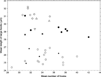
Figure 1 shows a scatterplot of blade length of large rostellar hooks, measured in micrometres, and hook number, from a variety of sources. As can be seen, in terms of blade length the pig isolates from Mexico and Poland clearly group together, and are quite distinct from isolates of sheep and Australian pig origin, both of which were characterized as the sheep strain (data not shown). Hook number was shown not to be a reliable character for ‘strain’ characterization.
Scatterplot of blade length of large rostellar hooks, measured in micrometres. The mean lengths for individual isolates in pigs are from this study; the means of individual isolates from 14 Australian mainland sheep, 2 UK horses, and 1 Egyptian camel are from unpublished data of the Hobbs et al. (Reference Hobbs, Lymbery and Thompson1990) study; the overall mean of 7 horse isolates is from Kumaratilake et al. (Reference Kumaratilake, Thompson and Eckert1986) of 6 elk isolates is from Thompsan et al. (2006), and of 21 camel isolates is from Eckert et al. (Reference Eckert, Thompson, Michael, Kumaratilake and El-Sawah1989); overall means of 29 camel strain isolates, and 78 sheep strain isolates from Iran are derived from both published and unpublished data from Harandi et al. (Reference Harandi, Hobbs, Adams, Mobedi, Morgan-Ryan and Thompson2002). Sheep, ○; horse, Δ; camel,  ; elk, ⋄; Mexican pig, ●; Polish pig,
 ; Australian pig, +.

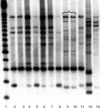
RFLP of rDNA ITS1 using four restriction enzymes showed identical banding patterns for Mexican pig and Polish pig isolates of E. granulosus (data not shown). These results were complemented by RAPD profiles obtained with the P01 primer (fig. 2) which also demonstrated that the Polish and Mexican pig isolates are identical and distinct from a range of other isolates of E. granulosus.
RAPD banding pattern using P01. Lanes: 1, Gibco 100 bp marker; 2 and 3, Australian sheep; 4 and 5, Iranian sheep; 6, Iranian goat; 7 and 8, Australian macropod; 9, Polish pig; 10 Mexican pig; 11, Australian pig; 12, UK horse; 13, USA moose.

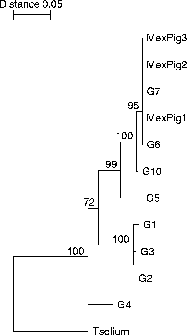
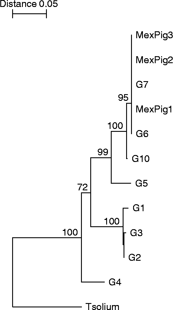
Figure 3 shows the neighbour-joining tree based on the alignment of the COI partial sequence. The phylogenetic analysis demonstrates that the Mexican and Polish pig isolates form a cluster together with the reference G7 pig isolate and G6 (camel strain).
Phylogenetic tree obtained for Echinococcus granulosus genotypes/isolates sequenced in the present study and by other authors at the COI locus. Numbers represent bootstrap support. G1, sheep strain; G2, Tasmanian sheep strain; G3, buffalo strain; G4, horse strain; G5, cattle strain; G6, camel strain; G7, pig strain; G10, cervid strain; MexPig, Mexican pig; Tsolium, Taenia solium. Sequence information for the COI locus of the E. granulosus genotypes was obtained from GenBank for comparison with the sequence data from our samples. Accession numbers for the COI sequences are as follows: G1 (AF297617); G2 (M84662); G3 (M84663); G4 (M84664); G5 (M84665); G6 (M84666); G7 (M84667); G10 (AF525457) and Taenia solium (AB086256).
Discussion
Our results support earlier studies suggesting that Echinococcus of pig origin is phenotypically and genetically distinct to Echinococcus maintained in other domestic host assemblages.
Samples of E. granulosus from pigs from Mexico were genetically identical to E. granulosus from Polish pigs and distinct from other major genotypes on the basis of both molecular and morphological characterization. These results strengthen the view that Echinococcus maintained in a cycle involving pigs and dogs is a distinct strain that is conserved genetically in different geographical areas. Maravilla et al. (Reference Maravilla, Thompson, Palacios-Ruiz, Estcourt, Ramirez-Solis, Mondragon-de-la-Pena, Moreno-Miller, Cardenas-Mejia, Mata-Miranda, Aguirre-Alcantara, Bonilla-Rodriguez and Flisser2004) recently characterized a single isolate of Echinococcus from a pig in Mexico and demonstrated identity at the COI locus to the pig strain of Echinococcus. The similarities between Echinococcus from Australian pigs and sheep has been discussed previously and demonstrates the role of the pig as an accidental host in regions where the sheep strain of E. granulosus predominates (Hobbs et al., Reference Hobbs, Lymbery and Thompson1990).
The recent application of molecular tools has helped to resolve many of the taxonomic issues concerning the status of species and strains in the genus Echinococcus, and the current situation has been reviewed extensively (Thompson & McManus, Reference Thompson, McManus, Eckert, Gemmell, Meslin and Pawlowski2001, Reference Thompson and McManus2002; McManus & Thompson, Reference McManus and Thompson2003). The present understanding of the status of Echinococcus species is a series of largely host-adapted species that are maintained in distinct cycles of transmission characterized by the principal intermediate hosts involved (Thompson, 2001; Thompson & McManus, Reference Thompson and McManus2002). The most widely distributed species is E. granulosus, which exists as a series of genetically distinct strains/genotypes, some of which are likely to warrant species status in the future, particularly those in pigs, camels and cervids (Harandi et al., Reference Harandi, Hobbs, Adams, Mobedi, Morgan-Ryan and Thompson2002; Thompson & McManus, Reference Thompson and McManus2002; Lavikainen et al., Reference Lavikainen, Lehtinen, Meri, Hirvelä-Koski and Meri2003; Obwaller et al., Reference Obwaller, Schneider, Walochnik, Gollackner, Deutz, Janitschke, Aspöck and Auer2004; Thompson et al., Reference Thompson, Boxell, Ralston, Constantine, Hobbs, Shury and Olson2006).
The present study has confirmed the occurrence of the pig strain outside Europe, and again raises the question of its taxonomic status. Our results and those of other workers support the close relationship of the cervid, camel and pig strains, which is also complemented by the morphological similarities of their adult, strobilar morphology (Thompson & Lymbery, Reference Thompson and Lymbery1988). Apart from hook dimensions, the characteristically long terminal proglottid seen by Sweatman & Williams (1963) in their worms of cervid origin is a feature shared by Echinococcus of cattle origin (E. ortleppi) as well as the camel and pig strains, which are all closely grouped genetically. It has been suggested that all three strains may belong to a single species (Thompson et al., 1995; Thompson & McManus, Reference Thompson, McManus, Eckert, Gemmell, Meslin and Pawlowski2001; Xiao et al., 2005). However, this may be simplifying the situation, particularly in view of recent comprehensive investigations on Echinococcus in cervids which support this largely sylvatic form as a distinct species (Lavikainen et al., Reference Lavikainen, Lehtinen, Meri, Hirvelä-Koski and Meri2003, Reference Lavikainen, Lehtinen, Laakonen, Agren, Oksanen and Meri2006; Thompson et al., Reference Thompson, Boxell, Ralston, Constantine, Hobbs, Shury and Olson2006). Consequently, it may be more realistic to consider the domestic pig and camel forms of Echinococcus as a single species (E. intermedius, as originally proposed by Lopez-Neyra & Soler Planas in 1943), or possibly as two distinct subspecies since they do not appear to occur sympatrically. This is because humans appear to be more susceptible to infection with the camel strain than they are to the pig strain (Thompson & McManus, Reference Thompson, McManus, Eckert, Gemmell, Meslin and Pawlowski2001). However, further studies are required to determine whether this reflects a parasite difference or differences in epidemiology and risk factors for human infection in endemic areas. Zacatecas state in north-central Mexico represents a well-known, localized focus for the pig–dog cycle of Echinococcus, and sanitary inspection in slaughterhouses is inadequate. However, this situation does not appear to have resulted in a risk to the human population of contracting cystic echinococcosis. Clearly, further epidemiological studies are required.