Introduction
Wake-up stroke (WUS) is characterized by an ischemic stroke, which occurs during sleep and accounts for between 14% and 29.6% of all ischemic strokes.Reference Fink, Kumar and Horkan1–Reference Moradiya and Janjua6 Acutely, the occurrence of a WUS results in a treatment dilemma as the onset of neurologic symptoms is considered to be the time of sleep onset, which is beyond 4.5 hours for intravenous (IV) tissue plasminogen activator (t-PA). Thus, historically WUS has been considered a contraindication for IV t-PA in the setting of acute ischemic stroke. Fortunately, contemporary research aims to assess the safety and efficacy of administering IV t-PA in patients with WUS using information from neuroimaging to estimate time of stroke onset. A potentially important risk factor for WUS is obstructive sleep apnea (OSA). Therefore, testing for OSA should take place in WUS patients and treatment of OSA in the acute and post-acute WUS settings should be considered.
The purpose of this review is to review the risk factors and potential mechanisms of WUS and subsequently describe treatments, which may be offered to those with WUS that might include treatments for OSA and thrombolytic therapy for acute ischemic stroke. This review will have particular emphasis on OSA and its role in propagating WUS, ongoing challenges in the acute treatment of WUS, and finally studies that have been completed to assess safety and efficacy of IV t-PA in patients with WUS.
Methods
The authors used PubMed (1966 to March 2018) to search for the Term “Wake-Up Stroke” cross-referenced with “pathophysiology,” “pathogenesis,” “pathology,” “magnetic resonance imaging,” “obstructive sleep apnea,” or “treatment.” These search parameters we used came up with 806 papers. The search was then narrowed down by screening the titles and abstracts to include the relevant papers on WUS, which included randomized controlled trials, original research on WUS pathophysiology, and assessment of WUS with neuroimaging. English language papers were reviewed and studies that focused only on acute Strokes with known-onset of symptoms were not reviewed. Also reviewed were pertinent papers from the Reference list of the above-matched manuscripts. Our review included assessment of the full text of 273 papers.
Risk Factors for WUS
Generally, ischemic stroke is most likely to occur in the early morning.Reference Tsai and Albers5 A meta-analysis of 31 studies of 11,816 stroke patients with known times of symptom onset found a circadian pattern of stroke onset with 55% of ischemic strokes occurring between 12:00AM and 6:00AM.Reference Elliott7 The increased risk of ischemic stroke in the early morning is attributable to many physiologic and disease factors. Much of this work follows the cardiology literature, as it is well known that myocardial infarction (MI) is more likely to occur in the early morning hours with about 40% of MI occurring in the morning.
In the morning, as persons begin to awaken and REM sleep becomes predominant, there is increased sympathetic nervous activity, Reference Somers, Dyken, Mark and Abboud8 which in turn stimulates the Renin-Angiotensin-Aldosterone system leading to elevations in blood pressure and heart rate. Further increasing the risk of WUS in the early morning is increased platelet aggregation and elevated levels of prothrombotic factors in the early morning.Reference Andrews, Gralnick, Merryman, Vail and Quyyumi9–Reference Wouters, Lemmens, Dupont and Thijs12 Platelet aggregation increases by as much as 27%–71% in the morning, which in part can be explained by increases in whole-blood platelet count and hematocrit as well as a doubling to tripling of catecholamines levels in the early morning.Reference Andrews, Gralnick, Merryman, Vail and Quyyumi9 These physiologic changes, which occur in sleep and in the early morning in normal individuals may explain at least in part, why ischemic stroke has a tendency to occur during sleep as platelet aggregation favors the formation of clots, specifically in the early morning. Physiologic changes favoring stroke during sleep may be enhanced in those with stroke risk factors. In hypertensive elderly patients, there are significant elevations in von Willebrand factor (vWF) early in the morning that may contribute to enhanced morning platelet aggregation.Reference Kario, Yano, Matsuo, Hoshide, Asada and Shimada10, Reference Goto, Sakai and Goto13, Reference Ikeda, Handa and Kawano14 Atrial fibrillation may also be associated with WUS, as the odds of a new atrial fibrillation diagnosis is nearly four-fold higher in patients with WUS compared to those having stroke in wakefulness (non-WUS).Reference Riccio, Klein and Pagani Cassara15, Reference Kim, Kim, Kwon, Kim and Kang16
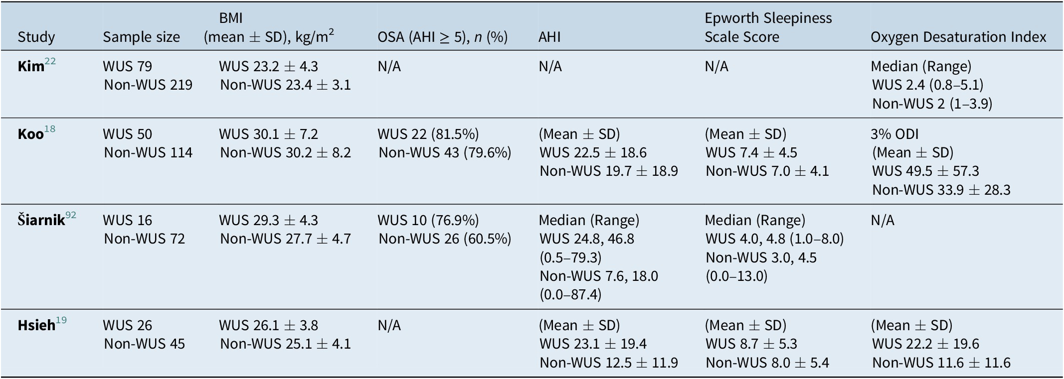
Disordered sleep may further predispose to WUS. There is some evidence that WUS is associated with severe obstructive sleep apnea (OSA) in men. Analysis of the Sleep Heart Health Study data of over 5,000 community-dwelling individuals showed that severe OSA with at least 20 apneas or hypopneas per hour of sleep is associated with an almost 3-fold ischemic stroke incidence over 8 years in men.Reference Redline, Yenokyan and Gottlieb17 No such increased stroke incidence was found in women. Consistent with this gender differentiation, another study showed that among 54 men with ischemic stroke who underwent polysomnography, nearly half with WUS compared to under 20% with non-WUS had severe OSA and frequent oxygen desaturation. Still in this study, the majority of both those with WUS and without WUS had OSA as defined by an apnea-hypopnea index (AHI) of ≥ 5, 81.5% with WUS versus 79.6% with non-WUS. The average AHI in WUS patients was reported to be 22.5 (26.9 in men, 9.8 in women), while the mean AHI in patients with non-WUS was 19.7 (17.9 in men, 22.8 in women).Reference Koo, Bravata and Tobias18 On the other hand, Hsieh et al. defined severe sleep-disordered breathing (SDB) as AHI ≥ 30, which was more prevalent in WUS patients compared to non-WUS patients (38.5% of WUS patients, 8.9% of non-WUS patients).Reference Hsieh, Lai, Liu, Hsieh and Hsu19 In OSA, increased cerebral blood flow occurs in the immediate post-apnea period and after about 15 seconds an approximately 20% decrease in cerebral blood flow occurs. At the same time oxygen desaturation following an apnea decreases partial pressure of oxygen in this decreased amount of blood going to the brain. The end result may be an ischemic stroke.Reference Zirak, Gregori-Pla and Blanco20 Additionally, intermittent hypoxia in OSA is also associated with blood hypercoagulability as both blood viscosity and platelet aggregation increase, while fibrinolytic activity decreases.Reference Bradley and Floras21 Indeed, other studies have shown that frequent nocturnal oxygen desaturation is more frequent among those with WUS compared to non-WUS. A study in a South Korean stroke unit showed that nearly 30% of persons with WUS compared to just 12% of those with non-WUS had frequent nocturnal desaturation.Reference Kim, Ko and Jeong22 Patients with WUS appear to have similar age and gender distribution as those with non-WUS.Reference Moradiya and Janjua6
Mechanisms of WUS
Analysis of International Stroke Trial data of over 17,000 acute ischemic strokes showed that WUS was more likely to occur by a lacunar mechanism and less likely to occur through a large vessel anterior circulation mechanism.Reference Moradiya and Janjua6 Our data also showed that WUS was more likely to occur through a small vessel mechanism, as 42.9% of 28 persons with WUS and just 14.0% of 44 persons with non-WUS had stroke occurring by small vessel mechanism.Reference Tanimoto , Mehndiratta and Koo23 In two independent stroke cohorts, we also found that persons with WUS had significantly higher low-density lipoprotein (LDL) than persons with non-WUS with large differences in each study of about 20 mg/dL.Reference Koo, Bravata and Tobias18, Reference Tanimoto , Mehndiratta and Koo23 In addition, to diabetes and hypertension, hyperlipidemia and specifically high LDL cholesterol is an independent risk factor for lacunar stroke.Reference Bezerra, Sharrett and Matsushita24 Patients with blockages of small arteries may be especially vulnerable to WUS in the setting of OSA, when near ischemic brain regions are further challenged with the severe hypoxia of OSA.
Ozdemir et al. reported in a case series that the presence of patent foramen ovale might increase the risk of cryptogenic strokes on awakening due to paradoxical emboli leading to systemic embolism.Reference Ozdemir , Beletsky, Hachinski and Spence25 Patients with severe OSA are about 3 times more likely to have a large patent foramen ovale than persons without OSA.Reference Shaikh, Jaye and Ward26 During an obstructive apnea, changes in intrathoracic pressure result in a transient increase in right-sided heart pressure, predisposing to paradoxical embolism.Reference Beelke, Angeli and Del Sette27 Additionally, persons with WUS are more likely than those with non-WUS to have right-to-left shunting.Reference Ciccone, Proserpio, Roccatagliata, Nichelatti, Gigli and Parati28
Neuroimaging in WUS
In WUS, the exact time of stroke onset is unknown. Patient with WUS presenting to an acute care facility beyond 4.5 hours of sleep onset are frequently considered ineligible for IV thrombolytic therapy, even though the time of onset may in fact have been within 4.5 hours.Reference Jauch, Saver and Adams29–32 Because of the unknown time of stroke onset in WUS, many studies have targeted neuroimaging as a way to approximate time of onset and potentially select patients for thrombolytic treatment.Reference Tsai and Albers5
Computed Tomography in WUS
Computed Tomography (CT) is uniformly used in clinical practice to exclude cerebral hemorrhage in patients with acute neurological symptoms, and also to guide the use of thrombolytic therapy in patients with hyperacute ischemic stroke.Reference Adams, Adams and Brott33, Reference Tomura, Uemura, Inugami, Fujita, Higano and Shishido34 Early changes seen on CT result from cerebral ischemia affecting water diffusion regulation in brain tissue leading to cytotoxic edema within a few minutes of an ischemic insult.Reference Alegiani, MacLean and Braass35 This edema occurs due to increased microvascular permeability, as the ischemic insult causes significant disruption of microvascular barriers.Reference Zoppo, Kummer and Hamann36 A few CT-based studies reported similarity in early ischemic changes observed in both WUS and non-WUS.Reference Huisa, Raman and Ernstrom37–Reference Todo, Moriwaki, Saito, Tanaka, Oe and Naritomi40 Roveri et al. compared early ischemic changes on CT imaging in WUS and non-WUS patients with similar clinical and demographic characteristics. There was a comparable degree of early ischemic change as evidenced by similar amounts of parenchymal hypoattenuation, blurring of the gray-white junction, and swelling as judged by the Alberta Stroke Program Early CT Score (ASPECTS), in the baseline CT scan in both groups. Follow-up CT scans in both groups also showed similarity in the extent of infarction. Based on these findings, it was presumed that the onset of WUS was shortly before, or even upon awakening.Reference Roveri, La Gioia, Ghidinelli, Anzalone, De Filippis and Comi38
The ability of CT-perfusion (CTP) to detect the extent of the ischemic penumbra following an ischemic event was evaluated in a few studies. For this purpose, different CTP parameters were used including cerebral blood flow (CBF), cerebral blood volume (CBV), mean transit time (MTT) and time-to-peak (TTP). CBV is the amount of blood present per brain tissue volume, while MTT is the average time the red blood cells spend in the capillary circulation between the arterial inflow and the venous outflow. TTP is defined as the time taken by the contrast material to achieve the best possible enhancement in the area of interest.Reference Khandelwal41 If the CBF is decreased, as in an ischemic event, but the CBV is stable or even increased, this indicates reversible ischemia. In contrast, if both CBF and CBV are decreased, this is considered a sign of irreversible ischemia.Reference Tomandl, Klotz and Handschu42, Reference Srinivasan, Goyal, Al Azri and Lum43 Using these parameters, Wintermark et al. stated that CTP could be used to draw the line between the area of ischemic infarction and the salvageable penumbra.Reference Wintermark, Flanders and Velthuis44 Campbell et al. used a different CTP approach in which they stated that the relative CBF is the optimal parameter in appreciating the size of the infarcted core of the ischemic tissue.Reference Campbell, Christensen and Levi45
Promising results were obtained in EXTEND, a multicentric randomized clinical trial, which randomized both WUS patients (if within 9 hours from the midpoint of sleep) and patients presenting 4.5–9.0 hours after the last known well time to IV alteplase or placebo. The investigators used both CTP imaging and perfusion-diffusion magnetic resonance imaging (MRI) techniques to determine the extension of the hypoperfused, yet salvageable brain areas. A favorable outcome was defined as a modified Rankin score (mRS) of 0–1, which was reported in 40 patients (35.4%) in the alteplase group versus 33 patients (29.5%) in the placebo group (adjusted risk ratio, 1.44; 95% CI 1.01–2.06; p = 0.04). However, symptomatic intracerebral hemorrhage occurred in 7 patients (6.2%) in the alteplase group versus 1 patient (0.9%) in the placebo group (adjusted risk ratio, 7.22; 95% CI 0.97–53.5; p = 0.05).Reference Ma, Campbell and Parsons46 Furthermore, a meta-analysis was conducted looking at the pooled data from EXTEND, Reference Ma, Campbell and Parsons46 ECASS-4: EXTENDReference Ringleb, Bendszus, Bluhmki, Donnan, Eschenfelder and Fatar47 and EPITHETReference Davis, Donnan and Parsons48 (EXTEND trial used CTP and MRI to judge the onset of stroke, ECASS-4: EXTEND and EPITHET trials used MRI only) to evaluate the efficacy of IV alteplase in acute ischemic stroke when used beyond the 4.5-hour window. Excellent functional outcome, defined as mRS of 0–1 at 90 days, occurred in 36% of the alteplase group and 29% of the placebo group, while functional independence, defined as mRS of 0–2 at 90 days, occurred in 49% of the alteplase group and 44% of the placebo group. Adverse events of symptomatic intracerebral hemorrhage occurred more in the alteplase group (5% alteplase group and <1% placebo group, odds ratio 9.7; 95% CI 1.23–76.55, p = 0.031).Reference Campbell, Ma and Ringleb49
Currently, TWIST is an ongoing open-label randomized controlled clinical trial that evaluates the efficacy of tenecteplase in patients presenting with WUS.50 Tenecteplase is a genetically modified alteplase molecule, which has higher specificity to fibrin, a longer half-life, and more rapid onset of action than alteplase.Reference Tsikouris and Tsikouris51, Reference Dunn and Goa52 This study is also assessing the ability of plain CT and CT angiography in recognizing WUS patients who are eligible to benefit from thrombolytic treatment with tenecteplase.50
As compared to CT, the use of MRI in ischemic strokes is more complicated and takes longer time to be done.Reference Vymazal, Rulseh, Keller and Janouskova53 Despite the broader availability of CT imaging as compared to MRI, the ability of CT to detect early ischemic changes in the brain is limited as the sensitivity of CT in recognizing early ischemic changes depends on the duration and severity of focal cerebral ischemia.Reference Kummer, Nolte, Schnittger, Thron and Ringelstein54 Another disadvantage of CT imaging carries the risk of radiational exposure.Reference Vymazal, Rulseh, Keller and Janouskova53
Magnetic Resonance Imaging (MRI) in WUS
Due to the limitations of CT in estimating stroke onset and evaluating the degree of damage, MRI has been used in different studies to perform these tasks.Reference Chalela, Kidwell and Nentwich55–Reference Schellinger, Thomalla and Fiehler57 MRI has the advantage of better tissue delineation than CT and facilitates the characterization of tissue areas at risk of infarction in the immediate time after an ischemic event (ischemic penumbra).Reference Albers, Thijs and Wechsler58–Reference Thomalla, Schwark and Sobesky60 Two MRI-based mismatch principles are currently followed to detect an ischemic penumbra: (a) perfusion weighted imaging (PWI)–diffusion-weighted imaging (DWI) mismatch and (b) DWI–fluid attenuated inversion recovery (FLAIR) mismatch.Reference Kurz, Advani, Behzadi, Eldoen, Farbu and Kurz61
PWI–DWI mismatch is based on the sequence of events taking place in the brain directly following an ischemic insult. In the few minutes after vessel occlusion, loss of blood supply causes irreversible damage to a core of infarcted tissue. There is also an ischemic penumbra surrounding this infarcted core, which is at risk for infarction but can still be salvaged.Reference Bang62 The infarction core is characterized by impaired diffusion of water molecules resulting from cytotoxic injury, edema and depleted cellular energy due to hypoperfusion, and this diffusion defect can be detected by DWI.Reference Bang62 On the other hand, PWI provides information about microcirculation in the capillary network.Reference Kim, Kang and Kim63, Reference Tatlisumak, Strbian, Abo Ramadan and Li64 Thus, it can capture the hemodynamic changes taking place in the ischemic tissue and the surrounding brain regions.Reference Neumann-Haefelin , Wittsack and Wenserski65
PWI-DWI mismatch refers to the difference between the area of infarcted brain (diffusion defect) and the area of the brain where perfusion is compromised (perfusion defect).Reference Jovin, Saver and Ribo66, Reference Lansberg, Straka and Kemp67 Heiss et al. stated that MRI-based PWI-DWI mismatch could be used to differentiate the area with irreversible damage from the surrounding area with reversible hypofunctionality with a comparable reliability to positron-emission tomography (PET).Reference Heiss and Sobesky68 Therefore, patients with a PWI-DWI mismatch are thought to potentially benefit from thrombolytic therapy in acute stroke settings. Fink et al. reported a similarity of PWI-DWI mismatch within 3 hours in both WUS and non-WUS. PWI-DWI mismatch was found in 82% of known-onset stroke patients and 73% of WUS patients.Reference Fink, Kumar and Horkan1 The same mismatch concept was used to guide mechanical and chemical thrombolytic therapy in a case report of two WUS patients, both of whom had severe deficits at onset, yet favorable ultimate outcomes, one being completely free of symptoms and the other having only mild language deficit.Reference Iosif, Oppenheim, Trystram, Domigo and Meder69
Several studies looked at the reliability of PWI–DWI mismatch in combination with DWI–FLAIR mismatch in identifying eligible patients with unknown-onset strokes to guide thrombolytic therapy.Reference Breuer, Schellinger and Huttner70–Reference Kang, Sohn and Hong73 Like DWI, MRI FLAIR sequencing leverages abnormalities in the properties of water to detect changes in cerebral tissue exposed to ischemia. DWI detects defects in diffusion of water within a few minutes after the ischemic event, while FLAIR detects cerebral edema over 3-6 hours. Based on these principles, the presence of DWI defect, without a matching FLAIR lesion, indicates that the ischemic infarction is of a recent onset, most likely < 4.5 hours.Reference Rubin and Barrett39, Reference Aoki, Kimura, Iguchi, Shibazaki, Sakai and Iwanaga74–Reference Thomalla, Rossbach and Rosenkranz76
Using these imaging concepts, Petkova et al. retrospectively studied FLAIR and DWI records of 130 patients with acute stroke of known onset. Sixty-three patients had their imaging done within the first 3 hours after the onset of stroke symptoms, while the remaining 67 patients underwent imaging more than 3 hours after the symptom onset. The sensitivity and specificity of DWI–FLAIR mismatch in identifying strokes, which occurred within 0–3 hours were 90% and 93%, respectively.Reference Petkova, Rodrigo and Lamy77 Other studies have shown that DWI-FLAIR mismatch has 62% sensitivity and 78% specificity in identifying stroke patients within 4.5 hours of symptom onset.
Several investigators have used this concept of DWI–FLAIR mismatch to guide thrombolytic therapy in WUS.Reference Aoki, Kimura and Iguchi78–Reference Mourand, Milhaud and Arquizan80 Outcomes in these patients in general were favorable and indicated that persons with WUS and DWI–FLAIR mismatch without contraindication to thrombolytic therapy may indeed benefit from this therapy without having excess risk of symptomatic hemorrhage.
MRI-Guided Clinical Trials
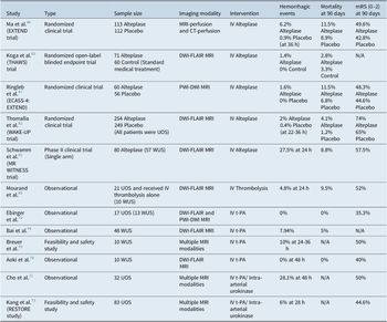
Recognizing the knowledge gap in acute therapy for WUS, investigators have carried out clinical trials to guide IV t-PA therapy in strokes with unknown symptom onset including WUS. Table 2 outlines details of these trials which are also commented on in the below section.
Studies assessing correlation between OSA and WUS.

BMI indicates body mass index.
Trials and observational studies assessing efficacy of MRI-guided thrombolytic therapy in WUS, Unknown-onset stroke (UOS) and/or 4.5–9 hours post stroke onset.

Sample size refers to UOS patients who underwent MRI/CT scanning and received IV thrombolysis. mRs indicates modified Rankin scale.
N/A, not applicable; WUS, wake-up strokes.
The MR WITNESS trial is a single arm safety trial, which included 183 patients who had acute ischemic stroke with unwitnessed symptom onset and were last known to be well 4.5–24 hours earlier. Safety outcomes included the primary outcome of symptomatic hemorrhage and a secondary outcome of brain edema risk. DWI–FLAIR mismatch was used to guide potential therapy. Patients were excluded if there was severe stroke with NIH Stroke Scale of > 25, a large infarction demonstrated on DWI (>100 cm3), or more than minimal FLAIR hyperintensity (> 1.15 signal intensity ratio). Eighty patients, including 57 WUS patients received IV t-PA. Symptomatic hemorrhage occurred in only one patient, and symptomatic cerebral edema occurred in only three subjects, both rates not different from symptomatic hemorrhage and edema rates observed in the ECASS-3 study. Ultimately, the trial successfully demonstrated that IV t-PA could safely be administered to acute ischemic stroke patients with unwitnessed symptom onset and last known well of 4.5–24 hours when quantitative DWI–FLAIR mismatch was present.Reference Schwamm, Wu and Song81
The MRI-Based Thrombolysis in Wake-Up Stroke (WAKE-UP) trial is a multicenter, randomized, double blinded, placebo-controlled clinical trial that involved patients with strokes of unknown onset, predominantly (about 90%) those patients with WUS. Eligible patients had ischemic stroke with unknown onset, a last known well time of greater than 4.5 hours, and DWI-FLAIR mismatch showing acute stroke on DWI with no hyperintensity shown on FLAIR imaging. Two hundred and fifty-four patients were randomized to receive IV t-PA and 249 patients were randomized to receive placebo. The alteplase group were more likely than the placebo group to have a favorable outcome (modified Rankin Scale of 0 or 1), 53.3% versus 41.8% (adjusted odds ratio of favorable outcome of 1.61; 95% CI 1.09–2.36 with a p = 0.02. Deaths (odds ratio 3.38; 95% CI 0.92–12.52, p = 0.07) and symptomatic hemorrhage (odds ratio 4.95; 95% CI 0.57–42.87; p = 0.15) occurred more commonly in the alteplase group, but not significantly so. The investigators concluded that IV t-PA is associated with favorable clinical 90-day outcome in patients with unknown-onset strokes when administration is guided by DWI-FLAIR mismatch. Unfortunately, the study had to be terminated early due to discontinuation of funding, thus limiting the interpretation of safety results which showed trends toward increases in symptomatic hemorrhage and mortality in the alteplase compared to placebo group.Reference Thomalla, Simonsen and Boutitie82
ECASS-4: EXTEND is a double-blinded randomized placebo-controlled clinical trial that used both PWI and DWI techniques to determine the presence of salvageable ischemic brain tissues in patients who presented 4.5–9 hours after the onset of the stroke or those who had the stroke symptoms upon waking up. Patients were randomized to either IV alteplase or placebo and the mRS distribution at 90 days did not show a significant difference between the two groups (odds ratio 1.20; 95% CI 0.63–2.27, p = 0.58). Also, the mortality rate at 90 days was not remarkably different between the two groups (11.5% alteplase and 6.8 placebo, p = 0.53) and single event of symptomatic intracerebral hemorrhage occurred in the alteplase group. However, it should not escape from notice that this trial has been terminated prematurely with a limited sample size of 119 patients (61 alteplase and 58 placebo).Reference Ringleb, Bendszus, Bluhmki, Donnan, Eschenfelder and Fatar47
The THAWS trial is a randomized controlled trial, which, similar to the WAKE-UP trial, used MRI-based thrombolysis in Japanese patients with stroke of unknown onset, and compared its efficacy to standard treatment.Reference Koga, Toyoda and Kimura83 The alteplase and standard care groups had similar rates for favorable outcome, defined as mRS of 0–1 at 90 days (relative risk 0.97; 95% CI 0.68–1.41, p = 0.892). However, it should be noted that there was comparable safety of alteplase use in stroke with unknown onset to that of standard care.Reference Koga, Yamamoto, Inoue, Asakura, Aoki and Kanzawa84
Therapy for OSA in WUS
There is a recognized high prevalence of OSA in ischemic stroke between 60% and 80%.Reference Johnson and Johnson85 Evidence further suggests that severe OSA and severe hypoxemia occur with increased frequency in patients with WUS compared non-WUS.Reference Koo, Bravata and Tobias18, Reference Kim, Ko and Jeong22 In WUS, OSA may be directly related to the mechanism of stroke and it may be particularly important in these patients to conduct polysomnography to rule out OSA. Furthermore, severe hypoxemia in OSA can only be detrimental in the setting of a new stroke and may in part account for neurologic worsening and poor functional and cognitive outcomes, which are known to occur at higher rates when OSA co-occurs with stroke.Reference Iranzo, Santamaria, Berenguer, Sanchez and Chamorro86–Reference Aaronson, Bennekom and Hofman88 Aaronson et al. reported in the results of their case-control study that stroke patients with OSA have a significant impairment of their attention, executive functions, visuoperception, psychomotor abilities, and intellectual functions when compared to stroke patients who did not have OSA.Reference Aaronson, Bennekom and Hofman88 Based on this, a randomized controlled trial evaluated the functional and cognitive outcomes of CPAP use for 4 weeks in stroke patients and compared these outcomes to those in stroke patients who received rehabilitation treatment but not CPAP. Taking into consideration that this trial did not include patients with OSA, the CPAP use in stroke patients was associated with a better cognitive outcome in attention and executive functioning.Reference Aaronson, Hofman and Bennekom89 For these reasons it is important to conduct polysomnography in all patients with ischemic stroke but perhaps particularly in those with WUS. Recent data shows that in persons with ischemic stroke and OSA, continuous positive airway pressure (CPAP) when used compliantly is associated with improvement in the one year modified Rankin Scale of nearly one point.Reference Bravata, Sico and Vaz Fragoso90 These data are for patients with ischemic stroke regardless of stroke timing. In WUS, treatment of OSA may be especially beneficial if the mechanism of stroke directly involved OSA.
The SAVE trial is an open-label randomized controlled trial comparing the use of CPAP combined with usual care to usual care alone in patients with moderate to severe OSA and either coronary artery disease or a cerebrovascular disease over a span of 3.7 years. The primary outcome of the study was death from cardiovascular events, stroke, MI or hospitalization due to unstable angina, heart failure or transient ischemic attacks. CPAP use did not decrease the risk of cardiovascular or cerebrovascular events including stroke (hazard ratio with CPAP 1.10, 95% CI 0.91–1.32, p = 0.34). The effectiveness of CPAP in affecting outcomes could have been reduced by the overall low CPAP usage in the intervention group of 3.3 hours per night.Reference McEvoy, Antic and Heeley91
Conclusion
WUS represents a commonly occurring treatment challenge in neurologic practice, as patients are often deemed ineligible for thrombolytic intervention since the onset of the ischemic event is unknown. However, recent studies demonstrate that MRI-based mismatch concepts; PWI–DWI and DWI–FLAIR, can be used to estimate the time of symptom onset and help guide thrombolytic therapy in eligible patients. Preliminary data suggests that appropriate patients with WUS can safely receive thrombolytic therapy when treatment decisions are guided by MRI-based mismatch concepts. However, definitive safety data are still needed. WUS also appears to be associated with severe OSA and severe hypoxemia and data in ischemic stroke in general show that therapy of OSA is associated with improved outcomes. Thus, it is particularly important in WUS patients to assess for OSA and to treat OSA if it is present.
Funding.
No funding was received in connection with this research.
Conflicts of Interest.
All the authors certify that they have no affiliations or involvement with any organization or entity with any financial interest (such as honoraria, educational grants, participation in speakers bureaus, membership, employment, consultancies, stock ownership, or other equity interest; and expert testimony or patent-licensing arrangements), or nonfinancial interest (such as personal or professional relationships, affiliations, knowledge or beliefs) in the subject matter or materials discussed in this manuscript.
Ethical Approval.
All procedures performed in studies involving human participants were in accordance with the ethical standards of the institutional review board and/or the national research committee and with the 1964 Helsinki Declaration and its later amendments, or comparable ethical standards.
Informed Consent This article does not contain any studies with human participants or animals performed by any of the authors.
Disclosures.
Mohamed Elfil, Mohamed Eldokmak, Alireza Baratloo, Nada Ahmed, Hardik P. Amin and Brian B. Koo do not have anything to disclose.